Abstract
Objective: To determine whether the presence of opaciication in the paranasal sinuses of children and adolescents without rhinosinusitis implies an increased risk of later development of upper respiratory tract symptoms.
Methods: This was a prospective study of a cohort of patients aged 0 to 18 years who underwent computerized tomography (CT) scans for indications unrelated to rhinosinusitis. Sinus opaciication was evaluated using an opaciication/development ratio score. The patients’ clinical progression was followed up using a questionnaire for 1 month after the scans.
Results: Fifty-six percent (56%) of the 106 patients enrolled in the study had opacity, the majority due to mucosal thickening. Intense opaciication was deined as “suspected” (score ≥ 15) and patients in this subset had a greater risk of developing symptoms during follow-up (odds ratio = 2.74; 95%CI 1.10-6.83) compared to those with no indings or discrete indings.
Conclusions: Intense incidental sinus opacity on CT indicates a risk of future development of a clinical respiratory condition.
J Pediatr (Rio J). 2011;87(5):433-8: Rhinosinusitis, computerized tomography, diagnosis.
O
RiginAlA
RtiCleCopyright © 2011 by Sociedade Brasileira de Pediatria
433
introduction
Symptomatic inlammation of the mucosal lining of the paranasal sinuses (PS), which is known as rhinosinusitis (RS), is highly prevalent and can be classiied as acute, recurrent or chronic.1 Acute RS (ARS) is one of the ive most common
indications for prescribing antibiotics2 and because of this it
is necessary to distinguish it from other conditions in order to avoid unnecessary use of these medications.
Diagnosis of RS is primarily clinical.1 Simple X-rays offer
very low accuracy and nowadays computerized tomography (CT) is the gold standard.3,4 It is unusual to use a CT scan
to diagnose ARS, but its indication for chronic RS cases
(CRS), which often involve obstructions such as nasal polyps and anatomic abnormalities, is well-established. If intraorbital or intracranial complications are suspected then a CT scan or a magnetic resonance image (MRI) with contrast is indicated.1,2,4,5
Mucosal thickening, luid levels and total opaciication of PS are all indings typical of RS,6 but can also be observed
in patients with colds, inluenza, rhinitis and allergic asthma at rates that vary from 33 to 88% of patients.7-11 Even in
people free from any respiratory disease whatsoever, these are often incidental indings.12-18
Clinical progression of incidental tomographic indings
in paranasal sinuses of asymptomatic individuals: cohort study
Severino A. de Araújo-neto,1 emílio C. e. Baracat2
1. Doutor, Faculdade de Ciências Médicas, Universidade Estadual de Campinas (UNICAMP), Campinas, SP, Brazil. Professor adjunto I, Universidade Federal da Paraíba (UFPB) e Faculdade de Ciências Médicas da Paraíba (FCM-PB), João Pessoa, PB, Brazil.
2. Doutor. Professor associado, Departamento de Pediatria, Faculdade de Ciências Médicas, UNICAMP, Campinas, SP, Brazil.
No conflicts of interest declared concerning the publication of this article.
Suggested citation: Araújo-Neto SA, Baracat EC. Clinical progression of incidental tomographic findings in paranasal sinuses of asymptomatic individuals: cohort study. J Pediatr (Rio J). 2011;87(5):433-8.
Figure 1 - Axial computerized tomography slices. A and B show the maxillary sinuses (m); C and D are the head slices that show the ethmoid (e), frontal (f) and sphenoid (esf.) sinuses
Many authors have stated that opacity has no clinical signiicance when there are no symptoms of RS.12,13,19
However, others claim that signiicant abnormalities merit clinical follow-up.12
The objective of this study is to investigate whether sinus abnormalities found in CT scans of asymptomatic children and adolescents are predictive of clinical progression to rhinosinusitis.
Methodology
This was a longitudinal cohort study. Informed consent was obtained from parents or guardians and the project was approved by the Research Ethics Committees at the teaching institutions involved.
Consecutive patients aged 0 to 18 years were recruited at a radiology department after referral for CT scans of the head for reasons other than RS. Patients were excluded if there was a suspicion of RS or clinical status suggestive of RS, if they had CRS or had been diagnosed with ARS less than 2 months previously, if they had suffered a recent head trauma, had had radiotherapy of the head or neck, or if they had cystic ibrosis or gastroesophageal relux disease. Patients were also excluded if their scans did not show the paranasal sinuses in their entirety or if they were lost to clinical follow-up.
A clinical score at admission (S5adm) was calculated for each patient by administering the S5 questionnaire,20 which
grades ive RS signs and symptoms on a scale from 0 to 3 points (head or face pain, daytime coughing, nighttime coughing, obstruction and runny nose). The inal score varies from zero to three and is calculated by summing the sub-scores and dividing by ive. The questionnaire’s authors20 deined S5 scores > 1 as positive for RS and
this was adopted as the cutoff point for excluding patients from the sample.
The technique used for head CT scans was the standard method used at the department, consisting of axial slices parallel to the orbitomeatal line, varying from 2 to 5 mm, at the posterior fossa, and from 5 to 10 mm, in the supratentorial region, depending on the size of the patient. Additionally, the scans performed on the study sample included two additional, more caudal, slices at the level of the maxillary sinuses (Figure 1), at an angle that did not bisect the plane of the eyes. Other sinuses were already covered by the slices for the head scan. The following helical CT machines were used: Tomoscan SR-4000 (Philips, Eindhoven, Holland), Helicat Flash (Elscint Company, Israel) and X-Vision (Toshiba, Tokyo, Japan). The images were saved as bitmap iles with levels of 0 to 400 HU and widths of 1,000 to 2,000 HU.
The CT scans were interpreted independently by two different radiologists with a minimum of 4 years’ experience
in the specialty, blind to the S5 scores. They indicated their opinions of the following items for each sinus: a) presence of opaciication; b) degree of opaciication; and c) type of opaciication (thickening, cyst/polyp, total or luid level opaciication). Disagreements were resolved by consensus.
The intensity of opaciication was quantiied by the opaciication/development ratio (ODR), which has been validated previously.21 Each sinus was scored as follows
for the opaciication component: a) 0 (zero) if normal or not yet developed; b) 1 (one) if < 2/3 of the area is opaque; c) 2 (two): if ≥ 2/3 of the area is opaque; and d) 3 (three) if opaciication is total. The opaciication score is the numerator of the ODR. For the development component, sinuses that are present score 3 (three) and absent sinuses score 0 (zero). The sum of the development scores is the denominator of the ODR. The percentage of opacity is therefore calculated as follows:
ODR = (total opaciication/total development) x 100. The result, which is in the range of zero to one, is multiplied by 100 to give an estimate of the percentage of the area of the PS that is opaque.18,21 Patients were
divided into two groups: “low probability” of opaciication (ODR < 15) and “suspected” opaciication (ODR ≥ 15). The cutoff point chosen was that which offered the best accuracy for RS.21
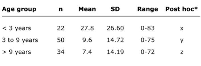
Age group n Mean SD Range Post hoc*
< 3 years 22 27.8 26.60 0-83 x
3 to 9 years 50 9.6 14.72 0-75 y
> 9 years 34 7.4 14.19 0-72 z
table 1 - Means, deviations and ranges of opacification/ development ratio by age group in a sample of patients given facial CT scans
SD = standard deviation. ANOVA: p = 0.003.
* The Dunnett test indicates that the youngest age group (x) is different (p < 0.05) from the other two (y and z), which, in turn, are not different from each other.
classiied as “one sinus affected” vs. “2 or more” vs. “all sinuses affected.”
The clinical follow-up score (S5fol) was recalculated weekly for 4 weeks after the scan by telephone interview using the same S5 questionnaire. The outcome was deined as negative if the patient scored S5fol ≤ 1 and positive if S5fol was greater 1 after any of the interviews.
SPSS version 13.0 was used to calculate statistics to a signiicance level of 5% (p < 0.05). The tests applied were as follows: the Mann-Whitney (MW) test and Kruskal-Wallis ANOVA were used to compare the means of two, or more, independent samples respectively; the Dunnett multiple comparisons test was used to supplement tests of the means of more than three independent samples in order to determine which was responsible for differences; the chi-square test or Fisher’s exact test were used for associations between categorical variables; Pearson’s coeficient and Spearman’s r were used to test for correlations between quantitative variables; odds ratios (OR) were used to study the cohort and signiicance was deined as when the 95% conidence interval (95%CI) does not pass through 1.
Results
A total of 129 patients were scanned. Twenty-three of these cases were excluded; ive because of imaging artifacts or because not all sinuses were shown and the other 18 because they were lost to follow-up. Fifty-seven (57) of the remaining 106 patients were female (53.8%). Age varied from 5 months to 18 years (mean = 6.8 years; standard deviation [SD] = 4.4 years). The most common indications for CT scans were epilepsy/convulsions (24.7%), delayed neuropsychomotor development (19.8%) and headaches (14.8%).
The maxillary and ethmoid sinuses were developed in all patients. The sphenoid sinus was developed in 77 (72.6%) and the frontal sinus in 33 patients (31.1%). Fifty-nine patients (55.7%) had some type of abnormal inding, most often in the maxillary sinuses (n = 46; 43.4%), followed by the ethmoid (n = 31; 29.2%) sphenoid (14/77; 18.2%), and frontal sinuses (1/33, 3.0%). The ODR scores varied from 0 to 83 (mean = 12.7; SD = 19.2). Seventy point seven percent (n = 75, 70.7%) of the sample had an ODR indicating low probability of opaciication (ODR < 15) and 29.3% (n = 31) had suspected ODR. Mucosal thickening alone was present in 72.9% of cases (n = 43/59). Cysts/ polyps and total opaciication were both present in seven patients (11.9%), and luid levels were observed in two patients (3.4%).
Opaciication was most intense in the under-3 age group, according to mean ODR scores, with a progressive reduction in the succeeding age groups (Table 1). Eighteen of the 69 patients who responded to the question about allergies were positive (26.1%). There were no signiicant
differences between patients with allergies and those free from allergies in terms of mean ODR (p = 0.247).
Anesthesia was required during the CT scan for 41 of the 106 patients (38.7%). Anesthesia use was determined by age, being used for 90.9% of patients less than 3 years old, for 36.0% of the 3-9 year-olds and for 8.8% of the over nines. A comparison of mean ODR scores indicated a signiicant difference between patients who were anesthetized (mean ODR = 18.8) and those who were not anesthetized (mean ODR = 8.8, p = 0.004).
In order to isolate the variables age and anesthesia statistically, the sample was divided into the subset that did not receive anesthesia (n = 65) from the population who did receive anesthesia (n = 41). When the means of the ODR scores were compared, the statistical difference between age groups were maintained in the total sample, but not in the no anesthesia group (p = 0.330 and p = 0.026, respectively).
Mean S5adm was 0.41 (SD = 0.32). There was no correlation between the S5adm (0 to 1) and ODR (0 to 100) scales (Spearman r = 0.077; p = 0.434). There was also no difference in mean S5adm between low probability and suspected ODR (p = 0.467). With regard to age, mean S5adm was greater in the 3 to 9 years group in relation to the other two (≤ 3 years and > 9 years, ANOVA: p = 0.013). Age was also analyzed by splitting patients under ive (n = 49) from those over 5 years old (n = 57), but there was no difference in S5adm (p = 0.629). Mean S5adm was greater among allergic patients (p = 0.032).
negative Positive
outcome outcome total
n % n % n
ODR < 15 57 79.2 15 20.8 72
ODR ≥ 15 18 58.1 13 41.9 31
Total 75 72.8 28 27.2 103
table 2 - Clinical outcome by “suspected” opacification/ development ratio (≥ 15) for patients followed-up for 4 weeks
ODR = opacification/development ratio.
Odds ratio for ODR > 15 against ODR < 15 = 2.74 (95%CI 1.10-6.83); chi-square = 4.875; p = 0.027.
Figure 2 - Distribution of percentages of positive outcomes (n = 28) by clinical follow-up week
The odds ratio for the likelihood of a member of the group with suspected ODR (ODR ≥ 15) having a positive outcome was 2.74 with relation to the low probability group (Table 2). There was no increase in the risk of a positive outcome related to opaciication of any speciic sinus, to type of abnormality (luid level/total opaciication vs. polyps/thickening), to extent of involvement (in two categories: one sinus vs. two or more sinuses) or related to asymmetrical involvement.
Discussion
Sinus opaciication is often detected in individuals with respiratory conditions other than RS. Kristo et al.7 studied
children with upper airway infections (UAI) using MRI and found opacity in 88%. Gwaltney et al.11 observed an elevated
rate of abnormal CT indings in adults with the common cold (up to 87% of maxillary sinuses). Kovalhuk et al.8 studied
children with asthma and allergic rhinitis using CT and found opaciication in 20%. Abnormal sinus indings may also be purely incidental. Havas et al. studied 666 CT scans of the heads of adults and found abnormalities in 42% of
them13. Sinus opacity was present in 41% of CT scans of
the temporal and orbital bones of patients aged less than 18 years who were studied by Lesserson et al.12 Manning
et al. studied children and adolescents using CT and MRI of the head and found abnormalities in the PS of 55%.19
A recent study by Hill et al.17 reported elevated igures,
with opacity in around 80% of asymptomatic children and adolescents.
Sinus abnormalities were observed in 55.7% of the patients in the present study, which is discretely higher than the majority of studies. In 73% of these cases, mucosal thickening was the only inding, while total opaciication of a cavity and luid level affected a minority. This predominance of discrete opacity in patients who do not have RS is the rule in published literature,12,13,17,19,22 as are the predominance
of opaciication of the maxillary (43%) and ethmoid (29%) sinuses and diffuse and bilateral opaciication that were observed here.13
Patients younger than 3 had signiicantly greater prevalence and intensity of opaciication than the older patients, which has also been shown in earlier studies.16,23
However, the effect of anesthesia on opaciication that was observed here, and has not been mentioned in prior studies, may have introduced bias into the relationship and must be investigated in greater depth in future research.
The indings reported here demand that a certain degree of opaciication be admitted in the radiological deinition of a “normal” sinus. According to Wald,24
radiological diagnostic criteria for RS should be limited to mucosal thickening of at least 4 mm, total opaciication or luid level. The least speciic of these criteria is mucosal thickening.25,26 Bhattacharia and Fried used the Lund
and Mackay score (LMS) for measuring opaciication and established that a cutoff of ≥ 4, which they called “high probability” of RS, offered the greatest diagnostic accuracy.22 An earlier study established the correlation
that LMS ≥ 4 corresponds to ODR ≥ 15 (here deined as “suspected” ODR).21 However, in the sample studied
here, 29% of the patients, none of whom had RS, had a “suspected” ODR score, indicating a relatively high rate of false-positive results using this cutoff point and emphasizing the low speciicity of tomography opaciication for diagnosing RS.
References
1. Rosenfeld MRI, Andes D, Bhattacharyya N, Cheung D, Eisenberg S, Ganiats TG et al. Clinical practice guideline: adult sinusitis.
Otolaryngol Head Neck Surg. 2007;137(3 Suppl):S1-31. 2. American Academy of Pediatrics. Subcommittee on Management
of Sinusitis and Committee on Quality Improvement. Clinical practice guideline: management of sinusitis. Pediatrics. 2001;108:798-808.
3. Zinreich SJ, Kennedy DW, Rosenbaum AE, Gayler BW, Kumar AJ, Stammberger H. Paranasal sinuses: CT imaging requirements for endoscopic surgery. Radiology. 1987;163:769-75.
4. McAlister WH, Parker BR, Kushner DC, Babcock DS, Cohen HL, Gelfand MJ et al. Sinusitis in the pediatric population. American College of Radiology. ACR Appropriateness Criteria. Radiology 2000;215Suppl:811-8.
5. Diretrizes Brasileiras de Rinossinusites. Rev. Bras. Otorrinolaringol. 2008;74(2 suppl.):6-59.
6. Wald ER. Radiographic sinusitis: illusion or delusion? Pediatr Infect Dis J. 1993;12:792-3.
In practice, radiologists should not make a diagnosis of RS without knowledge of the clinical picture. When patients are free from symptoms, opaciication intensity of ODR < 15, LMS of < 4 or mucosal thickening of < 4 mm are very unlikely to be evidence of RS.
In cases where clinical diagnosis is not clear cut and radiological indings of opacity confuse the picture even further, cultures of material collected from the sinus by direct puncture or nasal video endoscopy can deinitively rule out a diagnosis of RS.1,26 However, these procedures
are neither routine nor practical. Even if the opacity proves to be sterile, the suspicion still remains as to whether it is present because of physiological imbalances of drainage or aeration of the cavities and might yet facilitate proliferation of bacteria in the near future. This is the reason why the objective of this study was focused on the question of possible risk of clinical progression and was not restricted to criteria for interpreting what is normal and what is abnormal on a CT scan of the PS.
During clinical follow-up, 27% of the patients developed symptoms (positive outcome). Patients with ODR ≥ 15 (suspected) had a signiicantly higher chance of the outcome (OR = 2.74) when compared with those whose tomography indings were classed as low probability.
Two studies have investigated the behavior of incidental PS indings over time, but they only described imaging status and did not correlate it with patients’ clinical progress. Maly & Sundgren27 conducted a retrospective analysis of adults
who had been scanned twice with MRI for neurological reasons with a minimum interval of 4 months and found that in 90% of the patients the indings were either stable or improved at the second scan. In another study, children with short-duration respiratory symptoms (< 10 days) were scanned with MRI and 43% had positive indings. 2 weeks later a control MRI showed signiicant attenuation of sinus abnormalities, irrespective of whether symptoms had improved.7
As has been seen, allergic patients were at greater risk of the positive outcome, which may mirror exacerbations of their allergic conditions during follow-up. It is improbable that the allergies introduced any bias to the association between suspected ODR and the positive outcome, since no relationship was detected between allergy and ODR scores. Young age (< 5 years) and anesthesia also exhibited a tendency, although without signiicance, towards an increased risk of the positive outcome. The association with young age may be the result of the high incidence of inlammatory respiratory conditions among these children. The explanation for the role of anesthesia is not so clear cut. It is possible that statistical signiicance would have been achieved with a larger sample. Since both these factors also exhibited a relationship with higher ODR scores, the possibility that the risk of the positive outcome conferred by suspected ODR is merely the effect of a bias of association
with young age and/or anesthesia cannot be ruled out. This possible interference should be investigated in greater depth in future studies.
The study design imposed certain limitations of a methodological nature on this research. Some of the inclusion/exclusion criteria restricted the sample size and increased the time taken to complete the study, speciically, head trauma, previous history of RS and the prospective triage and exclusion of symptomatic patients. The exclusions due to lost follow-up also contributed. Furthermore, the extremely low expected monthly incidence of RS in an asymptomatic population meant that a patient who reached S5fol > 1 at any point would be withdrawn from follow-up irrespective of the duration of symptoms. Therefore, the “positive outcome” in this study indicates clinical deterioration, but not necessarily RS. Therefore, if the evidence reported here is to serve as a foundation for recommendations of clinical follow-up of asymptomatic children and adolescents with intense opaciication, it is also necessary to assess the beneits of those recommendations, whether for observation or preventative measures.
We preferred the ODR to the well-established LMS because the latter is of limited application with children since its scoring system does not take any account of absent sinuses.21,28 As the ODR is based on the proportion
of the total developed PS area that is opaque, it is less susceptible to interference from peculiarities of the growth and development of children and adolescents.
7. Kristo A, Uhari M, Luotonen J, Koivunen P, Ilkko E, Tapiainen T, et al. Paranasal sinus indings in children during respiratory infection evaluated with magnetic resonance imaging. Pediatrics. 2003;111(5 Pt 1):e586-9.
8. Kovalhuk LC, Rosário NA, Carvalho A. Inlammatory mediators, cell counts in nasal lavage and computed tomography of the paranasal sinuses in atopic children. J Pediatr (Rio J). 2001;77:271-8. 9. Talay F, Kurt B, Gurel K, Yilmaz F. Paranasal computed tomography
results in asthma patients: association between sinus sites and allergen types.Allergy Asthma Proc. 2008;29:475-9.
10. Turner BW, Cail WS, Hendley JO, Hayden FG, Doyle WJ, Sorrentino JV, et al. Physiologic abnormalities in the paranasal sinuses during experimental rhinovirus colds. J Allergy Clin Immunol. 1992;90(3 Pt 2):474-8.
11. Gwaltney JM Jr., Phillips CD, Miller RD, Riker DK. Computed tomographic study of the common cold. N Engl J Med. 1994;330:25-30.
12. Lesserson JA, Kieserman SP, Finn DG. The radiographic incidence of chronic sinus disease in the pediatric population. Laryngoscope. 1994;104:159-66.
13. Havas TE, Motbey JA, Gullane PJ. Prevalence of incidental abnormalities on computed tomographic scans of the paranasal sinuses. Arch Otolaryngol Head Neck Surg. 1988;114:856-9. 14. Bolger WE, Butzin CA, Parsons DS. Paranasal sinus bony anatomic
variations and mucosal abnormalities: CT analysis for endoscopic sinus surgery.Laryngoscope. 1991;101(1 Pt 1):56-64. 15. Glasier CM, Ascher DP, Williams KD. Incidental paranasal sinus
abnormalities on CT of children: clinical correlation. AJNR Am J Neuroradiol. 1986;7:861-4.
16. Glasier CM, Mallory GB Jr, Steele RW. Signiicance of opaciication of the maxillary and ethmoid sinuses in infants. J Pediatr. 1989;114:45-50.
17. Hill M, Bhattacharyya N, Hall TR, Lufkin R, Shapiro NL. Incidental paranasal sinus imaging abnormalities and the normal Lund score in children. Otolaryngol Head Neck Surg. 2004;130:171-5. 18. Araújo Neto S, Souza A, Pereira I, Baracat E. Alterações incidentais
dos seios da face na tomograia computadorizada do crânio e órbitas em crianças. Radiol Bras. 2005;38:245-50.
Correspondence:
Severino A. de Araújo Neto
Rua Maria Helena Rocha, 113, apto. 1102 CEP 58036-823 – João Pessoa, PB - Brazil Tel.: +55 (83) 3421.2985; +55 (83) 9946 9915 Fax: +55 (83) 3421.2985
E-mail: [email protected]
19. Manning SC, Biavati MJ, Phillips DL. Correlation of clinical sinusitis signs and symptoms to imaging indings in pediatric patients. Int J Pediatr Otorhinolaryngol. 1996;37:65-74.
20. Garbutt JM, Gellman EF, Littenberg B. The development and validation of an instrument to assess acute sinus disease in children. Qual Life Res. 1999;8:225-33.
21. Araújo Neto SA, Baracat EC, Felipe LF. A new score for
tomographic opaciication of paranasal sinuses in children. Braz J Otorhinolaryngol. 2010;76:491-8.
22. Bhattacharyya N, Fried MP. The accuracy of computed tomography in the diagnosis of chronic rhinosinusitis. Laryngoscope. 2003;113:125-9.
23. Diament MJ, Senac MO Jr., Gilsanz V, Baker S, Gillespie T, Larsson S. Prevalence of incidental paranasal sinuses opaciication in pediatric patients: a CT study. J Comput Assist Tomogr. 1987;11:426-31.
24. Wald ER. Management of sinusitis in infants and children. Pediatr Infect Dis J. 1988;7:449-52.
25. Lindbaek M, Kaastad E, Dølvik S, Johnsen U, Laerum E, Hjortdahl P. Antibiotic treatment of patients with mucosal thickening in the paranasal sinuses, and validation of cut-off points in sinus CT.
Rhinology. 1998;36:7-11.
26. Engels EA, Terrin N, Barza M, Lau J. Meta-analysis of diagnostic tests for acute sinusitis. J Clin Epidemiol. 2000;53:852-62. 27. Maly PV, Sundgren PC. Changes in paranasal sinus abnormalities
found incidentally on MRI.Neuroradiology. 1995;37:471-4. 28. Lund VJ, Kennedy DW. Staging for rhinosinusitis. Otolaryngol