Abstract
Objective: To assess iron deiciency or overload in infants with sickle cell disease in order to support the decision to recommend (or not) iron prophylactic supplementation in this population.
Methods: Cross-sectional and retrospective study with 135 infants below 2 years old (66 boys and 69 girls), 77 with SS and 58 with SC hemoglobin, born between 2005 and 2006 in Minas Gerais, Brazil. Indicators of possible iron deiciency were: mean corpuscular volume (MCV), mean corpuscular hemoglobin (MCH), transferrin saturation (TS), and ferritin. Blood transfusions had been given to 17 infants (12.6%, 95% conidence interval [95%CI] 7.0-18.2%) before laboratory tests were done.
Results: Ferritin and TS were signiicantly lower in SC infants (p < 0.001). When two indices were considered for the deinition of iron deiciency (low MCV or MCH plus low ferritin or TS), 17.8% of children (95%CI 11.3-24.3%) presented iron deiciency, mainly those with SC hemoglobin (p = 0.003). An analysis of infants who were not given transfusions (n = 118) showed that 19.5% presented iron deiciency. Fifteen infants (11.3%, 95%CI 5.9-16.7%) presented increased ferritin; the majority had been transfused.
Conclusions: Most infants with sickle cell disease do not develop iron deiciency, though some have a signiicant deicit. This study indicates that infants with sickle cell disease, mainly those with SC hemoglobin, may receive prophylactic iron; however, supplementation should be withdrawn after the irst blood transfusion.
J Pediatr (Rio J). 2011;87(5):405-11: Sickle cell disease, iron deiciency, ferritin, feeding and eating disorders of childhood, newborn screening.
O
RiginAlA
RtiCle Copyright © 2011 by Sociedade Brasileira de Pediatria405 introduction
Iron deiciency anemia is the most severe form of lack of iron and one of the many untoward consequences of this condition. It is also the most prevalent nutritional disease in the world.1 Iron deiciency affects individuals of all ages,
though some age groups are more susceptible, such as children and women of reproductive age.2 With the purpose
of lowering the prevalence of iron deiciency anemia, the Brazilian Ministry of Health carries out iron supplementation programs aimed at these groups.3
Sickle cell disease is the most common hereditary monogenic disease in the world, particularly within populations of African descent, and it is considered a public health problem in Brazil.4 The potential risk of iron
overload could justify excluding children with sickle cell disease from the aforementioned programs. Mechanisms that foster iron tissue storage, however, are not completely elucidated and do not seem to be similar to those in patients with other hereditary hemolytic anemias.5 Infants
Iron deiciency in Brazilian infants with sickle cell disease
Priscila C. Rodrigues,1 Rocksane C. Norton,2 Mitiko Murao,1 José N. Januario,3 Marcos B. Viana4
1. Mestre, Saúde da Criança e do Adolescente, Universidade Federal de Minas Gerais (UFMG), Belo Horizonte, MG, Brazil. Médica, Fundação Hemominas, Belo Horizonte, MG, Brazil.
2. Doutora, Saúde da Criança e do Adolescente, UFMG, Belo Horizonte, MG, Brazil. Professora associada, Departamento de Pediatria, UFMG, Belo Horizonte, MG, Brazil.
3. Mestre, Saúde da Criança e do Adolescente, UFMG, Belo Horizonte, MG, Brazil. Professor assistente, Departamento de Clínica Médica, UFMG, Belo Horizonte, MG, Brazil. Coordenador geral, Núcleo de Ações e Pesquisa em Apoio Diagnóstico (Nupad-UFMG), Belo Horizonte, MG, Brazil.
4. Doutor, Pediatria, Universidade Federal de São Paulo (UNIFESP). Professor titular, Departamento de Pediatria, UFMG, Belo Horizonte, MG, Brazil.
No conflicts of interest declared concerning the publication of this article. Financial support: Brazilian Ministry of Health, Brazil.
Suggested citation: Rodrigues PC, Norton RC, Murao M, Januario JN, Viana MB. Iron deficiency in Brazilian infants with sickle cell disease. J Pediatr (Rio J). 2011;87(5):405-11.
with sickle cell disease, nevertheless, are at risk of iron deiciency due to the accelerated growth that is typical of their age group.6 The paucity of studies providing
a more systematic assessment of the iron nutritional status in infants with sickle cell disease is evident. The heterogeneity found in the reported results seems to stem from the diversity of the groups studied in terms of age, sex, and transfusion status.6-14
Considering the high prevalence of iron deiciency anemia in the Brazilian population,15-18 the lack of knowledge about
this issue is rather worrying and raises the question: are infants with sickle cell disease at risk of iron deiciency or iron overload? The purpose of this study was to determine the prevalence of iron deiciency or overload in children with sickle cell disease, as a irst step to make recommendations about iron prophylactic supplementation for this particular group of infants.
Methods
This is a retrospective cross-sectional study that involved 135 infants diagnosed with sickle cell disease by the Newborn Screening Program of the State of Minas Gerais (Programa Estadual de Triagem Neonatal de Minas Gerais, PETN-MG), a Southeastern state in Brazil. They were followed up at Fundação Hemominas in Belo Horizonte.
The study population initially comprised all 160 children tracked by the PETN-MG from Jan 1, 2005 to Dec 12, 2006 and referred to the Hemocenter of Belo Horizonte for their irst appointment at about 2 months of age. All had FS or FSC hemoglobin (Hb) proiles when they were born. Twenty-ive children were excluded: 4 were later diagnosed with Sβ+-thalassemia, 5 were transferred to other hemocenters,
2 had their blood collected at privately-owned laboratories and 14 did not have their blood collected in time to research. Among the 135 infants studied, 17 (12.6%, 95% conidence interval [95%CI] 7.0-18.2%) were given at least one red blood cell transfusion. No children had received prophylactic iron supplementation.
Weight and height at the time of blood collection were transformed into z scores for height/age (HAZ), weight/age (WAZ), and weight/height (WHZ) and compared to the reference 2000 Center for Disease Control and Prevention growth charts in Epi-Info 3.5.1.19 A measuring rod was used
to gauge the height, with the infant in a supine position; a standard calibrated weight scale was used throughout the study.
Blood for the laboratory tests was drawn during the second hematologic visit at Fundação Hemominas, around the eighth month of age, or at any time thereafter if not collected at that point. A complete blood cell count, as well as mean corpuscular volume (MCV) and mean corpuscular hemoglobin (MCH), were determined by an automatic cell counter (Coulter T 890); reticulocyte count, by brilliant
cresyl blue dye; serum iron, total iron binding capacity, and transferrin saturation (TS), by modiied Goodwin method, using CELM E225, Bioclin Quibasa; and serum ferritin, by immunoturbidimetry, using Beckman Coulter Access 2. Reference values were those reported before.20,21 Children
with MCV < 70 l, MCH < 23 pg, TS < 12% and ferritin < 10 µg/L were considered to be potentially iron deicient. Increased ferritin concentration was deined as values above 142 µg/L.21
Student’s t test or Mann-Whitney test were respectively
used for comparisons of means between variables normally distributed or not. Kolmogorov-Smirnov statistic tested the normality of the distribution. Possible associations between nominal variables were assessed using the Fisher’s exact test. The statistical signiicance level for the alpha error was p ≤ 0.05.
The project was approved by the Universidade Federal de Minas Gerais (UFMG) Research Ethics Committee and by the Fundação Hemominas Ethics Committee, and was granted inancial support from the Brazilian Ministry of Health (FNS:172179850001/06-009).
Results
The study comprised 135 infants, 66 (48.9%) males and 69 (51.1%) females; 77 with SS hemoglobinopathy (57%) and 58 with SC hemoglobinopathy (43%). Age varied from 5.7 to 25.2 months, median 9.9 months.
Among these children, there was a pair of twins, 13 were premature (birth age lower than or equal to 37 weeks), and seven infants were classiied as low weight (birth weight lower than or equal to 2 500 g but born at term). There was no difference in weight (p = 0.93) and gestational age (p = 0.71) among children with SS vs. SC Hb.
A comparative analysis of the anthropometric data from these children with those from the population of reference demonstrated that only the WAZ score was signiicantly lower in the children under study (p < 0.001). No differences were found regarding WAZ, HAZ and WHZ scores among children with SS and SC Hb (p = 0.93, 0.59, and 0.61, respectively).
Results from hematological and biochemical tests are presented in Table 1; 15/77 SS vs. 2/58 SC infants had been transfused before tests (p = 0.007).
test n Mean Minimum 25th percentile Median 75th percentile Maximum
Hb (g/dL) 135 8.9 4.3 7.9 9.0 9.9 12.5
MCV* (l) 134 75.1 47.1 69.4 75.4 81.5 103.0
MCH* (pg) 134 23.5 14.0 21.4 23.9 25.8 32.1
WBC (x109/L) 135 14.1 6.7 10.4 12.8 16.6 38.3
Platelets (x109/L) 135 433.2 135.0 327.0 404.0 515.0 1,362.0
Reticulocytes (%) 133 7.7 0.6 2.2 4.6 12.5 28.0
Fetal Hb (%) 50 16.9 1.0 5.8 15.5 26.3 42.0
Serum iron* (µg/dL) 132 74.7 9.0 53.0 72.0 95.0 232.0
Ferritin (µg/L) 133 64.6 4.0 22.0 38.0 81.0 462.0
TS* (%) 132 20.4 2.4 15.1 20.2 25.7 44.1
table 1 - Laboratory tests for 135 infants with sickle cell disease identiied by the Newborn Screening Program of the State of Minas Gerais, Brazil (2005-2006)
Hb = hemoglobin; MCV = mean corpuscular volume; MCH = mean corpuscular hemoglobin; TS = transferrin saturation; WBC = white blood cell count. * Only these variables had a Gaussian distribution of values.
Test Transfused infants Non-transfused infants
(n = 17) (n = 118) p
Hb* (g/dL) 8.6 9.2 0.24
Fetal Hb* (%) 13.0 15.5 0.43
Reticulocytes* (%) 12.8 4.0 0.01
WBC* (x 109/L) 16.1 12.6 0.054
Platelets* (x 109/L) 440.0 400.5 0.69
MCV† (l) 82.7 74.1 0.001
MCH† (pg) 25.8 23.2 0.003
Serum iron† (µg/dL) 92.7 72.2 0.02
TS† (%) 26.5 19.5 0.002
Ferritin* (µg/L) 132.0 35.5 0.001
table 2 - Comparison between laboratory tests of infants who were given at least one red blood cell transfusion and of those who were not
Hb = hemoglobin; MCV = mean corpuscular volume; MCH = mean corpuscular hemoglobin; TS = transferrin saturation; WBC = white blood cell count. * Median values and Mann-Whitney test.
† Mean values and t test.
serum iron, TS, and ferritin) were all signiicantly lower in the non-transfused group (Table 2).
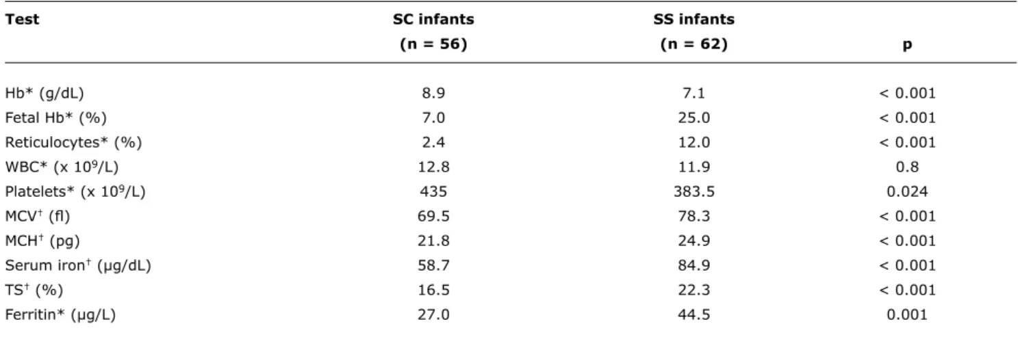
When only the non-transfused children were analyzed, there were no differences with regard to gender (p = 0.71), birth weight (p = 0.92) or gestational age (p = 0.8) between SS (n = 62) and SC children (n = 56). Regarding the hematological tests, SC children presented, as expected, total Hb levels higher than SS children, as well as lower fetal Hb concentration and lower reticulocyte count (p < 0.001, Table 3). White blood cell count was not different between SS and SC groups (p = 0.8), and platelet count was slightly
higher in SC children (p = 0.024). Indicators of possible iron deiciency were all signiicantly lower in SC infants (p ≤ 0.001, Table 3).
Test SC infants SS infants
(n = 56) (n = 62) p
Hb* (g/dL) 8.9 7.1 < 0.001
Fetal Hb* (%) 7.0 25.0 < 0.001
Reticulocytes* (%) 2.4 12.0 < 0.001
WBC* (x 109/L) 12.8 11.9 0.8
Platelets* (x 109/L) 435 383.5 0.024
MCV† (l) 69.5 78.3 < 0.001
MCH† (pg) 21.8 24.9 < 0.001
Serum iron† (µg/dL) 58.7 84.9 < 0.001
TS† (%) 16.5 22.3 < 0.001
Ferritin* (µg/L) 27.0 44.5 0.001
table 3 - Comparison between laboratory tests of infants who did not receive blood transfusion (n = 118), according to the type of hemoglobinopathy (SC or SS)
Hb = hemoglobin; MCV = mean corpuscular volume; MCH = mean corpuscular hemoglobin; TS = transferrin saturation; WBC = white blood cell count. * Median values and Mann-Whitney test.
† Mean values and t test.
Regarding the indicators of possible iron deiciency, the non-transfused group presented lower values for all variables, but the differences were statistically signiicant only for ferritin and MCV (p = 0.003 and 0.046, respectively).
Despite the restricted number of SC children who were given a transfusion (n = 2) in comparison with the non-transfused group (n = 56), there were statistically signiicant differences with respect to serum iron (p = 0.029) and TS levels (p = 0.036) and not signiicant differences with respect to MCV, MCH and ferritin levels, which were all lower in the non-transfused group.
The analysis of infants with MCV, MCH, ferritin and TS values lower than the reference values for their age group revealed that: a) 35/134 children (26.1%) were detected with MCV < 70 l; b) 54/134 (40.3%), with MCH < 23 pg; c) 16/133 (12%), with ferritin < 10 µg/L; and d) 20/132 (15.2%), with TS < 12%. The stratiication of these patients according to their Hb type revealed a higher prevalence of abnormal values for SC infants regarding MCV (p < 0.001), MCH (p < 0.001) and TS (p = 0.014), but no signiicant
differences with regard to ferritin (p = 0.11).
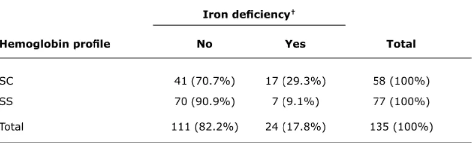
When deining as iron deicient those children who had the MCV or MCH lower than the reference limit for their age group and, simultaneously, had either of their iron kinetics tests (ferritin or TS) also below the reference limit, 24 children were considered to have iron deiciency (17.8%, 95%CI 11.3-24.3%).
Using the same criteria, SC infants had a signiicantly greater percentage of iron deiciency than SS infants (p = 0.003, Table 4). Hb proile (SS or SC) was not associated with prematurity or low birth weight (p = 0.81). Likewise, iron deiciency was not found to be associated
with prematurity and low birth weight in the present study (p = 0.76). Therefore, the aforementioned association of SC infants with iron deiciency was not confounded by other possible variables.
Among the 17 children who were given at least one transfusion, only one was iron deicient; among the 118 who did not receive a transfusion, 23 (19.5%) had iron deiciency (p = 0.31).
Considering children with ferritin concentration above 142 µg/L as having increased ferritin concentration,21
15/133 cases were detected (11.3%, 95%CI 5.9-16.7%; ferritin was not determined in two children): 13 were SS and 2 were SC (p = 0.024). Among children who received at least one transfusion, 8/17 (47.1%) had increased ferritin; on the other hand, when considering the 116 children who were not given a transfusion, 7 (6%) had increased ferritin (p < 0.001).
Discussion
The importance of newborn screening programs has been duly recognized ever since their implementation. In Brazil, the inclusion of screening for sickle cell disease, a pioneering undertaking by the State of Minas Gerais,4 represents an
important and historical landmark in healthcare for sickle cell patients and consolidated, in an indisputable manner, the need for further study and comprehension of this disease and its consequences during the lives of affected individuals.
Iron deiciency†
Hemoglobin proile No Yes total
SC 41 (70.7%) 17 (29.3%) 58 (100%)
SS 70 (90.9%) 7 (9.1%) 77 (100%)
Total 111 (82.2%) 24 (17.8%) 135 (100%)
* p = 0.003.
† Two indices were considered to define iron deficiency (low mean corpuscular volume or mean corpuscular
hemoglobin plus low ferritin or transferrin saturation).
table 4 - Association between iron deiciency and hemoglobinopathy (SC vs. SS infants*)
with sickle cell disease. The study population (n = 135) was superior to those of most other studies conducted abroad with similar purposes.6-11,13,14 All children were diagnosed
through the PETN-MG program, were followed up at a single service unit and had their tests done at a single laboratory, which allowed for sample uniformity. As this is a retrospective study, it was not possible to assess the dietary habits of participants, due to the heterogeneity of the data previously collected. The majority of other studies, likewise, did not assess dietary habits.6,10-12
Children with sickle cell disease, beginning at 2 years of age, show impaired somatic growth that affects weight more than height and is progressively aggravated up to the age of 18.22 There are studies that demonstrate differences
between SS and SC children, with the latter having a smaller deicit, which may be explained by the greater clinical severity of the former.22 In the present study, children with sickle
cell disease were, in their majority, full term infants with adequate weight for their gestational age. They did show some weight deicit, but no height deicit, when compared with the standard population. No signiicant difference was noticed in the anthropometric indices when the groups were separated as per hemoglobinopathy (SS vs. SC). Brazilian studies dealing with the growth and nutritional assessment of children with sickle cell disease found weight and height deicits over time,10,23 but children were generally older
than those of our study.
Patients with sickle cell disease quite often need blood transfusion to treat some acute clinical events. The small number of children who had been given a transfusion in the present study (12.5%) is surely explained by their age below two and by improvements of healthcare and monitoring in our Hematology Center. The “Hemominas card,” which is given to every patient, registers the basal Hb concentration and other clinical data. Accordingly, transfusion indications
are restricted to those strictly necessary and are based on the correlation between the clinical manifestations and the laboratory tests and not only upon the results of these tests.
There is great debate on which should be the most adequate method of diagnosing iron deiciency in individuals with sickle cell disease, due to characteristics that are inherent to the disease. The majority of currently used tests for that diagnosis have dificulties in their interpretation, be it in a combined or in an isolated approach. The presence of microcytosis may result from genetic alterations such as α-thalassemia, which affects 30 to 35% of the Hb S population.7,15 In our Hematology Center, 30% of SS children
have –
α
3.7 deletions.24Serum iron presents circadian variations and depends on dietary conditions. On the other hand, an increased iron binding capacity and a decreased TS seem to be good indicators of iron deiciency.14,25 Low serum ferritin is also
a good indicator,7 but even if it is within the normal range,
iron deiciency may not be ruled out, since inlammatory and infectious processes, which are rather common events in sickle cell disease, may increase ferritin concentration above the reference values.14 Bone marrow evaluation of
iron, an invasive method, may not be reliable for body iron assessment.26 Free erythrocyte protoporphyrin has
limited value in diagnosing iron deiciency in patients with sickle cell disease, as the igures are increased due to reticulocytosis.25 Similarly, serum transferrin receptor
and erythropoietic drive, thus obscuring concomitant iron deiciency.14
The present study suggests that 17.8% of the children were iron deicient when a combined criterion of low MCV or MCH and a simultaneous low ferritin or TS was used. Other studies have found a prevalence from 8.5 up to 100%,6-10,12-14 depending on the age of the children, the
deinition for iron deiciency, and the economic status of the country where the study was done. A Jamaican study assessed 141 patients younger than 5 years (121 SS and 20 SC) and found iron deiciency anemia in 8.5% of the children.12 Similar to our results, when the groups were
separated, SC children showed a higher prevalence of iron deiciency (42%) than SS children (5.8%). The single study that was not able to ind iron deiciency was a North American study assessing 104 SS children with an average age of 7.3 years, much higher than that of the present study. Findings were attributed to the improvement of the dietary conditions among the African American population over the years.11
A recent review of Brazilian studies has shown that the median prevalence of anemia in children below 5 years of age is 53% and may be higher in infants.15 In the present study,
children were all below 2 years, the age with the greater risk for the development of iron deiciency. Though with a chronic hemolytic anemia, these patients experience the same nutritional hindrances as other Brazilian children in the same age group. In the presence of iron deiciency, anemia can be aggravated in these children, and the possibility of overloading the cardiovascular system, which leads to poor tissue oxygenation and hinders their performance of activities and growing, is a threat.22
The negative consequences of anemia in long-term neurocognitive development are well known. Most of the studies dealing with iron-deicient children have found an association between iron deiciency and hindered motor and cognitive development, in addition to behavioral problems.27,28 Children with sickle cell disease face a greater
risk of having learning disabilities than their siblings or peers who do not have the disease, probably due to cerebral strokes, silent or not, caused by the characteristic recurrent cerebral vaso-occlusive episodes. Other mechanisms may also be involved, such as chronic anemia and reduced pulmonary function, which would lead to chronic tissue hypoxia and its ensuing consequences.29,30
Given the impact of iron deiciency anemia on somatic and cognitive development, and the lack of evidence that would contraindicate iron supplementation in infants with sickle cell disease, an individualized assessment is suggested. On the other hand, considering the small number of children with high ferritin levels in the present study and the strong association of ferritin with blood transfusions, it may be inferred that there is low risk in the use of fortiied lours and of iron supplementation for infants with sickle cell
disease, mainly for those with SC disease. Many of these infants were found to have iron deiciency in our study. Our reasoning is that it may be inadequate to hold back iron supplementation for these infants fearing iron accumulation, because, in an already complex condition that often limits proper development, allowing for the addition of one more aggravating factor seems to be unwise.
The limitations of this study are: a) its cross-sectional design, which does not allow for a longitudinal evaluation of the laboratory data of the children with sickle cell disease during the irst three years of life; b) the retrospective collection of data, which does not allow for a homogenous laboratorial assessment at pre-determined ages. A prospective longitudinal study has been devised to ascertain whether these conclusions really are applicable for children with sickle cell disease; c) the absence of a control group without sickle cell disease. Such a group could add to the study, but its absence does not preclude valid conclusions, given the fact that the prevalence of anemia in Brazilian infants is consistently very high all over the country, as stated before.15,16,18
In conclusion, infants with sickle cell disease, mainly those with the SC type, may have iron deicit. Iron supplementation may be given and withdrawn after the irst blood transfusion. Iron kinetics tests are useful in doubtful cases and should be interpreted on the basis of clinical and hematological data. Longitudinal studies are needed to conirm these recommendations.
Acknowledgements
The authors would like to thank the Brazilian Health Ministry for the inancial support, the Núcleo de Ações e Pesquisa em Apoio Diagnóstico from Universidade Federal de Minas Gerais (Nupad-UFMG) and Fundação Hemominas for the logistic support they provided to this study, and the medical students Paola G. Giostri, Daniel C. Discacciati, Filipe C. R. de Souza and Maria A. G. Rocha for their contributions.
References
1. World Health Organization. Iron deiciency anaemia. Assessment, prevention, and control. A guide for programme managers.
http://whqlibdoc.who.int/hq/2001/WHO_NHD_01.3.pdf. Access: 12/2/2010.
2. Coutinho GG, Goloni-Bertollo EM, Bertelli EC. Iron deiciency anemia in children: a challenge for public health and for society.
Sao Paulo Med J. 2005;123:88-92.
4. Fernandes AP, Januario JN, Cangussu CB, de Macedo DL, Viana MB. Mortality of children with sickle cell disease: a population study. J Pediatr (Rio J). 2010;86:279-84.
5. Koren A, Fink D, Admoni O, Tennenbaum-Rakover Y, Levin C.
Non-transferrin-bound labile plasma iron and iron overload in sickle-cell disease: a comparative study between sickle-cell disease and beta-thalassemic patients. Eur J Haematol. 2010;84:72-8. 6. Vichinsky E, Kleman K, Embury S, Lubin B. The diagnosis of iron
deiciency anemia in sickle cell disease. Blood. 1981;58:963-8. 7. Nagaraj Rao J, Sur AM. Iron deiciency in sickle cell disease. Acta
Paediatr Scand. 1980;69:337-40.
8. Okeahialam TC, Obi GO. Iron deiciency in sickle cell anaemia in Nigerian children. Ann Trop Paediatr. 1982;2:89-92.
9. Davies S, Henthorn J, Brozovic M. Iron deiciency in sickle cell anaemia. J Clin Pathol. 1983;36:1012-5.
10. Pellegrini Braga JA, Kerbauy J, Fisberg M. Zinc, copper and iron and their interrelations in the growth of sickle cell patients. Arch Latinoam Nutr. 1995;45:198-203.
11. Stettler N, Zemel BS, Kawchak DA, Ohene-Frempong K, Stallings VA. Iron status of children with sickle cell disease.J Parenter Enteral Nutr. 2001;25:36-8.
12. King L, Reid M, Forrester TE. Iron deiciency anaemia in Jamaican children, aged 1-5 years, with sickle cell disease.West Indian Med J. 2005;54:292-6.
13. Mohanty D, Mukherjee MB, Colah RB, Wadia M, Ghosh K, Chottray GP, et al. Iron deiciency anaemia in sickle cell disorders in India.
Indian J Med Res. 2008;127:366-9.
14. Lulla RR, Thompson AA, Liem RI. Elevated soluble transferrin receptor levels relect increased erythropoietic drive rather than iron deiciency in pediatric sickle cell disease.Pediatr Blood Cancer. 2010;55:141-4.
15. Jordão RE, Bernardi JLD, Barros-Filho AA. Prevalência de anemia ferropriva no Brasil: uma revisão sistemática. Rev Paul Pediatr. 2009;27:90-8.
16. Monteiro CA, Szarfarc SC, Mondini L. Tendência secular da anemia na infância na cidade de São Paulo (1984-1996). Rev Saude Publica. 2000;34:62-72.
17. Norton RC, Figueredo RC, Diamante R, Goulart EM, Mota JA, Viana MB, et al. Prevalence of anemia among school-children from Rio Acima (State of Minas Gerais, Brazil): use of the standardized prevalence method and evaluation of iron deiciency. Braz J Med Biol Res. 1996;29:1617-24.
18. Silva DG, Francheschini SC, Priore SE, Ribeiro SMR, Szarfarc SC, Souza SB, et al. Anemia ferropriva em crianças de 6 a 12 meses atendidas na rede pública de saúde do município de Viçosa, Minas Gerais. Rev Nutr. 2002;15:301-8.
Correspondence: Marcos Borato Viana
Departamento de Pediatria da UFMG Av. Alfredo Balena, 190 - Sala 267
CEP 30130-100 - Belo Horizonte, MG - Brazil Tel./Fax: +55 (31) 3409.9770
E-mail: [email protected]
19. Kuczmarski RJ, Ogden CL, Guo SS, Grummer-Strawn LM, Flegal, KM, Mei Z, et al. 2000 CDC Growth Charts for the United States: Methods and development. National Center for Health Statistics. Vital Health Stat. 2002;11:1-18.
20. Dallman PR, Siimes MA. Percentile curves for hemoglobin and red cell volume in infancy and childhood. J Pediatr. 1979;94:26-31. 21. Siimes MA, Addiego JE Jr, Dallman PR. Ferritin in serum: diagnosis
of iron deiciency and iron overload in infants and children. Blood. 1974;43:581-90.
22. Stevens MC, Maude GH, Cupidore L, Jackson H, Hayes RJ, Serjeant GR. Prepubertal growth and skeletal maturation in children with sickle cell disease.Pediatrics. 1986;78:124-32.
23. Silva CM, Viana MB. Growth deicits in children with sickle cell disease. Arch Med Res. 2002;33:308-12.
24. Belisário AR, Rodrigues CV, Martins ML, Silva CM, Viana MB. Coinheritance of α-thalassemia decreases the risk of cerebrovascular disease in a cohort of children with sickle cell anemia. Hemoglobin. 2010;34:516-29.
25. Koduri PR. Iron in sickle cell disease: a review why less is better.
Am J Hematol. 2003;73:59-63.
26. Natta C, Creque L, Navarro C. Compartmentalization of iron in sickle cell anemia - an autopsy study. Am J Clin Pathol. 1985;83:76-8.
27. Grantham-MacGregor S, Arni C. A review of studies on the effect of iron deiciency on cognitive development in children.J Nutr. 2001;131:649S-68S.
28. Beard JL. Why iron deiciency is important in infant development.
J Nutr. 2008;138:2534-6.
29. Armstrong FD, Thompson RJ, Wang W, Zimmerman R, Pegelow CH, Miller S, et al. Cognitive functioning and brain magnetic resonance imaging in children with sickle cell disease. Pediatrics.
1996;97:864-70.