Prevalence of gastroesophageal reflux disease in
patients with idiopathic pulmonary fibrosis*
Prevalência da doença do refluxo gastroesofágico em pacientes com fibrose pulmonar idiopática
Cristiane Dupont Bandeira, Adalberto Sperb Rubin,
Paulo Francisco Guerreiro Cardoso, José da Silva Moreira,Mirna da Mota Machado
Abstract
Objective: To determine the prevalence of gastroesophageal reflux disease (GERD) and to evaluate its clinical presentation, as well as the esophageal function profile in patients with idiopathic pulmonary fibrosis (IPF).
Methods: In this prospective study, 28 consecutive patients with IPF underwent stationary esophageal manometry, 24-h esophageal pH-metry and pulmonary function tests. All patients also completed a symptom and quality of
life in GERD questionnaire. Results: In the study sample, the prevalence of GERD was 35.7%. The patients were
then divided into two groups: GERD+ (abnormal pH-metry; n = 10) and GERD− (normal pH-metry; n = 18). In
the GERD+ group, 77.7% of the patients presented at least one typical GERD symptom. The pH-metry results showed that 8 (80%) of the GERD+ group patients had abnormal supine reflux, and that the reflux was exclusively
in the supine position in 5 (50%). In the GERD+ and GERD− groups, respectively, 5 (50.0%) and 7 (38.8%) of the
patients presented a hypotensive lower esophageal sphincter, 7 (70.0%) and 10 (55.5%), respectively, presenting lower esophageal dysmotility. There were no significant differences between the groups regarding demographic characteristics, pulmonary function, clinical presentation or manometric findings. Conclusions: The prevalence of GERD in the patients with IPF was high. However, the clinical and functional characteristics did not differ between the patients with GERD and those without.
Keywords: Pulmonary fibrosis; Gastroesophageal reflux; Prevalence; Manometry; Esophageal pH monitoring.
Resumo
Objetivo: Determinar a prevalência da doença do refluxo gastroesofágico (DRGE) e sua apresentação clínica, além
do perfil funcional do esôfago em pacientes com fibrose pulmonar idiopática (FPI). Métodos: Foram avaliados
prospectivamente 28 pacientes com FPI. Os pacientes foram submetidos à esofagomanometria estacionária, pHme-tria de 24 h e testes de função pulmonar, assim como responderam a questionários sobre sintomas e qualidade de
vida em DRGE. Resultados: A prevalência de DRGE foi de 35,7%. Os pacientes foram divididos em dois grupos:
grupo DRGE+ (pHmetria anormal; n = 10), e grupo DRGE− (pHmetria normal; n = 18). No grupo DRGE+, 77,7% dos pacientes apresentavam pelo menos um sintoma típico de DRGE. Nesse grupo de pacientes, 8 (80%) apre
-sentaram refluxo em posição supina, e 5 (50%) apre-sentaram refluxo exclusivamente nessa posição. Nos grupos DRGE+ e DRGE−, respectivamente, 5 (50,0%) e 7 (38,8%) dos pacientes apresentaram hipotonia do esfíncter esofágico inferior, assim como 7 (70,0%) e 10 (55,5%) apresentaram hipomotilidade esofágica. Não houve dife -renças significativas entre os grupos quanto a características demográficas, função pulmonar, apresentação clínica
ou achados manométricos. Conclusões: A prevalência de DRGE nos pacientes com FPI foi elevada. Entretanto, as
características clínicas e funcionais não diferiram significativamente entre os pacientes com e sem DRGE.
Descritores: Fibrose pulmonar; Refluxo gastroesofágico; Prevalência; Manometria; Monitoramento do pH esofágico.
* Study carried out in the Gastrointestinal Motility Laboratory of the Pereira Filho Ward of the Santa Casa Hospital in Porto Alegre in collaboration with the Postgraduate Program in Pulmonology of the Universidade Federal do Rio Grande do Sul – UFRGS, Federal University of Rio Grande do Sul – Porto Alegre, Brazil.
Correspondence to: Cristiane Dupont Bandeira. Rua Júlio da Silva Rodrigues, 190/102, Carianos, CEP 88047-616, Florianópolis, SC, Brasil.
Tel 55 48 8846-0508. Email: [email protected] Financial support: None.
of the Santa Casa Hospital in Porto Alegre, at the Lung Transplant Outpatient Clinics of the Santa Casa Hospital in Porto Alegre and the General Hospital in Porto Alegre and at the Pulmonology Clinic of these two institutions. Between 2004 and 2008, we evaluated 34 patients who met the American Thoracic Society (ATS) and European Respiratory Society (ERS)(3) criteria for the
diag-nosis of IPF. Of those 34, 6 were excluded for the following reasons: death before the comple-tion of the study (2 patients); concomitant lung cancer (1 patient); previous repair of hiatal hernia (1 patient); withdrawal from the study (1 patient); and loss to follow-up (1 patient). Therefore, 28 patients completed the evaluation. In17 (60.3%) of those 28 patients, the histolog-ical diagnosis of IPF was confirmed by surghistolog-ical open lung biopsy, whereas, in the remaining 11, the diagnosis was based on the ATS/ERS criteria for a diagnosis of IPF in the absence of open lung biopsy(3): exclusion of other known causes
for interstitial lung disease; abnormal pulmonary function testing results (evidence of restricted or impaired gas exchange); HRCT findings of bibasilar reticular abnormalities with minimal ground-glass opacities; and a clinical history consistent with the diagnosis.
After giving written informed consent, patients completed a general questionnaire on respiratory and digestive symptoms, as well as the Quality of Life Scale for Gastroesophageal Reflux Disease (GERD-QoL), modified and validated for use in Brazil. The GERD-QoL comprises 11 ques-tions, and points from 0 to 5 are used in order to respond to 10 of these questions as follows: 0, no symptoms; 1, symptoms that do not affect the daily routine; and so on, progressively, up to 5, which indicates constant symptoms that affect activities of daily living. The last question, also rated from 0 to 5, refers to patient satisfac-tion with the present situasatisfac-tion.(10) Therefore, the
maximum score is 55.
Patients were submitted to pulmonary func-tion tests, and the values of FVC, FEV1, the FEV1/FVC ratio, TLC and corrected DLCO were analyzed. All tests were performed using a Collins/ GS Plus device (Warren E. Collins, Inc., Braintree, MA, USA), in accordance with the guidelines of the Brazilian Thoracic Association.(11)
Esophageal manometry was performed using a computerized stationary manometry system, consisting of a catheter with 4 distal radial and
Introduction
Idiopathic pulmonary fibrosis (IPF) is a chronic and progressive interstitial disease characterized by the histological pattern of usual interstitial pneumonia. It is esti-mated that the annual incidence of IPF is approximately 10.7 cases/100,000 men and 7.4 cases/100,000 women, with a prevalence of up to 20.2 cases/100,000 population.(1) This
disease typically evolves to severe respiratory failure and can be accompanied by other comorbidities. The mean survival of the indi-viduals affected is approximately 3 years after diagnosis, and, among the treatment options available, none seems to substantially affect the progression of the disease.(2-4) The etiology of
IPF is unknown. Various hypotheses have been investigated, and, among possible risk factors, smoking, environmental conditions, genetic predisposition, viral infections and chronic aspi-ration secondary to gastroesophageal reflux (GER) are considered.(5,6)
Since the end of the XIX century, when pulmonary complications were observed in patients with diffuse esophageal dilation, GER has been associated with respiratory symp-toms.(7) In 1971, Pearson & Wilson found 6 cases
(4.2%) of pulmonary fibrosis of unknown cause in a case series of 143 patients with achalasia.(8)
Interest in the association between IPF and GER was renewed after one group of authors(5)
reported that the prevalence of GER disease (GERD), frequently asymptomatic, in patients with IPF and in patients with interstitial lung disease other than IPF (control group), all of whom were submitted to 24-h pH-metry, was 94% and 50%, respectively. Subsequently studies also suggested the existence of this asso-ciation.(6,9)
The objective of this study was to deter-mine the prevalence of GERD and to evaluate its clinical presentation, as well as the esophageal function profile (as measured by 24-h esopha-geal pH-metry and esophaesopha-geal manometry), in a group of patients with IPF. In addition, we aimed at comparing the patients with GERD and those without regarding clinical characteristics.
Methods
trodes, either a single electrode (Zinectics; Medtronic-Synetics, Skorlunde, Denmark) or a pair of electrodes (Alacer Biomédica, São Paulo, Brazil), connected to a portable computerized recorder (Digitrapper MK III; Synectics Medical) capable of registering one pH measurement every 4 s over a 24-h period. The distal pH-metry electrode was placed 5 cm above the proximal border of the LES, which had been previously located via esophageal manometry. When the dual-electrode catheter was used, the distance between the two electrodes was 15 cm. Prior to each test, the electrodes were calibrated in buffer solutions (Alacer Biomédica) at pH 7 and pH 1. Patients were instructed to discontinue the use of antacids and prokinetic agents 7 days prior to the test, as well as to fast for 4 h before the test. A log was provided for recording meal start and end times, as well as time spent in the supine position and any symptoms. At the end of the 24-h period, the patients returned to the labora-tory for removal of the catheter and analysis of the data recorded (Esophogram; Synectics Medical). In addition, a graphic, descriptive report showing the results of the test was issued. The analysis was based on the table devised by Johnson and the scoring system developed by DeMeester.(14,15)
Tests in which the DeMeester score was higher than 14.7 were considered abnormal. Only data provided by the distal electrode were used in the 4 longitudinal channels axially distributed at
intervals of 5 cm (Synectics Medical, Stockholm, Sweden). We used a system of continuous perfusion involving a low-compliance capil-lary pneumohydraulic pump (Mui Scientific, Mississauga, Ontario, Canada), connected to external pressure transducers. Pressures were registered by a digital computerized poly-graph (PC Polygraf HR; Synetics Medical), with graphic records being made in real time. The analysis was performed using specific software (Polygram; Synectics Medical). All procedures were performed after a fast of at least 4 h. The tone, position, extent and relaxation of the lower esophageal sphincter (LES) and upper esophageal sphincter (UES) were analyzed, as were the morphology, amplitude and duration of the esophageal contractions generated by the sequence of 10 swallows of 5 mL of water at 30-s intervals. The results were based on the criteria for normality obtained in our labora-tory,(12) which are similar to those found in the
literature.(13) The length of the esophagus was
defined as the distance between the proximal border of the LES and the distal border of the UES. The criteria for the definition of the primary and secondary esophageal disorders were based on the literature.(14)
In order to perform pH-metry, we used an external reference electrode connected to a semi-disposable catheter with antimony
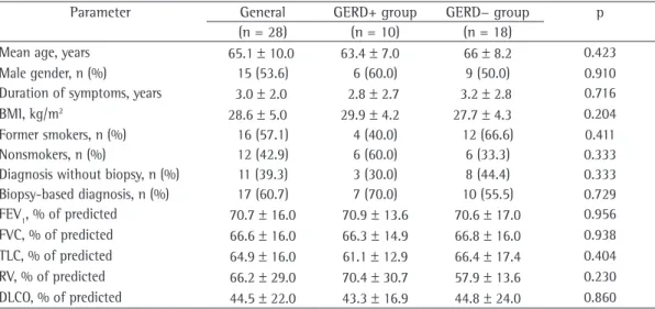
elec-Table 1 - Demographic data and pulmonary function test results in the two study groups, by esophageal pH-metry findings.
Parameter General GERD+ group GERD− group p
(n = 28) (n = 10) (n = 18)
Mean age, years 65.1 ± 10.0 63.4 ± 7.0 66 ± 8.2 0.423
Male gender, n (%) 15 (53.6) 6 (60.0) 9 (50.0) 0.910
Duration of symptoms, years 3.0 ± 2.0 2.8 ± 2.7 3.2 ± 2.8 0.716
BMI, kg/m2 28.6 ± 5.0 29.9 ± 4.2 27.7 ± 4.3 0.204
Former smokers, n (%) 16 (57.1) 4 (40.0) 12 (66.6) 0.411
Nonsmokers, n (%) 12 (42.9) 6 (60.0) 6 (33.3) 0.333
Diagnosis without biopsy, n (%) 11 (39.3) 3 (30.0) 8 (44.4) 0.333
Biopsy-based diagnosis, n (%) 17 (60.7) 7 (70.0) 10 (55.5) 0.729
FEV1, % of predicted 70.7 ± 16.0 70.9 ± 13.6 70.6 ± 17.0 0.956
FVC, % of predicted 66.6 ± 16.0 66.3 ± 14.9 66.8 ± 16.0 0.938
TLC, % of predicted 64.9 ± 16.0 61.1 ± 12.9 66.4 ± 17.4 0.404
RV, % of predicted 66.2 ± 29.0 70.4 ± 30.7 57.9 ± 13.6 0.230
DLCO, % of predicted 44.5 ± 22.0 43.3 ± 16.9 44.8 ± 24.0 0.860
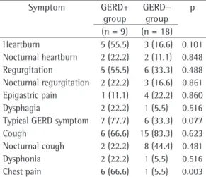
Cough was the most common symptom (in 15 patients; 83.3%). The comparison of the demographic and spirometric characteristics, as well as of the symptom questionnaire results, revealed no significant differences between the groups, with the exception of chest pain, which was more common in the GERD+ group patients (Tables 1 and 2). The mean GERD-QoL score in
the GERD+ and GERD− groups was, respectively,
17.2 and 7.8, this difference being of borderline significance (p = 0.058; Figure 1).
analysis, since only two patients were submitted to dual-electrode pH-metry.
The statistical analysis included the compar-ison of data related to the patients with GERD (abnormal pH-metry) and those without GERD (normal pH-metry). The chi-square test and the Student’s t-test were used. The level of signifi-cance was set at 5%.
Results
Of the 28 adult patients included in the study (aged between 49 and 80 years; mean, 64.3 years), 15 (53.6%) were male and 13 (46.4%) were female. The mean duration of symptoms attributed to IPF was 3.2 years, and the most common symptoms were dyspnea on exer-tion and cough. The mean body mass index of the group as a whole was 28.6 kg/m2. Most of
the patients (57.1%) were smokers or former smokers. In 10 patients (35.7%), the 24-h esophageal pH-metry detected the presence of abnormal acid reflux. The clinical characteris-tics of the 28 patients studied can be seen in Table 1. The patients were divided into two groups, according to the esophageal pH-metry results: pathological acid reflux group (GERD+;
n = 10) and normal pH-metry group (DRGE−;
n = 18).
In the GERD+ group, there was a predomi-nance of males, the mean age was over 60 years, the duration of symptoms was longer than 2 years, and 40.0% of the patients were former smokers. Most patients (70.0%) already received some type of treatment for IPF, especially corti-costeroids, and 40.0% already used proton pump inhibitors. The mean corrected DLCO was 43.3% of predicted. Most (77.7%) presented at least one typical GERD symptom, and 66.6% reported cough. The results of the GERD-QoL indicated that the quality of life in this group was moder-ately impaired by the presence of GER.
In the GERD− group, approximately half of
the patients were male, the duration of symp-toms was longer than 3 years, and most patients were former smokers who received some type of treatment, usually prednisone alone or in combination with an immunosuppressant. In this group, only 4 patients (22.2%) used proton pump inhibitors. The mean corrected DLCO was similar to that found in the GERD+ group. Even with normal pH-metry results, 6 patients (33.3%) presented at least one typical GERD symptom.
Table 2 - Results of the general questionnaire on symptoms in the two groups studied.
Symptom GERD+
group
GERD−
group p
(n = 9) (n = 18)
Heartburn 5 (55.5) 3 (16.6) 0.101
Nocturnal heartburn 2 (22.2) 2 (11.1) 0.848
Regurgitation 5 (55.5) 6 (33.3) 0.488
Nocturnal regurgitation 2 (22.2) 3 (16.6) 0.861
Epigastric pain 1 (11.1) 4 (22.2) 0.860
Dysphagia 2 (22.2) 1 (5.5) 0.516
Typical GERD symptom 7 (77.7) 6 (33.3) 0.077
Cough 6 (66.6) 15 (83.3) 0.623
Nocturnal cough 2 (22.2) 8 (44.4) 0.481
Dysphonia 2 (22.2) 1 (5.5) 0.516
Chest pain 6 (66.6) 1 (5.5) 0.003
GERD: gastroesophageal reflux disease. Results expressed as n (%).
p = 0.058
Mean score in the GERD+ and GERD− groups
Sy
m
ptom
s
core
55
45
35
25
15
5 0
17.29
7.81
GERD+ group GERD− group
Figure 1 - Symptom score on the Quality of Life Scale for Gastroesophageal Reflux Disease in the GERD+
group (abnormal pH-metry; n = 10) and GERD− group
Studies have shown that GER is implicated in the pathogenesis of many lung diseases, including asthma, chronic cough, pneumonia and obstructive sleep apnea.(21-24) In addition,
GER has been studied as a possible etiologic or complicating factor of interstitial lung diseases, such as systemic sclerosis and IPF.(21)
Furthermore, there is a proposal to include a new histological pattern associated with GER, the so-called centrilobular fibrosis (CLF) pattern, in the group of interstitial pneumonias.(25) The
CLF pattern was identified alone or in combina-tion with the pattern of nonspecific pneumonia in 21.0% and 84.0%, respectively, of a case series of 28 patients with systemic sclerosis. In patients with CLF alone and treated exclusively with intensive antireflux therapy, the lung disease remained stable for a period of 1 year.(26)
In the present study, the prevalence of GERD was 35.7%, and 77.7% of the patients presented at least one typical GERD symptom. Although this prevalence rate is high in relation to that observed in the general population, most studies using esophageal pH-metry in IPF have demon-strated even higher prevalence rates. This can be explained by the variability among the selection criteria of each study, as well as by the epidemio-logical characteristics of GERD. The performance of the pH-metry test can also be a source of variations, especially if the sphincters are not located via esophageal manometry. In addition, In the esophageal manometry of the GERD+
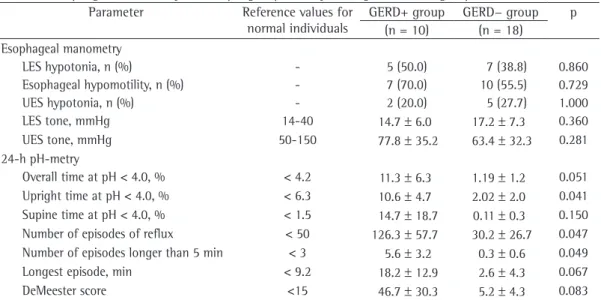
patients, esophageal hypomotility was the most common finding (in 70.0%), followed by LES
hypotonia. In the GERD− group, esophageal
hypomotility occurred in 50.0% of the patients, whereas LES hypotonia occurred in 38.3%.
In the 24-h esophageal pH-metry, 80.0% of the GERD+ patients presented reflux in the supine position, and 30.0% of those patients presented reflux exclusively in that position. There were significant differences between
the GERD+ and GERD− groups regarding the
percent of upright time at pH < 4, number of episodes of GER and number of GER episodes longer than 5 min. The pH-metry findings are shown in Table 3.
Discussion
The prevalence of GERD has been shown to be high, according to population-based studies, ranging from 10% to 20% in western countries and reaching 5% in eastern countries.(17)
Approximately 18% of healthy individuals in the United States have heartburn at least once a week.(18) In Brazil, a study conducted
in 22 cities detected the presence of heartburn one or more times a week in 11.3% of a sample of 13,000 individuals.(19) In the city of Pelotas,
located in the state of Rio Grande do Sul, Brazil, the prevalence of heartburn at least once a week was 18.2%.(20)
Table 3 - Esophageal manometry and esophageal pH-metry findings in the two groups studied.
Parameter Reference values for
normal individuals
GERD+ group GERD− group p
(n = 10) (n = 18)
Esophageal manometry
LES hypotonia, n (%) - 5 (50.0) 7 (38.8) 0.860
Esophageal hypomotility, n (%) - 7 (70.0) 10 (55.5) 0.729
UES hypotonia, n (%) - 2 (20.0) 5 (27.7) 1.000
LES tone, mmHg 14-40 14.7 ± 6.0 17.2 ± 7.3 0.360
UES tone, mmHg 50-150 77.8 ± 35.2 63.4 ± 32.3 0.281
24-h pH-metry
Overall time at pH < 4.0, % < 4.2 11.3 ± 6.3 1.19 ± 1.2 0.051
Upright time at pH < 4.0, % < 6.3 10.6 ± 4.7 2.02 ± 2.0 0.041
Supine time at pH < 4.0, % < 1.5 14.7 ± 18.7 0.11 ± 0.3 0.150
Number of episodes of reflux < 50 126.3 ± 57.7 30.2 ± 26.7 0.047
Number of episodes longer than 5 min < 3 5.6 ± 3.2 0.3 ± 0.6 0.049
Longest episode, min < 9.2 18.2 ± 12.9 2.6 ± 4.3 0.067
DeMeester score <15 46.7 ± 30.3 5.2 ± 4.3 0.083
patients were definitely diagnosed with IPF based on the ATS/ERS criteria for making a diagnosis of IPF in the absence of open lung biopsy, those criteria having a specificity of up to 96.0%.(3) However, the fact that biopsy was
not performed in all cases might have been an unfavorable factor for the analysis of the results as a whole. In most studies of IPF, the clinical manifestations presented by the patients were not sufficient to distinguish patients with pathological GER from those without it, since approximately half of the patients do not report typical symptoms, such as heartburn or regur-gitation.(5-7,9,28,29) In the present study, 77.7% of
the patients with pathological GER presented at least one typical symptom. Cough, the most common extraesophageal respiratory symptom of GERD,(19) is present in most patients,
regard-less of the presence of GERD. Within the context of IPF, it is practically impossible to determine whether cough is due to the lung disease or whether it is caused or exacerbated by GER. In addition, cough itself can cause GER, due to increased intra-abdominal pressure, leading to a cough-reflux-cough cycle.(24) Although some
GERD− group patients presented GERD symp
-toms, the impact was small, according the quality of life scale. However, no investigation aimed at clarifying the diagnosis of the causal agent of those symptoms was performed. The differential diagnosis of GERD can include peptic disease, functional dyspepsia, cholelithiasis, ischemic cardiomyopathy and achalasia.(27)
The most common esophageal manometry finding was esophageal hypomotility, followed
by LES hypotonia. Even the GERD− group
patients presented hypomotility (55.5%) and LES hypotonia (38.8%). These findings are recognized as the most common abnormali-ties associated with respiratory symptoms and inefficacious esophageal motility in GERD, constituting some of the factors that make acid clearance difficult, and this favors GER.(19) In a
study of patients with mixed connective tissue disease,(28) the occurrence of HRCT findings of
interstitial lung disease was significantly greater among the patients with esophageal dilation and among those with severe esophageal motor dysfunction. One group of authors,(29) evaluating
lung transplant candidates with advanced lung disease, found that the prevalence of abnormal manometry results was high (76%) and that the number of patients in the present study is
small, greatly limiting the scope of the conclu-sions regarding the true prevalence of GERD in this population.
The gold standard for the diagnosis of GERD is pH-metry, which has sensitivity and specifi-city of 96.0%.(27) In one study,(5) 16 (94%) of
the 17 patients with IPF presented abnormal pH-metry, of whom 11 had acid reflux at the distal and proximal electrodes, 4 had acid reflux exclusively at the distal electrode and 1 had acid reflux exclusively at the proximal electrode. In the control group, consisting of 8 patients with other interstitial lung diseases, 50% presented acid reflux. Most patients, however, did not report any GERD symptoms. It is of note that, in the present study, with only 17 patients with IPF, the technique used do perform pH-metry did not include previously locating the lower esophageal sphincter via manometry. More recently, one group of authors, studying 65 patients with IPF, found the prevalence of abnormal acid reflux to be 76% at the distal electrode and 63% at the proximal electrode.(6) Although only 47% of the
patients evaluated in that study had classical GERD symptoms, 78% presented at least one symptom suggestive of GERD, and the authors found no correlation between IPF severity and GERD. In the analysis of the pH-metry results in the present study, due to the reduced number of patients submitted to dual-electrode pH-metry, we chose to use only data obtained from the distal electrode. It is desirable that dual-elec-trode pH-metry be performed in patients with extraesophageal manifestations of GERD. However, due to the positioning of the proximal electrode, it is a procedure that is susceptible to errors, and there is no consensus on the standard of normality for acid reflux at the proximal elec-trode. In addition, the combination of pH-metry and impedance monitoring seems to be the most appropriate diagnostic resource to study this type of patient in the future, since it provides data on non-acid reflux.
was more impaired due to esophageal hypomo-tility, which is more common is these patients.
Acknowledgments
The authors would like to thank the Santa Casa Hospital in Porto Alegre and the Postgraduate Program in Pulmonology of the Federal University of Rio Grande do Sul.
References
1. American Thoracic Society. Idiopathic pulmonary fibrosis: diagnosis and treatment. International consensus statement. American Thoracic Society (ATS), and the European Respiratory Society (ERS). Am J Respir Crit Care Med. 2000;161(2 Pt 1):646-64.
2. Rubin AS, Moreira JS, Porto NS, Irion KL, Moreira RF,
Scheidt B. Fatores prognósticos em fibrose pulmonar idiopática. J Pneumol. 2000;26(5):227-34.
3. American Thoracic Society; European Respiratory Society. American Thoracic Society/European Respiratory Society International Multidisciplinary Consensus Classification of the Idiopathic Interstitial Pneumonias. This joint statement of the American Thoracic Society (ATS), and the European Respiratory Society (ERS) was adopted by the ATS board of directors, June 2001 and by the ERS Executive Committee, June 2001. Am J Respir Crit Care Med. 2002;165(2):277-304. Erratum in: Am J Respir Crit Care Med. 2002;166(3):426.
4. Verma S, Slutsky AS. Idiopathic pulmonary fibrosis--new
insights. N Engl J Med. 2007;356(13):1370-2.
5. Tobin RW, Pope CE 2nd, Pellegrini CA, Emond MJ, Sillery J, Raghu G. Increased prevalence of gastroesophageal reflux in patients with idiopathic pulmonary fibrosis. Am J Respir Crit Care Med. 1998;158(6):1804-8.
6. Raghu G, Freudenberger TD, Yang S, Curtis JR, Spada C, Hayes J, et al. High prevalence of abnormal acid gastro-oesophageal reflux in idiopathic pulmonary fibrosis. Eur Respir J. 2006;27(1):136-42.
7. Kennedy JH. “Silent” Gastroesophageal Reflux: An Important but Little Known Cause of Pulmonary Complications. Dis Chest. 1962;42(1):42-5.
8. Pearson JE, Wilson RS. Diffuse pulmonary fibrosis and hiatus hernia. Thorax. 1971;26(3):300-5.
9. Patti MG, Tedesco P, Golden J, Hays S, Hoopes C, Meneghetti A, et al. Idiopathic pulmonary fibrosis: how often is it really idiopathic? J Gastrointest Surg. 2005;9(8):1053-6; discussion 1056-8.
10. Velanovich V, Vallance SR, Gusz JR, Tapia FV, Harkabus MA. Quality of life scale for gastroesophageal reflux disease. J Am Coll Surg. 1996;183(3):217-24.
11. Sociedade Brasileira de Pneumologia e Tisiologia. Diretrizes para Testes de Função Pulmonar. J Pneumol. 2002;28(Suppl 3):S1-S238.
12. Barros IJ, Felicetti JC, Camargo JJ, Cardoso PF. Parâmetros de normalidade para esofagomanometria. J Pneumol. 1995;21(Suppl 1):S19.
13. Richter JE, Wu WC, Johns DN, Blackwell JN, Nelson
JL 3rd, Castell JA, et al. Esophageal manometry in 95 healthy adult volunteers. Variability of pressures with age and frequency of “abnormal” contractions. Dig Dis Sci. 1987;32(6):583-92.
35% of the patients with fibrosis presented LES hypotonia. In another study,(6) however,
esopha-geal motility and LES tone were normal in most of the 65 patients with IPF evaluated.
Some authors have suggested that patients with more severe disease, or delayed diag-nosis, have a higher prevalence of GERD due to greater changes in respiratory mechanics asso-ciated with advanced fibrosis. The decrease in lung compliance results in an increase in nega-tive intrapleural pressure during inhalation, and this increased negative intrapleural pressure, transmitted directly to the esophagus, could contribute to dysfunction of this organ as a whole or of the LES.(6) In the present study, the
patients were evaluated at different times in the natural history of the disease, and the mean time since diagnosis was 3 years. However, the degree of lung function impairment was not signifi-cantly greater in the GERD+ group patients.
Of the 10 patients with pathological GER, 8 presented reflux in the supine position, a finding that had also been previously reported by others.(5,9) In nocturnal reflux, the gastric content
that refluxes is cleared more slowly by the distal esophagus, since there is no contribution of the force of gravity, as well as since swallowing is less frequent and saliva production is low. In addi-tion, the basal tone of the UES and the defensive cough reflex are reduced, making it possible for the gastric acid to reflux freely, which suggests that, based on the combination of these factors, GER is a possible pathophysiological mecha-nism for the onset of pulmonary fibrosis.(5) In a
recent case report of 4 patients with IPF treated exclusively with antireflux therapy, pulmonary function test results stabilized or improved after the patients received the appropriate treatment for GERD and none of them presented exacerba-tions during the follow-up period.(30)
23. Santos LH, Ribeiro IO, Sánchez PG, Hetzel JL, Felicetti JC, Cardoso PF. Evaluation of pantoprazol treatment response of patients with asthma and gastroesophageal reflux: a randomized prospective double-blind placebo-controlled study. J Bras Pneumol. 2007;33(2):119-27. 24. Richter JE. Review article: extraoesophageal
manifestations of gastro-oesophageal reflux disease. Aliment Pharmacol Ther. 2005;22 Suppl 1:70-80. 25. de Carvalho ME, Kairalla RA, Capelozzi VL, Deheinzelin
D, do Nascimento Saldiva PH, de Carvalho CR.
Centrilobular fibrosis: a novel histological pattern of idiopathic interstitial pneumonia. Pathol Res Pract. 2002;198(9):577-83.
26. de Souza RB, Borges CT, Capelozzi VL, Parra ER, Jatene FB, Kavakama J, et al. Centrilobular fibrosis: an underrecognized pattern in systemic sclerosis. Respiration. 2009;77(4):389-97.
27. Streets CG, DeMeester TR. Ambulatory 24-hour esophageal pH monitoring: why, when, and what to do. J Clin Gastroenterol. 2003;37(1):14-22.
28. Fagundes MN, Caleiro MT, Navarro-Rodriguez T, Baldi
BG, Kavakama J, Salge JM, et al. Esophageal involvement and interstitial lung disease in mixed connective tissue disease. Respir Med. 2009;103(6):854-60.
29. Fortunato GA, Machado MM, Andrade CF, Felicetti JC, Camargo JJ, Cardoso PF. Prevalence of gastroesophageal reflux in lung transplant candidates with advanced lung disease. J Bras Pneumol. 2008;34(10):772-78. 30. Raghu G, Yang ST, Spada C, Hayes J, Pellegrini CA.
Sole treatment of acid gastroesophageal reflux in idiopathic pulmonary fibrosis: a case series. Chest. 2006;129(3):794-800.
14. Nasi A, Michelson N, editors. Manometria e pHmetria
esofágicas. São Paulo: Roca; 2001.
15. Johnson LF, Demeester TR. Twenty-four-hour pH monitoring of the distal esophagus. A quantitative measure of gastroesophageal reflux. Am J Gastroenterol. 1974;62(4):325-32.
16. Johnson LF, DeMeester TR. Development of the 24-hour intraesophageal pH monitoring composite scoring system. J Clin Gastroenterol. 1986;8 Suppl 1:52-8. 17. de Barros SG. Gastroesophageal reflux
disease--prevalence, risk factors and challenges... [Article in Portuguese]. Arq Gastroenterol. 2005;42(2):71. 18. Aguero GC, Lemme EM, Alvariz A, Carvalho BB, Schechter
RB, Abrahão L Jr. Prevalence of supraesophageal manifestations in patients with gastroesophageal erosive and non-erosive reflux disease [Article in Portuguese]. Arq Gastroenterol. 2007;44(1):39-43.
19. Moraes-Filho JP, Chinzon D, Eisig JN, Hashimoto CL,
Zaterka S. Prevalence of heartburn and gastroesophageal reflux disease in the urban Brazilian population. Arq Gastroenterol. 2005;42(2):122-7.
20. Nader F, da Costa JS, Nader GA, Motta GL. Prevalence
of heartburn in Pelotas, RS, Brasil: population-based study [Article in Portuguese]. Arq Gastroenterol. 2003;40(1):31-4.
21. Ing AJ. Interstitial lung disease and gastroesophageal reflux. Am J Med. 2001;111 Suppl 8A:41S-44S. 22. Palombini BC, Villanova CA, Araújo E, Gastal OL, Alt DC,
Stolz DP, et al. A pathogenic triad in chronic cough: asthma, postnasal drip syndrome, and gastroesophageal reflux disease. Chest. 1999;116(2):279-84.
About the authors
Cristiane Dupont Bandeira
Masters Student in Pulmonology. Postgraduate Program in Pulmonology, Universidade Federal do Rio Grande do Sul – UFRGS, Federal University of Rio Grande do Sul – Porto Alegre, Brazil.
Adalberto Sperb Rubin
Professor. Postgraduate Program in Pulmonology, Universidade Federal do Rio Grande do Sul – UFRGS, Federal University of Rio Grande do Sul – Porto Alegre, Brazil.
Paulo Francisco Guerreiro Cardoso
Associate Professor of Thoracic Surgery. Department of Surgery, Fundação Faculdade Federal de Ciências Médicas de Porto Alegre – FFFCMPA, Federal Foundation School of Medical Sciences of Porto Alegre, Porto Alegre, Brazil.
José da Silva Moreira
Adjunct Professor of Pulmonology. Universidade Federal do Rio Grande do Sul – UFRGS, Federal University of Rio Grande do Sul – Porto Alegre, Brazil.
Mirna da Mota Machado