Refining the Laparoscopic Retroperitoneal Lymph Node
Dissection for Testicular Cancer
Frederico R. Romero, Andrew Wagner, Fabio A. Brito, Michael Muntener, Guilherme C.
Lima, Louis R. Kavoussi
James Buchanan Brady Urological Institute, Johns Hopkins Medical Institutions, Baltimore,
Maryland, USA
ABSTRACT
Since its initial description, the laparoscopic retroperitoneal lymph node dissection has evolved considerably, from a purely diagnostic tool performed to stage germ cell testicular cancer to a therapeutic operation that fully duplicates the open technique. Herein, we describe the current technique employed at our institution, along with illustrations of all surgical steps, and delineate the refinements of the technique over time.
Key words: testicular neoplasms; lymph node excision; laparoscopy; diagnostic techniques, surgical Int Braz J Urol. 2006; 32: 196-201
INTRODUCTION
Retroperitoneal lymph node dissection (RPLND) has been used for diagnosis and treatment of clinical stage I and II nonseminomatous germ cell tumors (NSGCT), and as a salvage therapy for bulky metastatic germ cell testicular tumors following cisplatin-based chemotherapy.
Since the first description of open RPLND in 1902 (1), the surgical technique has undergone sev-eral modifications in an effort to decrease morbidity and enhance oncological efficacy (2). The laparoscopic RPLND (L-RPLND) has similarly evolved. Initially used purely as a diagnostic tool, L-RPLND, when prerly performed, has developed into a therapeutic op-eration that strictly adheres to established oncologic principles and fully duplicates the open technique (3). The objective of the present article is to il-lustrate in detail the laparoscopic technique employed
at our institution, outlining the refinements of L-RPLND over time.
SURGICAL TECHNIQUE
After the induction with general endotracheal anesthesia, a nasogastric tube and Foley catheter are inserted. Patients are placed in a modified flank-up position using a jelly role to slightly elevate the ipsi-lateral side. They are then taped securely to the oper-ating room table across the chest, hips, and legs. This allows the surgeon to role the patient into a completely lateral position during the procedure if necessary. Ab-domen and flank are prepped and draped in a sterile fashion.
cm below the xiphoid process (Figure-1). All ports are placed under direct vision and sutured to the skin with 2-0 silk sutures to avoid inadvertent removal during the procedure.
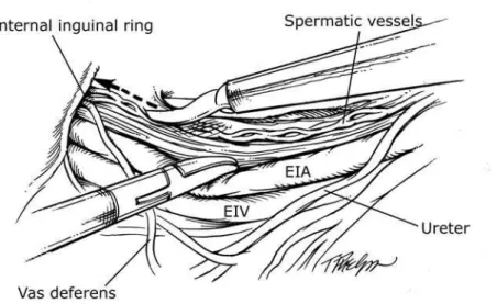
To begin the dissection on either side, the co-lon is reflected medially by incising the line of Toldt. Care is taken not to damage the delicate mesenteric vasculature. The spermatic vessels are first identified, then dissected free of surrounding tissues distally to-ward the internal inguinal ring (Figure-2). At the area of the internal ring, sharp and blunt dissection is nec-essary, often using monopolar cautery, to liberate the most distal aspect of the vessels, along with the non-absorbable stitch from the previous radical inguinal orchiectomy. Great care is necessary to avoid injury to the adjacent iliac vessels during this dissection. The spermatic vessels, along with all contiguous lymphatic tissue, are sharply dissected and brought up to their origin (renal hilum on the left, and inferior vena cava on the right), where they are clipped and transected.
The removal of the retroperitoneal lymph node packets is performed in a modified template fashion. The borders of our dissection are shown in Figure-3. Dissection over the aorta is halted at the level of the inferior mesenteric artery to avoid damaging the hy-Figure 1 – Port site placement during laparoscopic
retroperito-neal lymph node dissection.
pogastric plexus of nerves, which can result in impaired ejaculation. A right-sided dissection necessitates com-plete mobilization of the duodenum and head of pan-creas medially using sharp dissection and the strict avoidance of thermal energy (Kocher maneuver). This will expose the inferior vena cava (IVC) and aorta (Ao).
Figure 3 – A) Template dissection limits for right-sided tumors consist of ureter (lateral), midpoint of aorta (medial), bifurcation of iliac vessels (inferior), and renal hilum (superior). B) Template dissection limits for left-sided tumors consist of ureter (lateral), mid-point of vena cava (medial), bifurcation of iliac vessels (distal), and renal hilum (superior).
Both right and left dissections generally pro-ceed in a similar fashion. First, a split/roll technique is used to gather all precaval/preaortic and lateral lym-phatic tissues up to the renal hilum, exposing the re-nal vein and rere-nal artery (Figure-4). The lateral nodal tissues are lifted and separated from the underlying
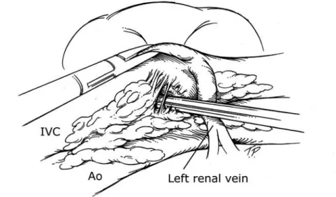
psoas fascia. The interaortocaval tissue is then dis-sected. An atraumatic grasper is used to gain addi-tional exposure to this area by lifting the vena cava and aorta, allowing the nodal packet to be gently teased off the surface of the great vessels (Figure-5). We liberally use 5 and 10 mm titanium clips to
se-cure vasculature and lymph structures. Finally, retro-caval/retroaortic dissection is performed to remove the only remaining lymphatic tissue, again taking care to clip all lymphatic channels and lumbar vessels en-countered (Figure-6). The sympathetic chains are identified and carefully dissected free (Figure-7).
Figure 5 – Interaortocaval dissection is performed with meticulous ligation of all lumbar and lymphatic vessels encountered. The right renal artery and left renal vein are always identified during the superior portion of this dissection and should not be confounded with lumbar vessels. IVC = inferior vena cava, Ao= aorta.
Figure 6 – Retrocaval dissection is facilitated by rolling and retracting the vena cava with an atraumatic grasper, to allow access to the retrocaval space. All lymphatic and lumbar vessels encountered are carefully ligated and divided. IVC = inferior vena cava, Ao = aorta.
Meticulous lymphatic ligation minimizes the risk of postoperative lymphocele. Venous bleeding is often encountered and should first be controlled us-ing direct pressure. A laparoscopic laparotomy pad (standard pad cut into a 3 cm wide strip for insertion through the trocar) is frequently used for this pur-pose and most venous bleeding will stop with several minutes of continuous pressure. To gain control of arterial bleeding, especially that from the aorta, we resort to using clips, bipolar cautery, or if the bleed-ing is directly from the aorta, intracorporeal suturbleed-ing using 3-0 monofilament, nonabsorbable suture.
Retroperitoneal lymph nodes and spermatic cord are placed into a 10 cm specimen bag, removed from the abdomen, and sent for pathologic analysis. At the end of the procedure, intraabdominal pressure is lowered to 5 mm Hg to evaluate active bleeding. Port sites are closed endoscopically under direct vi-sion using 0-polyglactin suture.
COMMENTS
We have been performing L-RPLND for tes-ticular cancer since 1992 at our institution. Originally, the procedure was used in a diagnostic fashion to pro-vide pathologic staging information for clinical Stage I NSGCT. In 1999, we retrospectively reviewed our first 29 patients undergoing L-RPLND for clinical Stage I NSGCT. We demonstrated that the procedure provided useful pathologic information with minimal short and long-term morbidity. This data supported the notion that L-RPLND was a feasible, minimally invasive surgical alternative to observation or open RPLND (4). With time and additional laparoscopic experience, we began to perform L-RPLND on post-chemotherapy patients who required resection of re-sidual retroperitoneal masses. In 2002, we reported the results of 7 such patients. Again, L-RPLND was considered feasible in this situation, though extremely challenging due to the chemotherapy-induced retro-peritoneal fibrosis (5).
After our initial experience, it was apparent that we could perform a dissection that mirrored that of the open procedure. Therefore, our objectives for L-RPLND evolved from a diagnostic to a therapeutic
intervention, although because approximately 50% of pathologic stage II patients relapse (6), we con-tinue to use 2 cycles of chemotherapy when positive nodes are discovered. In 2003, we evaluated the long-term oncologic efficacy of our patients. There were no abdominal recurrences, however 1 of 15 (6.6%) patients with pathologic stage I disease had biochemi-cal recurrence with a median follow-up of 5.8 years. Though our numbers were relatively small we con-cluded that cancer control appeared similar, and L-RPLND offered minimal morbidity compared with the open procedure (7).
As of May 2005, we have performed a total of 92 L-RPLND for testicular cancer. Seventy-six (82.6%) patients underwent the complete template dissection as described above, and sixteen (17.4%) patients underwent an abbreviated dissection due to positive lymph nodes found on frozen section. Me-dian age was 30.5 years-old (range 15 to 45). Sev-enty-seven (83.7%) patients underwent L-RPLND for clinical stage I or II NSGCT of the testis, and 15 (16.3%) for residual retroperitoneal mass following chemotherapy. Right and left-sided modified unilat-eral template dissection were performed in 49 (53.3%) and 40 (43.5%) patients, respectively. Three (3.2%) patients underwent bilateral dissection. Intraoperative complications occurred in 10 (10.8%) patients: cavotomy (5.4%), injury to the renal hilum (3.1%), transection of the external iliac artery (1%), and gall-bladder lesion (1%). Open conversion rate was 5.4%. The median estimate blood loss was 300 mL (range 50 to 4500), and median length of hospital stay was 2 days (range 1 to 71).
conva-lescence). It remains to be seen, however, if this pro-cedure will become commonplace as it requires ad-vanced laparoscopic skill and patience. Furthermore, with improvements in chemotherapeutic regimens, RPLND may be less commonly indicated in the fu-ture. A prospective randomized trial comparing the morbidity and oncologic outcomes of laparoscopic and open RPLND would be the ideal method of fully evaluating the L-RPLND in this setting.
ACKNOWLEDGEMENT
Timothy Phelps, MS, FAMI (Department of Arts as Applied to Medicine, Johns Hopkins Univer-sity) made the illustrations.
CONFLICT OF INTEREST
None declared.
REFERENCES
1. Roberts JB: Excision of lumbar lymphatic nodes and spermatic vein in malignant diseases of the testicle. Ann Surg. 1902; 36: 539-49.
2. Donohue JP: Evolution of retroperitoneal lym-phadenectomy (RPLND) in the management of non-seminomatous testicular cancer (NSGCT). Urol Oncol. 2003; 21: 129-32.
3. Allaf ME, Bhayani SB, Link RE, Schaeffer EM, Varkarakis JM, Shadpour P, et al.: Laparoscopic ret-roperitoneal lymph node dissection: duplication of open technique. Urology. 2005; 65: 575-7.
4. Nelson JE, Chen RN, Bishoff JT, Oh WK, Kantoff PW, Donehower RC, et al.: Laparoscopic retroperitoneal lymph node dissection for clinical stage I nonseminomatous germ cell testicular tumors. Urol-ogy. 1999; 54: 1064-7.
5. Palese MA, Su LM, Kavoussi LR: Laparoscopic ret-roperitoneal lymph node dissection after chemotherapy. Urology. 2002; 60: 130-4.
6. Williams SD, Stablein DM, Einhorn LH, Muggia FM, Weiss RB, Donohue JP, et al.: Immediate adjuvant chemotherapy versus observation with treatment at relapse in pathological stage II testicular cancer. N Engl J Med. 1987; 317: 1433-8.
7. Bhayani SB, Ong A, Oh WK, Kantoff PW, Kavoussi LR: Laparoscopic retroperitoneal lymph node dissec-tion for clinical stage I nonseminomatous germ cell testicular cancer: a long-term update. Urology. 2003; 62: 324-7.
8. Peschel R, Gettman MT, Neururer R, Hobisch A, Bartsch G: Laparoscopic retroperitoneal lymph node dissection: description of the nerve-sparing technique. Urology. 2002; 60: 339-43; discussion 343.
9. Albqami N, Janetschek G: Laparoscopic retroperito-neal lymph-node dissection in the management of clini-cal stage I and II testicular cancer. J Endourol. 2005; 19: 683-92; discussion 692.
10. Mariano MB, Tefilli MV: Laparoscopic retroperito-neal lymphadenectomy after chemotherapy for stage IIB testicular tumors. Int Braz J Urol. 2001; 27: 527-34.
Accepted after revision: October 10, 2005
Correspondence address:
Dr. Frederico R. Romero
600 North Wolfe Street Suite 161 Jefferson Street Bldg.
Baltimore, MD, 21287-8915, USA Fax: + 1 410-502-7711