Virtual Computed Tomography Cystoscopy in Bladder
Pathologies
Halil Arslan, Kadir Ceylan, Mustafa Harman, Yuksel Yilmaz, Osman Temizoz, Saban Can
Departments of Radiology and Urology, Yuzuncu Yil University School of Medicine, Van, Turkey
ABSTRACT
Objective: Assessed the usefulness of virtual cystoscopy performed with multidetector computed tomography (CT) in
patients with different urinary bladder pathologies compared to the conventional cystoscopy.
Materials and Methods: Eighteen patients with different bladder pathologies, which consisted of 11 tumors, 3 diverticula,
2 trabecular changes and 2 stones, were assessed with conventional cystoscopy and virtual CT cystoscopy. The results of virtual CT cystoscopy were compared with the findings of conventional cystoscopy. We determined the detection rate and positive predictive value of CT imaging based virtual cystoscopy in the diagnosis of urinary bladder lesions.
Results: CT scanning was well tolerated by all patients, and no complications occurred. Images in 16 (88%) of the 18
virtual cystoscopic examinations were either of excellent or good quality. All tumors except one, 2 trabecular changes and 2 stones were characterized with similar findings in the both of methods. The masses ranged from 0.4 to 7.0 cm in diameter. While conventional cystoscopy could not evaluate interior part of the diverticulum, virtual CT cystoscopy could demon-strate clearly within it. There were no false-positive findings in our series.
Conclusion: Virtual CT cystoscopy is a promising technique to be used in the detection of bladder lesions. It should be
considered especially at the evaluation of bladder diverticula. In the future, it may be possible or even advantageous to incorporate into the imaging algorithm for evaluation of bladder lesion.
Key words: bladder; cystoscopy; tomography, spiral computed; tumors; diverticula; stones Int Braz J Urol. 2006; 32: 147-54
INTRODUCTION
Bladder pathologies are consisted of the im-portant group of genitourinary tract diseases. The most common complaints in bladder disease are micro-scopic and macromicro-scopic hematuria, disuria and other voiding symptoms. All these symptoms may be re-lated to inflammatory, neoplastic, stones, neurologic, obstructive or congenital abnormalities. Urogram, sonography (US), computed tomography (CT), mag-netic resonant imaging (MRI) and some other radio-logical modality have been used for a long time in all these pathologies. However, conventional cystoscopy
adequate bladder distention and thin-slice scanning, must be satisfied. Therefore, negative findings on CT warrant performance of conventional cystoscopy in patients with bladder pathology (1-4).
Recently, three-dimensional computer-ren-dering techniques with rapid image acquisition have led to the development of virtual-reality imaging. With commercially available software, virtual reality im-aging allows interactive intraluminal navigation through any hollow viscus, simulating conventional cystoscopy. This technique of virtual endoscopy has been applied to many organs, including the colon, bronchus, stomach, and bladder (3-6).
Currently, most authors have been studied with virtual cystoscopy about the bladder tumor. Few reports are found in the literature regarding different bladder pathology such as diverticulum or inflamma-tory pseudotumor based virtual cystoscopy of the uri-nary bladder (7,8).
The purpose of this study was to evaluate the usefulness of virtual cystoscopy using a volume ren-dering algorithm performed with multidetector CT in patients with different urinary bladder pathology compared with the gold standard that is, conventional cystoscopy and to determine the modality’s detection rate and positive predictive value.
MATERIALS AND METHODS
Eighteen patients (mean age 56 ± 11 years, range 40 to 72 years) were referred from the urology department because of the different urinary bladder disease, which consisted of 11 tumors, 3 diverticula, 2 trabecular changes and 2 stones for this study. We carried out both conventional and virtual cystoscopy in all patients. Time interval between conventional and CT cystoscopy ranged from zero to 7 days. Each patient had various clinical histories. Most of the pa-tients presented painless hematuria or dysuria. Con-ventional cystoscopies were carried out with unaware of virtual cystoscopic findings. The conventional cys-toscopies were performed with rigid 21F cystoscope (Storz, Germany) with a field of view of 30 degrees in all patients under general or local anesthesia. Vir-tual cystoscopic examinations were started with ob-taining adequate bladder distention in supine
posi-tion. Helical CT was performed with 4 channel CT scanner (Somatom Sensation 4, Siemens Medical Systems, Erlangen, Germany), in single breath hold, with 1 mm collimation, 1 mm reconstruction interval and 3 mm thickness. Other scanning parameters were as follows: 1 mm reconstruction interval, mAs 153, and 120 kV, feed/rotation 5 mm. The scanning time was only 8-12 second. Prior to the scan, adequate fill-ing of the bladder with approximately 250-450 mL of air was required. At the same time, IV 100 mL contrast medium was administered in all patients by a power injector at a rate of 2.0-2.5 mL/s for possible extravesical invasion of the tumor or some other pa-thology. The patients were then turned to the prone position, and CT of the bladder was repeated with use of the same parameters after a repeated scout view was obtained. Additional bladder distention with ap-proximately 80-120 mL of air was necessary in some of the patients, since repositioning led to leakage of some of the insufflated gas from the bladder.
The data were downloaded to an independent workstation (Leonardo; Siemens Medical Systems) equipped with software for interactive intraluminal navigation. Using multiplanar reformation from source images, a central observation point was de-fined in the middle of the lumen of the bladder. The camera for virtual cystoscopy was placed in the cen-ter of the bladder lumen and thereafcen-ter was advanced to each quadrant in turn. When a possible abnormal-ity was discovered, it was fully evaluated from vari-ous angles.
defined as a lesion when it was wider at the base. A lesion was characterized as wall thickening when there was elevation of the bladder wall without a dis-crete mass. The quality of each CT image was also evaluated in terms of the residual urine, which may obscure the bladder mucosa, and the degree of dis-tention. Complications due to CT cystoscopy were recorded.
Three radiologists (HA,MH,OT) blinded to the findings of conventional cystoscopy, indepen-dently interpreted the images prospectively, and any discrepant readings were resolved by consensus. The results of virtual CT cystoscopy were compared with the findings of conventional cystoscopy, which is considered the standard. The lesions that were not prospectively identified at CT cystoscopy were ret-rospectively evaluated for visibility on transverse and virtual images. The pathology report in each patient with bladder tumor was also reviewed for further cor-relation. Using conventional cystoscopy as the gold standard, we analyzed them to determine the detec-tion rate of CT imaging-based virtual cystoscopy in the diagnosis of urinary bladder lesions.
RESULTS
CT scanning was well tolerated by all pa-tients, and no complication occurred. Images in 16 (88%) of the 18 virtual cystoscopic examinations were of excellent or good quality, with adequate bladder distention and minimum residual urine. Images in 2 examinations were suboptimal due to either
moder-ate residual urine or inadequmoder-ate bladder distention. Tumoral lesions were seen in one of them. However, in other patient, a smaller-than-4 mm polypoid tu-moral lesion could not be detected.
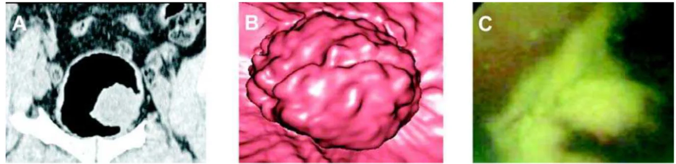
On conventional cystoscopy, 11 tumors were found in 18 patients. These masses ranged from 0.4 to 7.0 cm in diameter (mean, 1.5 cm). Out of 11 poly-poid lesions, 8 were larger than 5 mm, and 3 were 5 mm or smaller. Eight out of the polypoid lesions were larger than 5 mm, and 3 were 5 mm or smaller. One of the polypoid lesions was calcified. Six out of the 11 lesions were located on the lateral wall (Figures-1 and 2); 2 on the posterior wall; 2 on the anterior wall and 1 in the bladder neck. All lesions were diagnosed as transitional cell carcinoma in the pathology reports. All these tumors had been described by the virtual cistoscopy with nearly similar findings in size local-ization and surface of the tumor except one lesion, which was smaller than 5 mm 90% of the tumors were diagnosed by virtual cystoscopy as compared to con-ventional cystoscopy. Mucosal thickness and trabeculations were also seen in the virtual CT cys-toscopy and the appearance was similar in both mo-dalities (Figure-3). Bladder stone could not differen-tiate the tumor or polyp without adjustment of the lowest and highest point of the density value in the volume-rendering method in two patients (Figure-4). Three diverticula were diagnosed, but the interior of the diverticula could not be evaluated by conventional cystoscopy. Their lumens were easily detected by virtual cystoscopy. In three diverticula, virtual CT cystoscopies were superior to conventional
Figure 1 – 50-year-old man with transitional cell carcinoma obtained in area toward left wall shows polypoid lesion. A) Coronal
cystoscopy in demonstration of the interior of the di-verticula (Figure-5).
Transverse section and virtual CT images were complementary in lesion detection and charac-terization. Although areas of wall thickening and tra-beculation were seen on the virtual images, they were more conspicuous on the transverse views. However,
Figure 3 – 71 year-old man with trabeculation because of the prostate hypertrophy. A) Coronal multiplanar reconstruction image.
B) Magnified virtual CT cystoscopy appearance. C) General trabecular appearance of mucosal surface in virtual CT cystoscopy. Virtual CT cystoscopy shows mucosal thickness and trabeculation similar with the conventional cystoscopy.
Figure 4 – 55 year-old man with solitary bladder stone. A) Transverse section. B) Virtual CT cystoscopy appearance. C) Conventional
cystoscopy. Stone could be easily differentiated from the surrounding tissue in virtual CT cystoscopy appearance after the adjustment threshold value of density.
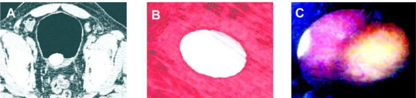
Figure 2 – 60 year-old man with primary urinary bladder cancer. A) Transverse section. B) Virtual CT cystoscopy appearance.
C) Conventional cystoscopy. Virtual CT cystoscopic image focused on polyp with 12 mm located near left urethral orifice. Internal urethral orifice can be identified only polyp with 12mm located near left urethral orifice. Internal urethral orifice can be identified in lower midportion.
Con-the walls of hollow organs. A promising advantage of this imaging modality is that views not possible in conventional endoscopic examination can be created. The volumetric data obtained with helical CT or MR imaging are computer-rendered to generate three-di-mensional images, and with commercially available software, intraluminal navigation through any hollow viscus is possible. There are two main techniques for the reconstruction of virtual image. One of them is volume rendering and the other is surface-rendering algorithm. Of the different three-dimensional render-ing techniques available, the perspective volume ren-dering provides more information because the entire data set is incorporated (15-19). We used a volume-rendering algorithm in this study.
Virtual endoscopy has been most widely ap-plied to imaging of the colon and many investigators report its feasibility in the depiction of colorectal polyps (20,21). After the first report of virtual cys-toscopy, in the study by Vining at al., there have been a lot of studies on the utility of virtual cystoscopy of the bladder. The urinary bladder is a good candidate for virtual cystoscopy because of its simple luminal morphology, its relatively small volume, and the ab-sence of involuntary peristalsis. Therefore, a virtual cystoscopic rendering of the bladder takes a short time to navigate and does not require great skill on the part of the operator (4-6,18). On the other hand, ac-cording to a study by Kim et al., virtual cystoscopy was found superior than multiplanar reconstruction and source CT images for lesion detection in the con-trast material-filled bladder (22). However, most
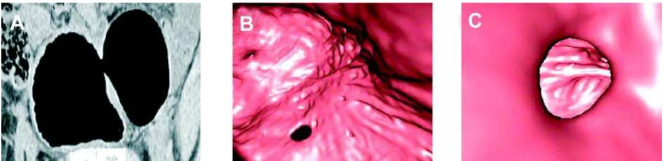
stud-Figure 5 – 45 year-old men with virtual CT cystoscopic images of bladder wall diverticulum. A) Sagittal multiplanar reconstruction
image. B) Virtual CT cystoscopy appearance of the neck of the diverticulum. C) Interior appearance of the diverticulum. Diverticulum could be evaluated both of the multiplanar reconstruction images and virtual CT cystoscopy, but could not in the conventional cystos-copy.
sidering conventional cystoscopy to be the gold stan-dard, we found the following diagnostic values for the identification of bladder lesions on virtual cys-toscopy. In 17 out of 18 patients (94.4%), lesions were detected by using virtual CT cystoscopy.
COMMENTS
Several imaging techniques are available for use in the detection of bladder pathology. US, uro-gram, CT, MRI and conventional cystoscopy could be used in the bladder disease. Conventional cystos-copy was accepted as a gold standard in bladder (3,5). However, there are several disadvantages of the con-ventional cystoscopy. It is often difficult to perform adequately when exploring the anterior bladder wall or a diverticulum cavity. Primary intradiverticular carcinomas are rare, but diagnosis is often difficult with conventional method (6,9,10,11). There are some contraindications for the conventional cystoscopy such as bacteriuria, acute cystitis, urethritis, prostati-tis, obstructive prostatic hypertrophy, and stricture or rupture of the urethra. Marked hematuria is another factor that limits the technical success of cystoscopy, thereby decreasing its reliability. On the other hand, cystoscopy is performed in general or local anesthe-sia and it is an invasive and uncomfortable procedure for patients, and complications such as infections, uretral or bladder perforation, scarring, and stricture of the urethra have been observed (3,6,12-14).
ies were performed in bladder tumor and previous stud-ies have focused solely on known bladder lesion. There have been no enough studies on different pathologies. We carried out this study on different bladder patholo-gies and evaluated the capabilities of virtual cystos-copy, such as diverticula, trabeculation and stone.
As a minimally invasive procedure, virtual CT cystoscopy provides many advantages as compared to conventional cystoscopy. The virtual CT cystoscopy images could be stored in file and the lesion could be compared in follow up period with based images. The size of a tumor is measured objectively. Access to the anterior bladder wall or the lumen of a diverticulum is not restricted in virtual cystoscopy because various software reconstruction tools can be used and the tu-mor can be easily detected (23). Patients with a severe urethral stricture or marked prostatic hypertrophy, who may be poor candidates for conventional cystoscopy, can safely undergo virtual CT cystoscopy. It is also indicated for patients who are at risk of complications such as hemorrhage, perforation, infection, or pain, and for the examination of young patients (3,5,6). In our group, diverticula were very well examined by virtual CT cystoscopy while conventional cystoscopy could not evaluate the interior of lesions. However, we had some difficulties in bladder stone using virtual cystos-copy. It was very difficult to differentiate the polyp without setting the threshold density value. This also showed us that axial images and virtual cystoscopy images should be evaluated together.
Two techniques have been used to obtain the CT source data for reconstructed virtual cystoscopic images, scanning the bladder that has been filled with either air or contrast material. Both methods have some advantages and disadvantages when compared with one another. Most previous studies have been chosen to scan the air-filled bladder. However, vir-tual cystoscopy of the air-filled bladder is inherently invasive because catheterization is required to intro-duce air into the bladder. Supine and prone examina-tion is another disadvantage of the air-filled bladder method. On the other hand, filling the bladder with IV contrast material has been easily achieved in many studies. In this method, there is no need for examina-tion in prone and supine posiexamina-tion. Therefore, this means lesser radiation and cost (3,22). However, urine
and contrast could not be mixed properly with this method for virtual cystoscopy. This is one of the dis-advantages. Secondly, IV contrast application is man-datory and this is another difficulty. Waiting for blad-der filling and inadequate distention is another dis-advantage. In addition to these disadvantages, pos-sible scheduling problems may arise in a busy CT practice because of the repeated patient positioning and scanning required (3,4,5). In our study, like many others, we used air-filled bladder for virtual cystos-copy. If there is already a Foley catheter inside the bladder, the air-filled bladder method might be pref-erable. However, when there is no catheter and a IV contrasted examination has already been planned, the second method can be used for virtual cystoscopy.
How-ever, it was reported that urethra could also be evalu-ated by virtual cystoscopy (24).
In conclusion, virtual CT cystoscopy is a prom-ising technique for tumor and some other bladder le-sions, such as diverticula. Virtual CT cystoscopy is likely superior to demonstrate the interior part of the diverticulum. Adequate bladder distention and analy-sis of virtual images are required for optimal evalua-tion. This minimally invasive method can be of value for screening, primary diagnosis and surveillance of bladder lesions. Virtual CT cystoscopy may be indi-cated as a clinical routine when conventional cystos-copy is contraindicated or restricted in feasibility and interpretation or there is risk of hemorrhage, perfora-tion, or pain especially in young patients. In the future, it may be possible or even advantageous to incorpo-rate into the imaging algorithm for evaluation of blad-der lesion through continued development and advance-ment of hardware and software. To determine the clini-cal value of virtual CT cystoscopy in the different blad-der pathology, however, larger prospective studies in the general patient population are necessary.
REFERENCES
1. Carter HB: Basic Instrumentation and Cystoscopy. Walsh PC, Retik BA,Vaughan ED, Wein AJ (eds.). Campbell’s Urology, Saunders, Philadelphia. 8 ed., 2002; vol. 1, pp. 111-21.
2. Bernhardt TM, Schmidl H, Philipp C, Allhoff EP, Rapp-Bernhardt U: Diagnostic potential of virtual cys-toscopy of the bladder: MRI vs CT. Preliminary re-port. Eur Radiol. 2003; 13: 305-12.
3. Kim JK, Ahn JH, Park T, Ahn HJ, Kim CS, Cho KS: Virtual cystoscopy of the contrast material-filled blad-der in patients with gross hematuria. AJR Am J Roentgenol. 2002; 179: 763-8.
4. Vining DJ, Zagoria RJ, Liu K, Stelts D: CT cystos-copy: an innovation in bladder imaging. AJR Am J Roentgenol. 1996; 166: 409-10.
5. Song JH, Francis IR, Platt JF, Cohan RH, Mohsin J, Kielb SJ, et al.: Bladder tumor detection at virtual cys-toscopy. Radiology. 2001; 218: 95-100.
6. Lammle M, Beer A, Settles M, Hannig C, Schwaibold H, Drews C: Reliability of MR imaging-based virtual cystoscopy in the diagnosis of cancer of the urinary bladder. AJR Am J Roentgenol. 2002; 178: 1483-8.
7. Zantl N, Beer A, van Randenborgh H, Hartung R: Vir-tual endoscopy of the urinary tract. Urologe A. 2002; 41: 552-8.
8. Sakamoto Y, Tanaka H, Kawabata G: Inflammatory pseudotumor of the urinary bladder diagnosed using 3D-CT cystoscopy. Hinyokika Kiyo. 2003; 49: 587-90. 9. Durfee SM, Schwartz LH, Panicek DM, Russo P: MR
imaging of carcinoma within urinary bladder diver-ticulum. Clin Imaging. 1997; 21: 290-2.
10. Stephenson WT, Holmes FF, Noble MJ, Gerald KB: Analysis of bladder carcinoma by subsite. Cystoscopic location may have prognostic value. Cancer. 1990; 66: 1630-5.
11. Baniel J, Vishna T: Primary transitional cell carcinoma in vesical diverticula. Urology. 1997; 50: 697-9. 12. Bavetta S, Olsha O, Fenely J: Spreading sepsis by
cys-toscopy. Postgrad Med J. 1990; 66: 734-5.
13. Mosbah A, Kane A, Zhani R, Hattab C: Iatrogenic ure-thral strictures of the male urethra. Acta Urol Belg. 1990; 58: 87-93.
14. Golomb J, Waizbard E, Iellin A, Merimsky E: Recur-rent bladder perforation in chronic irradiation cystitis. J Urol. 1986; 92: 47-8.
15. Blezek DJ, Robb RA: Evaluating virtual endoscopy for clinical use. J Digit Imaging. 1997; 10(Suppl 1): 51-5. 16. Rubin GD, Beaulieu CF, Argiro V, Ringl H, Norbash AM, Feller JF, et al.: Perspective volume rendering of CT and MR images: applications for endoscopic im-aging. Radiology. 1996; 199: 321-30.
17. Calhoun PS, Kuszyk BS, Heath DG, Carley JC, Fishman EK: Three-dimensional volume rendering of spiral CT data: theory and method. Radiographics. 1999; 19: 745-64.
18. Yazgan C, Fitoz S, Atasoy C, Turkolmez K, Yagci C, Akyar S: Virtual cystoscopy in the evaluation of blad-der tumors. Clin Imaging. 2004; 28: 138-42.
19. Hopper KD, Iyriboz AT, Wise SW, Neuman JD, Mauger DT, Kasales CJ: Mucosal detail at CT virtual reality: surface versus volume rendering. Radiology. 2000; 214: 517-22.
20. Hara AK, Johnson CD, Reed JE, Ahlquist DA, Nelson H, MacCarty RL, et al.: Detection of colorectal pol-yps with CT colography: initial assessment of sensi-tivity and specificity. Radiology. 1997; 205: 59-65. 21. Chen SC, Lu DS, Hecht JR, Kadell BM: CT
colonography: value of scanning in both the supine and prone positions. AJR Am J Roentgenol. 1999; 172: 595-9.
and source CT images with contrast material-filled bladder for detecting lesions. AJR Am J Roentgenol. 2005; 185: 689-96.
23. Prando A: CT-virtual endoscopy of the urinary tract. Int Braz J Urol. 2002; 28: 317-22.
24. Chou CP, Huang JS, Wu MT, Pan HB, Huang FD, Yu CC, et al.: CT voiding urethrography and virtual ure-throscopy: preliminary study with 16-MDCT. AJR Am J Roentgenol. 2005; 184: 1882-8.
Accepted after revision: January 25, 2006
Correspondence address: Dr. Halil Arslan
YYÜ Týp Fakültesi Hastanesi Radyoloji Anabilim Dalý
Maras. Cad, 65200, Van, TURKEY Fax: + 90 432-2167519
E-mail: drhalilarslan@hotmail.com
EDITORIAL COMMENT
CT-cystoscopy has been shown to be a very accurate technique since it is able to detect lesions larger than 0.5 cm and is able to show mucosal ab-normalities as small as 2 mm.
CT-cystoscopy can be obtained either with gas-filled bladder or with contrast-material-filled bladder. Usually the sensitivity of this technique is higher for the detection of polypoid lesions in com-parison with sessile lesions. In our institution we rou-tinely evaluate the axial images together with virtual images since this combination, allows a significant increase in the overall sensitivity of this technique.
Virtual cystoscopy can also be obtained us-ing magnetic resonance imagus-ing (MR-cystoscopy). MR-cystoscopy has some advantages over CT-cys-toscopy since there is no need for bladder catheter-ization or intravenous injection of contrast material, but has lower spatial resolution (better for lesions larger than 1 cm in diameter).
In this report, 18 patients with bladder pa-thologies were evaluated by CT-cystoscopy using dis-tension of the bladder with gas after bladder cath-eterization. All patients were evaluated in both prone
and supine position. The size of detected lesions ranged from 0.4 to 7.0 cm in diameter and there were no false-positive findings.
Bladder tumors can be noninvasively diag-nosed using CT-cystoscopy or MR-cystoscopy, since both give comparable views to conventional cystos-copy. Virtual cystoscopy is helpful in cases where conventional cystoscopy is inconclusive or cannot be performed. One of the strengths of this technique is to add diagnostic information to conventional cys-toscopy in the evaluation of bladder diverticula. Tu-mor within bladder diverticulum with narrow lumen can be easily demonstrated by virtual endoscopy.
Our goal for the future is to improve spatial resolution of CT-cystoscopy, reduce the radiation dose to the patient and provide useful information in order to allow conventional cystoscopy guided by the ct-cystoscopic findings.