Ectopic Adrenal Tissue in the Spermatic Cord in Pediatric
Patients: Surgical Implications
Roberto Mendez, Manuel G. Tellado, Ivan Somoza, Jorge Liras, A. Sanchez-Abuin, Ernesto
Pais, Diego Vela
Pediatric Urology, Department of Pediatric Surgery, Children’s Hospital Teresa Herrera, Complexo
Hospitalario Juan Canalejo, A Coruna, Spain
ABSTRACT
Objective:To study the incidence and relevance of ectopic adrenal tissue in pediatric patients who underwent groin surgical explorations.
Materials and Methods: We studied 1120 patients with groin surgical explorations during a period of 8 consecutive years. Patients’ clinical data and histological findings were analyzed.
Results:We found ectopic adrenal tissue in 13 patients in 1120 groin surgical exploration (1.16%). Of the 13 cases, 5 were diagnosed as having undescended testes, 6 inguinal hernia and 2 communicating hydrocele. Median age at diagnosis was 5.6 years. Histological sections showed adrenal cortical tissue with no medulla present.
Conclusion:Based on the clinical implications of those adrenal rests it is mandatory the removal of this ectopic tissue whenever encountered during surgical interventions in the groin region in children.
Key words: adrenal glands; aberrant tissue; spermatic cord; child
Int Braz J Urol. 2006; 32: 202-7
INTRODUCTION
Ectopic adrenal tissue along the spermatic cord is a rare diagnosis in childhood. Since Morgagni´s first description in 1740, only 95 additional cases have been reported in the available English lit-erature, most of them in pediatric patients. Overall incidence in different studies varies from 1% to 9.3% of the patients who underwent groin surgical explo-rations (Table-1). Adrenal cortex rests may undergo hyperplasia in patients with increased adrenocortico-tropic hormone (ACTH) production and potentially malignant neoplasm.
We report thirteen cases of accessory adrenal located in the spermatic cord. The clinical and
surgi-cal implications of this uncommon anomaly are dis-cussed and a through review of the literature is made.
MATERIAL AND METHODS
From April 1997 to March 2005, we studied 1120 pediatric patients under 14 years of age, who underwent surgical groin exploration for undescended testes, communicating hydrocele or inguinal hernia. We reviewed the clinical charts of patients encoded as having ectopic adrenal in the database of our de-partment.
carried out using standard procedure opening the ex-ternal oblique fascia and dissecting the spermatic cord elements. No surgical efforts of more aggres-sive cranial dissection of the spermatic cord were made.
Data about operation performed, age, gender and pathological characteristics of the nodules were recorded. All the pathological tissue founded along the spermatic cord were analyzed by the pathology department of our hospital and confirmed as ectopic adrenal cortical rests using the hematoxylin-eosin stain.
Data were registered using Microsoft Excel® database and analyzed using SPSS® 11.0 software. The chi-squared statistical test was used for compari-son of incidence between diagnostic groups with p value < 0.001.
RESULTS
We found ectopic adrenal tissue in 13 patients of 1120 groin surgical exploration (1.16%). All the cases were boys with none detected in girls. No cases of bilateral nodules were demonstrated in patients sub-mitted for bilateral procedures. Of the 13 cases, 5 were diagnosed as having undescended testes, 6 inguinal hernia and 2 communicating hydrocele. Median age at diagnosis was 5.6 years (range 2 to 10 years). There was no statistically significant difference concerning the incidence in the diagnostic groups studied (p = 0.37).
All the nodules were located along the spermatic cord in proximity to the deep inguinal ring embedded in the cremasteric fibers near the deferens. The appearance of the ectopic adrenal tis-sue was similar in all the patients: a small in size (< 3 mm) bright yellowish soft nodule clearly dif-ferent in color and consistency from the fat (Fig-ure-1).
Histological sections of the excised nodules showed adrenal cortical tissue consisting of three lay-ers of adrenal cortex (glomerulosa, fasciculata and reticularis) with no medullary tissue present (Figure-2). Two cases of focal calcifications were observed (Figure-3).
Figure 1 – Intraoperative photograph during orchidopexy for criptorchidism. Arrow and forceps tip marks the well-defined yellow nodule.
Figure 2 – Histologic section with lipid rich cortical cells clearly seen with no medullary tissue (HE, X150).
COMMENTS
Aberrant adrenal tissue is not a rare finding near the adrenal gland proper but the occurrence of ectopic adrenal tissue in structures around the sper-matic cord and testis is rather rare. In 1740, Morgagni first described yellowish nodules resembling adre-nal tissue adjacent to the main glands (1). Since then, several accounts have been published locating ec-topic adrenal tissue in various sites, most frequently
in relation to the kidney, but less than 100 cases have been reported near the genital structures (Table-1). Of these, approximately 80 cases have been de-scribed in male genital structures in childhood (2-34). Instead this low rate of incidence, probably this entity is more frequent than we thought before, and likely many cases like these have not been reported. The lower incidence in girls is not easy to explain but may reflect differences in underlying diagnosis (29).
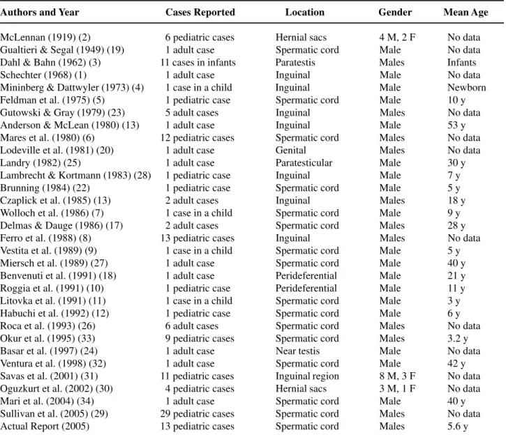
Table 1 – Cases of adrenal rests in the structures around the testis reported in the available literature.
Authors and Year Cases Reported Location Gender Mean Age
McLennan (1919) (2) 06 pediatric cases Hernial sacs 4 M, 2 F No data Gualtieri & Segal (1949) (19) 01 adult case Spermatic cord Male No data Dahl & Bahn (1962) (3) 11 cases in infants Paratestis Males Infants
Schechter (1968) (1) 01 adult case Inguinal Male No data
Mininberg & Dattwyler (1973) (4) 01 case in a child Inguinal Male Newborn Feldman et al. (1975) (5) 01 pediatric case Spermatic cord Male 10 y Gutowski & Gray (1979) (23) 05 adult cases Inguinal Males No data Anderson & McLean (1980) (13) 01 adult case Inguinal Male 53 y Mares et al. (1980) (6) 12 pediatric cases Spermatic cord Males No data Lodeville et al. (1981) (20) 01 adult case Genital Males No data
Landry (1982) (25) 01 adult case Paratesticular Male 30 y
Lambrecht & Kortmann (1983) (28) 01 pediatric case Inguinal Male 7 y Brunning (1984) (22) 01 pediatric case Spermatic cord Male 5 y Czaplick et al. (1985) (13) 02 adult cases Inguinal Males 18 y Wolloch et al. (1986) (7) 01 case in a child Spermatic cord Male 9 y Delmas & Dauge (1986) (17) 02 adult cases Spermatic cord Males 28 y Ferro et al. (1988) (8) 13 pediatric cases Inguinal Males No data Vestita et al. (1989) (9) 01 case in a child Spermatic cord Male 5 y Miersch et al. (1989) (27) 01 adult case Spermatic cord Male 40 y Benvenuti et al. (1991) (18) 01 adult case Perideferential Male 21 y Roggia et al. (1991) (10) 01 pediatric case Perideferential Male 11 y Litovka et al. (1991) (11) 01 case in a child Spermatic cord Male 3 y Habuchi et al. (1992) (12) 01 pediatric case Spermatic cord Male 6 y Roca et al. (1993) (26) 06 adult cases Spermatic cord Males No data Okur et al. (1995) (33) 09 pediatric cases Spermatic cord Males 3.2 y
Basar et al. (1997) (24) 01 adult case Near testis Male No data
Embriologically the adrenal develops from 2 primordia: the cortex arises from the mesoderm and the medulla from ectoderm of the neural crest. The primitive cortex is formed during the 4th and 5th weeks from mesothelial cells formed between the mesentery root and the developing gonad that prolif-erate, separate and condense in the mesenchyma of the dorsal abdominal wall. Another group of cells from the same area is added to this later to form the defini-tive cortex. Cells from the neural crest invade the primitive cortex to form the medulla. Encapsulation of the medulla occurs late in fetal development (7,10,13). It is generally accepted that these adrenal rests were due to mechanical separation and displace-ment of portions of cortical tissue during migration and descent of the sex glands in the male embryonic development. They also may have a multiple primor-dial origin from pluripotent cells in the original loca-tions (13,14). Some heterotopic tissue remains in the area of the adrenal gland near the kidney, but others may migrate with the genitalia descent to the pelvis and scrotum. Some authors estimated that these rests may be present in 50% of newborns but most of them become atrophic by adult life (1,15). Other organs in which an accessory adrenal has been found are the colon, pancreas, retroperitoneum, liver, broad liga-ment and celiac plexus (7,16).
The pathologic appearance of this tissue is characteristic. The findings consisted of a thin yellow-ish nodule 1 to 5 mm in diameter embedded in cremas-teric fibers (13). Adrenal rests situated far from the original gland are composed entirely of cortical adre-nal tissue with no evidence of medullary cells found in these rests, but the more proximal may contain me-dulla. Usually a capsule of connective tissue with small blood vessels can be seen surrounding these nodules (3). Of the three cortical layers, predominate the fasciculata and glomerulosa. The reticularis layer is usually seen only in older children (6).
Most cases of ectopic adrenal tissue in sper-matic cord have been found incidentally during sur-gical procedures (like herniotomy, orchiopexy, etc) in the inguinoscrotal region (17,18). Examples of heterotopic tissues in autopsies have been reported in adults and children usually underneath the capsule of the kidney (1-3,6,16).
The clinical implications of those rests are essential in the surgical approach of the patients. Some authors cite a compensatory functional hyper-trophy of these tissues in rats and human beings in which both adrenals were extirpated (1,6). In pa-tients who have undergone bilateral adrenalectomy due to pathologic ACTH production, compensatory hyperplasia of the ectopic adrenal tissue may be re-sponsible for the recurrence of the disease (7,10). Another clinical aspect is the possibility of forma-tion and development of malignant diseases in the ectopic adrenal cells (13,19-21,34). Although the occurrence of neoplasm in ectopic adrenal nodules is far from common, pheochromocytoma, Leydig cell’s tumor and adrenal adenoma has been reported (21,29,35).
Based on these facts, we think that removal of ectopic adrenal tissue in the spermatic cord would be warranted whenever encountered during surgical operations in inguinal region. It is very easy to excise the adrenocortical ectopic tissue during the groin sur-gery; however, meticulous dissection of the spermatic cord should not be performed in order to avoid the damage of the spermatic vessels and deferens. The nodule is usually embedded in the cremasteric fibers of the spermatic cord, very close to the deferens and attached to the hernia sac and it is very simple to have it dissected free without vascular injury. The lesion of the deferens has not been reported in the litera-ture. In agreement with others authors, we consider that it is also important for urologist to keep in mind the possibility that a nodule around the spermatic cord may be ectopic adrenal tissue. It is reasonable to ex-cise this nodule without jeopardizing the viability of the spermatic cord structures.
CONFLICT OF INTEREST
None declared.
REFERENCES
2. McLennan A: On the presence of adrenal rests in the walls of hernial sacs. Surg Gynec Obst. 1919; 29: 387. 3. Dahl EV, Bahn RC: Aberrant adrenal cortical tissue near the testis in human infants. Am J Pathol. 1962; 40: 587-598.
4. Mininberg DT, Dattwyler B: Ectopic adrenal tumor presenting as torsion of the spermatic cord in a new-born infant. J Urol. 1973; 109: 1037-8.
5. Feldman AE, Rosenthal RS, Shaw JL: Aberrant adre-nal tissue: an incidental finding during orchiopexy. J Urol. 1975; 113: 706-8.
6. Mares AJ, Shkolnik A, Sacks M, Feuchtwanger MM: Aberrant (ectopic) adrenocortical tissue along the sper-matic cord. J Pediatr Surg. 1980; 15: 289-92. 7. Wolloch Y, Ziv Y, Dintsman M: Accessory adrenal: an
incidental finding during orchiopexy. Panminerva Med. 1986; 28: 47-9.
8. Ferro F, Bosman C, Caterino S et al. Ectopia corticosurrenale nel cordone spermatico: Solo una curiositá anatomica? Acta Urol Ital. 1988; 5: 415-7. 9. Vestita G, Veneziani P, Manghisi D, Scavelli V, Sorino
F, Gabrieli G, et al.: A rare occurrence of adrenal ectopy im the spermatic cord. A short clinical note. G Chir. 1989; 10: 499-500.
10. Roggia A, Marandola P, Broggini P, Bono P, de Francesco O, Rovati L: Ectopic adrenal cortex tissue in the spermatic cord: clinico-surgical implications. Arch Esp Urol. 1991; 44: 1165-6.
11. Litovka VK, Zhurilo IP, Khudiakov AE: Atopic adre-nal tissue in the testicle of a small child with cryp-torchism. Khirurgiia (Mosk). 1991; 8: 164.
12. Habuchi T, Mizutani Y, Miyakawa M: Ectopic aber-rant adrenals with epididymal abnormality. Urology. 1992; 39: 251-3.
13. Anderson JR, Ross AH: Ectopic adrenal tissue in adults. Postgrad Med J. 1980; 56: 806-8.
14. O´Crowley CR, Martland HS: Adrenal heterotopia rests and so-called Grawitz tumors. J Urol. 1943; 50: 756-68. 15. Czaplicki M, Bablok L, Kuzaka B, Janczewski Z: Heterotopic adrenal tissue. Int Urol Nephrol. 1985; 17: 177-81.
16. Nelson AA: Accessory adrenal cortical tissue. Arch Pathol 1939; 27: 955-9.
17. Delmas V, Dauge MC: Accessory adrenals in the sper-matic cord. Apropos of 2 cases. Ann Urol (Paris). 1986; 20: 261-4.
18. Benvenuti S, Sepich CA, Cecchi M, Castagna M, Viacava P, Fiorentini L: Peri-deferential ectopic adre-nal gland. Apropos of a case. J Urol (Paris). 1991; 97: 107-8.
19. Gualtieri T, Segal AD: Case of adrenal type tumor of spermatic cord. J Urol. 1949; 61: 949-5.
20. Lodeville D, Zaroli A, Lampertico P: Adenomatoid tumor of the male genital tract: report of three cases, one associated with adrenal cortical rest. Pathologica. 1981; 73: 629-37.
21. Vela Navarrete R, Barat A, Berrocal A, Lopez de Alda A, Quezada F: Testicular adrenal rests tumor: a diffi-cult diagnosis. Actas Urol Esp. 1990; 14: 146-8. 22. Bruning H, Kootstra G, Walther FJ, Arends JW:
Ec-topic adrenocortical tissue along the spermatic cord. Z Kinderchir. 1984; 39: 269-70.
23. Gutowski WT 3rd, Gray G Jr: Ectopic adrenal in in-guinal hernia sacs. J Urol. 1979; 121: 353-4.
24. Basar M, Erdogan S, Aydoganli L, Basar H, Kulacoglu S, Akalin Z: Aberrant adrenal cortical tissue adjacent to immature testis. Arch Ital Urol Androl. 1997; 69: 141-2.
25. Landry GS: Case for diagnosis: ectopic adrenal tissue in testicular adnexa. Mil Med. 1982; 147: 679-80. 26. Roca Suarez A, Alvarez Ossorio JL, Del Toro Bacerra
JA, Maximiano Vasquez R, Gordon Laporte R: Adre-nal ectopia in the spermatic cord. Actas Urol Esp. 1993; 17: 584-7.
27. Miersch WD, Vogel J, Molitor D, Winter P: Ectopic adrenal cortex tissue. Z Urol Nephrol. 1989; 82: 487-90.
28. Lambrecht W, Kortmann KB: Incidence and signifi-cance of accessory adrenal tissue in the inguinal re-gion in childhood. Chirurg. 1983; 54: 39-41.
29. Sullivan JG, Gohel M, Kinder RB: Ectopic adrenocor-tical tissue found at groin exploration in children: in-cidence in relation to diagnosis, age and sex. BJU Int. 2005; 95: 407-10.
30. Oguzkurt P, Oz S, Kayaselcuk F: Ectopic adrenal tis-sue: an incidental finding during inguinoscrotal opera-tions in children. Hernia. 2002; 6: 62-3.
31. Savas C, Candir O, Bezir M, Cakmak M: Ectopic adrenocortical nodules along the spermatic cord of children. Int Urol Nephrol. 2001; 32: 681-5.
32. Ventura L, Leocata P, Hind A, Greco I, Ventura T: Ectopic adrenal tissue in the spermatic cord. Case re-port and review of the literature. Arch Ital Urol Androl. 1998; 70: 15-8.
33. Okur H, Kucukaydin M, Kazez A, Kontas O: Ectopic adrenal tissue in the inguinal region in children. Pediatr Pathol Lab Med. 1995; 15: 763-7.
EDITORIAL COMMENT
Heterotopic or ectopic adrenal cortical tis-sue (EACT) are found in the upper abdomen or any-where along the path of descent of the gonads. The locations where EACT can be found are: celiac axis area (32%); broad ligament (23%); adnexa of the testes (7.5%); kidney (subcapsular upper pole) (0.1%-6%) and spermatic cord (3.8%-9.3%) (1,2). These anatomic locations can be explained on an embryologic basis given the close spatial relation-ship between the developing kidneys and adrenal glands. There are even bizarre anatomic sites where one can find these EACT, such as: placenta, liver, lung and intracranial cavity (1). Usually these adre-nal rests are found incidentally during inguiadre-nal op-erations and present macroscopically as bright yel-low small nodules (1-5mm in diameter) and micro-scopically as lipid rich cortical cells without a med-ullary component (1-3). These rests have some clini-cal significance as they may undergo marked hy-perplasia in conditions associated with excessive ACTH production, and occasionally may give rise to neoplasms. The overall incidence of EACT in different studies varies from 1% to 9.3% in pediat-ric patients. These big series in the literature stick out the importance of recognizing and removing these EACT, whenever encountered, owing to the clinical relevance of these ectopies (2-5).
REFERENCES
1. Lack EE. Heterotopic and Accessory Adrenal Tissues In: Lack EE, (ed.), Tumors of the Adrenal Gland and Extra-Adrenal Paraganglia. Fascicle 19, third series. Washington, DC: Armed Forces Institute of Pathol-ogy. 1997; pp. 34-5.
2. Mares AJ, Shkolnik A, Sacks M, Feuchtwanger MM: Aberrant (ectopic) adrenocortical tissue along the sper-matic cord. J Pediatr Surg. 1980; 15: 289-92. 3. Savas C, Candir O, Bezir M, Cakmak M: Ectopic
adrenocortical nodules along the spermatic cord of children. Int Urol Nephrol. 2001; 32: 681-5.
4. Sullivan JG, Gohel M, Kinder RB: Ectopic adrenocor-tical tissue found at groin exploration in children: in-cidence in relation to diagnosis, age and sex. BJU Int. 2005; 95: 407-10.
5. Ferro F, Bosman C, Casterino S et al. Ectopia corticosurrenale nel cordone spermatico: Solo una curiositá anatomica? Acta Urol Ital 5: 415-417, 1988.
Dr. Patrícia S. de Matos
Department of Pathology, School of Medicine, State University of Campinas São Paulo, Brazil
35. Abe T, Matsuda H, Shindo J, Nonomura K, Koyanagi T: Ectopic pheochromocytoma arising in the spermatic cord 5 years after removal of bilateral carotid body
tumors and adrenal pheochromocytomas. Int J Urol. 2000; 7: 110-1.
Accepted after revision: January 11, 2006
Correspondence address: Dr. Roberto Méndez
Department of Paediatric Surgery
Complexo Hospitalario Universitario de Santiago Avda. Choupana s/n.