Fisioter. Mov., Curitiba, v. 27, n. 4, p. 565-572, out./dez. 2014 Licenciado sob uma Licença Creative Commons DOI: http://dx.doi.org.10.1590/0103-5150.027.004.AO08
[T]
Evaluation of clinical and radiographic measures
and reliability of the quadriceps angle measurement
in elderly women with knee osteoarthritis
1[I]
Avaliação das medidas radiográicas e clínicas e coniabilidade
do ângulo do quadríceps em idosas com osteoartrite de joelhos
[A]
Mateus Ramos Amorim[a], Sueli Ferreira da Fonseca[b], Arthur Nascimento Arrieiro[c], Wellington Fabiano Gomes[d], Ana Cristina Rodrigues Lacerda[e]
[a] Physiotherapist graduated at Universidade Federal dos Vales do Jequitinhonha e Mucuri (UFVJM), Diamantina, MG - Brazil, e-mail: [email protected]
[b] Physiotherapist graduated at Universidade Federal dos Vales do Jequitinhonha e Mucuri (UFVJM), Diamantina, MG - Brazil, e-mail: [email protected]
[c] MSc, Universidade Federal dos Vales do Jequitinhonha e Mucuri (UFVJM), Programa Multicêntrico de Ciências Fisiológicas, Diamantina, MG - Brazil, e-mail: [email protected]
[d] PhD, professor, Universidade Federal dos Vales do Jequitinhonha e Mucuri (UFVJM), Departamento de Fisioterapia, Diamantina, MG - Brazil, e-mail: [email protected]
[e] PhD, professor, Universidade Federal dos Vales do Jequitinhonha e Mucuri (UFVJM), Departamento de Fisioterapia, Diamantina, MG - Brazil, e-mail: [email protected]
[R]
Abstract
Introduction: Knees osteoarthritis (OA) is a complex degenerative disease with intra-articular changes affecting the amplitude of the quadriceps angle (Q). To measure this variable, it is necessary to use reli-able protocols aiming at methodological reproducibility. The objective was to evaluate the intra-examiner and inter-examiner reliability of clinical and radiographic measures of the Q angle and to investigate the
relationship between the degree of OA and the magnitude of this angle in the elderly. Materials and methods: 23 volunteers had the Q angle measured by two evaluators at 48-h interval. Clinical measurements were collected by using the universal goniometer in the same position adopted in the radiographic examination.
Results: The intra-examiner reliability was good (0.722 to 0.763) for radiographic measurements and low (0.518 to 0.574) for clinical assessment, while inter-examiner reliability was moderate (0.634) for radio-graphic measurements and low (0.499) to the clinics. The correlation analysis between the radioradio-graphic val-ues with the OA classification showed no correlation between them (p = 0.824 and r = -0.024). Conclusion: Clinically, it is suggested that the radiographic examination is preferable to evaluate the Q angle of elderly women with knee osteoarthritis. Moreover, the magnitude of this angle did not correlate with the degree of impairment of OA in this population.
Keywords: Reliability. Q angle. Osteoarthritis. Goniometry. Physical Therapy. [B]
Resumo
Introdução: A osteoartrite (OA) de joelhos é uma doença degenerativa complexa com alterações intra-articula-res que comprometem a amplitude do ângulo do quadríceps (Q). Para mensuração dessa variável, é necessária a utilização de protocolos que apresentem confiabilidade, visando reprodutibilidade metodológica. Objetivo: Avaliar a confiabilidade intra e interexaminadores das medidas clínicas e radiográficas do ângulo Q e verificar se existe relação entre o grau de OA e a magnitude deste ângulo em idosas. Materiais e métodos: 23 voluntá-rias, tiveram o ângulo Q mensurado por 2 avaliadores, com intervalo de 48 horas. As medidas clínicas foram coletadas por meio do goniômetro universal na mesma posição adotada no exame radiográfico. Resultados: A confiabilidade intraexaminador foi boa (0,722–0,763) nas medidas radiográficas e baixa (0,518–0,574) nas medidas clínicas, enquanto a confiabilidade interexaminadores foi moderada (0,634) nas medidas radiográfi-cas e baixa (0,499) nas clíniradiográfi-cas. A análise da correlação entre os valores radiográficos com a classificação da OA não demonstrou correlação entre os mesmos (p = 0,824 e r = -0,024). Conclusão: Clinicamente, sugere-se que o exame radiográfico seja preferível para avaliação do ângulo Q de idosas com OA de joelhos. Além disto, a magnitude deste ângulo não se relacionou com o grau de acometimento da OA nesta população. [K]
Palavras-chave: Confiabilidade. Ângulo Q. Osteoartrite. Goniometria. Fisioterapia.
Introduction
Brazil is currently going through a phenome-non known as population aging (1). Along with the changes on the population's age structure, epide-miological changes, characterized by diseases and risk factors related to lifestyle, have also been ob-served (2). Currently, there's an estimative of 17.6 million of elderly in Brazil (3) and osteoarthritis (OA) is among the group of common diseases found in this population.
OA is a chronic-degenerative disease affecting both articular cartilage and other peri-articular structures, affecting women more than men (4, 5). Among the main characteristics of the disease we can list pain, stiffness and loss of physical function (4). The knee joint is the most commonly affected weight-bearing
joints (6). In Brazilian population, the prevalence of knee OA was found in 37% of seniors surveyed (7).
The misalignment of the knee joint in the fron-tal plane is one of the main predisposing factors for the onset and progression of OA (8, 9). According to Hinman et al. (10), alignment variations larger than 5° — both in varus and in valgus — are sufficient to influence the functionality of the knee joint. It is believed that the presence of osteophytes, the nar-rowing of intra-articular and also the sclerosis of sub-chondral bone (11) affect the space arrangement of the bone structures adjacent to the knee joint. These lesions could lead to a consequent loss of joint bio-mechanics that compromises the amplitude of the quadriceps angle (Q).
measurement in elderly women with knee osteoarthritis 567
in the frontal plane. This angle is formed between the line of action of the quadriceps muscle and the direction of the patellar tendon (13). Thus, one may infer that the Q-angle measurement is an essential part of the evaluation of knee joint diseases, with a normal average of 15-20° in women (14). The angle measure may derive from measurements performed by X-rays (15) and clinical exams (16) or by photo-grammetry (17), with the first two methods using the goniometer. The manual goniometer is a widely-used technique in clinical practice of the physiotherapists, in order to evaluate the range of motion. Among the advantages of this methodology we find the low cost of the instrument and its easy-to-measure property (18). The evaluation of the Q-angle is continuously employed in physical therapy practice, in individuals with the patellofemoral syndrome (16, 19).
For clinical rehabilitation and research in physi-cal therapy, the study on the measures’ reliability is essential for ensuring the consistency of the data needed to monitor the treatment of patients (18, 20). The study of this property is important to esti-mate the error of a measurement, i.e., how the value obtained varies from the actual value (21). In other words, it refers to the reproducibility and consistency of measurement (22).
Several tests have been developed in order to assess the reliability of the Q-angle measurement (19, 23, 24). In clinical evaluation, imaging tests such as radiography can be considered as the gold standard for this analysis (25). However, the reli-ability of measurements of this angle has not been studied in elderly women with knee OA. Therefore, we propose controlled clinical and radiographic examinations, in order to assess the reliability of Q-angle measurements and investigate a possible relationship between the degree of OA and the magnitude of the Q-angle in this population. The objectives of this study were to evaluate the intra-examiner and inter-intra-examiner reliability of clinical and radiographic measures of Q-angle in elderly women with knee OA and to investigate the rela-tionship between the degree of OA and the Q-angle magnitude in this population.
The choice for studying this population was due to a greater prevalence, incidence, severity and func-tional impact of knee osteoarthritis within this group of individuals, what could lead to important limita-tions and burden the public health service with long rehabilitation process (25).
Materials and methods
The project was approved by the Ethics Commitee of the Federal University of Jequitinhonha and Mucuri Valleys (049/09) and fulfills Resolution 196/96, of National Health Council concerning the Human Research Code of Ethics. This was a blinded study, where the Q-angle measurement in elderly women with knee OA was assessed by clinical and radiographic exams.
Subjects
To evaluate data reliability, two previously trained examiners (1 and 2) — so called experienced — were the study subjects. A pilot study was conducted for the measured variables, with the purpose of training the researchers. The achievement of the two evaluation methods of the Q-angle was made independently, with an interval of 48 hours to assess the inter-examiner reliability and immediately repeated after the previous examiner, evaluating the intraobserver reliability and assessing the reliability of inter-examiners’ measures.
Examiners were blinded from measures under-taken by the previous examiner and there was no marking of anatomical landmarks during assess-ments, in order to avoid any influence on measure -ments between the examiners. Data collected on the first day of evaluation were used for the analysis of inter-examiner reliability. Examiners did not have access to previous records by the end of data col-lection. The forms of both examiners were distinct, not allowing the comparison of the measurements previously performed.
Sample
A sample calculation (26) was performed based on the dependent variables of the study (clinical and radiographic measures of Q-angle), allowing an oscil-lation of 10% around the mean of the data obtained in the study. The sample needed for the study included 21 volunteers and 42 knees for evaluation.
knee at 30o and tibia 30 upright. The knee flexion
was kept during the test by using a goniometer, fixed at 30° (11). Despite submitting weightbearing on the knees, the voluntaries were told to leave the quad-riceps femoris muscle relaxed while maintaining an appraiser alignment of the hip. The feet of the par-ticipants were placed in Romberg position, i.e., the medial edges of the foot touching each other (27).
To be included in the study, the volunteers should be aged 60 years and above, have a diagnosis of bilat-eral osteoarthritis based on radiographic criteria of Kellgren and Lawrence (28), be able to return to the same spot assessment after 48 hours to a reassess-ment, have not undergone any surgical procedures on the lower limbs and have no recent trauma in the knees. In this case, 24 elderly women were included in the study, and, therefore, 48 knees were investigated.
Initially, the volunteers answered a questionnaire containing structured data, such as: name, age and phone numbers; and questions about the inclusion criteria mentioned above. At that moment, there was a measure of participants’ body weight and height.
Clinical and radiographic measures of Q-angle of the elderly were held in Santa Casa de Caridade de Diamantina/MG and in the Outpatient Physiotherapy Care of UFVJM respectively.
Radiographic measure
For this condition, the radiographic views were used for the OA classification. The knees in weight-bearing and semiflexion emphasized the narrowing of the joint space (11) and put the patella within the trochlear groove (24).
To evaluate the radiographically-derived Q-angle measurement, the tibial tubercle, the midpoint of the patella and the femoral shaft axis were identified on radiographs by two examiners.
The Q-angle was the one formed by the goniom-eter's fixed arm, aligned with the bisecting line of the femur and, by the movable arm, aligned with the tibial tuberosity marking, with the fulcrum located at the center of the patella.
Clinical measure
The data for clinical analysis were collected regardless of the radiographic examination. The
position adopted for clinical measure was simi-lar to that used for radiographic examination. An examiner maintained the hip alignment while the other conducted the measurement. The center of the patella was initially defined as the midpoint of a line that brought the most medial and the most lateral region of the patella, determined through the patella edges’ palpation. Furthermore, the most prominent region of the tibial tuberosity was de-marcated, by palpation, after the voluntaries’ posi-tioning. Thereafter, the bisection line of the femur was determined by aligning the goniometer's fixed arm from the center of the patella until the anterior superior iliac spine (14).
Statistical analysis
The SPSS software version 18.0 for Windows was used for statistical analysis. The intra and inter-examiner reliability was assessed by the Intraclass Correlation Coefficient (ICC), which measures the internal consistency between two variables or fac-tors. The magnitude of the association was defined according to the Cronbach alpha coefficient as (≤ 0.6), low (0.6 to ≤ 0.7) moderate (0.7 to ≤ 0.8) good, (0.8 to ≤ 0.9) and very good (≥ 0.9) excellent (29). The Shapiro-Wilk normality test was used. Afterwards, the Spearman correlation test was used to verify the existence of a correlation between the degree of OA and the magnitude of the Q-angle. The confidence interval was 95% (p < 0.05).
In the descriptive analysis, Q-angle values above 20° were considered valgus, while the ones below 15° were considered varus.
Twenty-three elderly women were evaluated and one of them showed no radiographic diagnosis of knee bilateral OA, being excluded from the analysis. Both knees (n = 46) were considered in the analy -sis of reliability data, since the volunteers had bilat-eral OA. All measurements obtained by the examin-ers on the first day (n = 92) were considered for the descriptive and correlation analysis.
Results
measurement in elderly women with knee osteoarthritis 569
Table 1 - Characterization of the studied population
Variable N (23)
Age (years ± SD) 67.30 ± 4.06
BMI (kg/m ± SD) 29.58 ± 3.81
Radiographic level of the knee OA (%)
Level 1 26
Level 2 26
Level 3 22
Level 4 26
Note: BMI = Body Mass Index; SD = Standard Deviation; OA = Osteoarthritis.
requirements of Kellgren and Lawrence (28), were included on the study (Table 1).
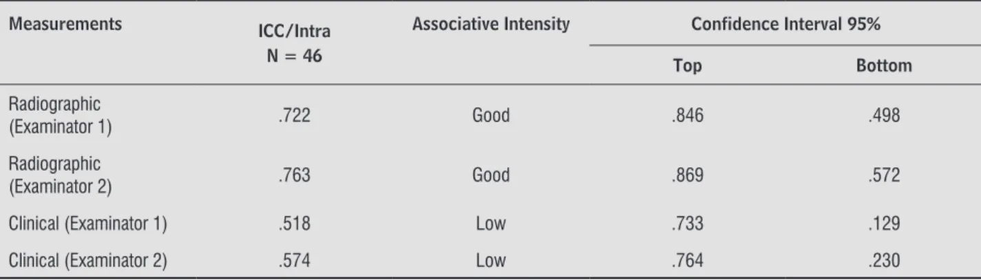
The intexaminer reliability was good for ra-diographically-derived measures and low for clinical measures (Table 2).
The inter-examiner reliability was moderate for clinical measures (Table 3).
Later, it was found that the data did not show normal distribution and, since the radiographic measurements
showed better reproducibility, it was analyzed the cor-relation between the values obtained by these measures and the radiographic severity of OA, with no correlation observed between them (p = 0.824 and r = -0.024).
The results obtained by the descriptive analysis of all radiographically-derived values showed a preva-lence of: (varus = 93% and normal values = 3%) for examiner 1 and (varus = 83%, normal = 13% and valgus = 1%) for examiner 2.
Table 2 - Intra-examiner reliability obtained through the ICC
Measurements ICC/Intra N = 46
Associative Intensity Confidence Interval 95%
Top Bottom
Radiographic
(Examinator 1) .722 Good .846 .498
Radiographic
(Examinator 2) .763 Good .869 .572
Clinical (Examinator 1) .518 Low .733 .129
Clinical (Examinator 2) .574 Low .764 .230
Note: ICC = Intraclass Correlation Coeficient.
Table 3 - Inter-examiner reliability obtained through the ICC
Measurements ICC/Intra N = 46
Associative Intensity Confidence Interval 95%
Top Bottom
Radiographic .634 Moderate .798 .339
Clinical .499 Low .723 .095
some of the volunteers in the study were obese (48% had a BMI > 30 kg/m²) (32), we believe that this fact probably made it difficult to palpate bony structures necessary for clinical assessment of the quadriceps angle, thus influencing the reliability of clinical measurement. Furthermore, the positioning of the volunteers did not influence the method’s in -tensity of association, since the attitude during the two analyses was similar. Thus, the physical therapist, as a competent professional, should seek all neces-sary information on the follow up treatment of pa-tient under his∕her responsibility, using radiographic measures to quadriceps angle analysis, especially in obese individuals (33).
The low reliability values found for the two mea-sures appear to be related to the distance and the arrangement of referential anatomical points for the Q-angle measurement. This fact may have hindered the placement of the goniometer arms, considering that the conformation of the anatomical structures involved such as the muscle mass of the quadriceps femoris muscle is affected by the posture. In paral-lel, the placement of multiple joint complexes seems to alter the measure (17). The study by Chacur et al. (34), evaluating the Q-angle through clinical measurements, found a high prevalence of valgus and excessive Q-angle in obese women with knee OA. Given that the increased Q-angle would encour-age more pressure on the lateral compartment of the joint femoro-patellar (35), providing greater weight bearing in one compartment of the knee and leading to early OA (36), a possible relation-ship between the disease severity and the magni-tude of the Q-angle was assessed, but there was no correlation between them in the present study. Moreover, the descriptive analysis of the voluntaries demonstrated a high prevalence of varus (> 80%) rather than valgus (< 1%) in the study population. Thus, we believe that other biomechanical and/ or anthropometric factors beyond varus, such as decreased Q-angle, overweight, muscle weakness of the quadriceps femoris or thickness of the articular cartilage (37, 38) may have influenced the degree of the study voluntaries’ OA.
There are limitations in this study which should be interpreted in light of the proposed objectives. It should be mentioned that the data reliability will only work on the positions adopted during the mea-sures and they cannot be extrapolated to other posi-tions taken or other imaging tests.
Discussion
The results of this study indicated good reliabil-ity for measures of quadriceps angle, derived radio-graphically, while clinical measurements obtained in the same position showed low reliability. In the comparison of inter-examiners of the evaluation methods, a moderate agreement for radiographic measurement and low agreement for the clinic mea-surement were verified. The clinical relevance of this result lies in the ease-to-measure property of data on outpatient routine, since the radiographic examination is usually employed in the diagnosis and on the monitoring of knee osteoarthritis. Then, it was evaluated the relationship between radio-logical disease severity and the magnitude of the Q-angle. No correlation between them was observed in the present study.
These findings showed a greater reliability than those from Greene et al. (15) which reported low intra and inter-examiner clinically and radiographi-cally-derived Q-angle measurement in asymptomatic individuals. Instead, Caylor et al. (30) found, on the clinical measurement of the Q-angle, very good intra-examiner reliability and moderate inter-intra-examiner reliability in individuals with anterior knee pain. This fact may be resulted from the positions adopted by these studies, which may differ from the present one. In this case, the use of different postural assessment methods for Q-angle measuring should be cautious, since methodologies have already shown existing dif-ferences between each other (17).
According to Moncrieff and Livingston (9) the characteristics of reliability will depend on the evalu-ators’ ability to correctly repeat the marking of ana-tomical landmarks used to define the measures of interest. Therefore, the greater ease of the examiner on identifying anatomical structures — necessary for Q-angle measurement on radiographs — may have influenced the reliability of the measure. Although the radiographic examination is considered the gold standard for measuring the Q-angle, the difficult vi -sualization of the anterior tibial tuberosity on radio-graphs may have affected the association strength of the method, so much so that the intensity found was not superior to good. The anatomical accident had insufficient bone overlap to generate remarkable opacity to the ionizing radiation.
measurement in elderly women with knee osteoarthritis 571 9. Moncrieff MJ, Livingston LA. Reliability of a
digital-photographic-goniometric method for coronal-plane lower limb measurements. J Sport Rehabil. 2009;18(2):296-315.
10. Hinman RS, May RL, Crossley KM. Is there an alterna-tive to the full-leg radiograph for determining knee joint alignment in osteoarthritis? Arthritis Rheum. 2006;55(2):306-13.
11. Davies AP, Glasgow MMS. Imaging in osteoarthritis: a guide to requesting plain X-rays of the degenerate knee. Knee. 2000;7(3):139-43.
12. Hungerford DS, Barry M. Biomechanics of the patello-femoral joint. Clin Orthop Relat Res. 1979;(144):9-15.
13. Horton MG, Hall TL. Quadriceps femoris muscle angle: normal values and relationships with gender and selected skeletal measures. Phys Ther. 1989; 69(11):897-901.
14. Akinbo SRA, Tella BA, Jimo OO. Comparison of bilat-eral quadriceps angle in asymptomatic and symptom-atic males with unilateral anterior knee pain [inter-net]. J Pain Symptom Contr Palliat Care. 2008;8(1).
15. Greene CC, Edwards TB, Wade MR, Carson EW. Reli-ability of the quadriceps angle measurement. Am J Knee Surg. 2001;14(2):97-103.
16. Smith TO, Davies L, O'Driscoll ML, Donell ST. An evalua-tion of the clinical tests and outcome measures used to assess patellar instability. Knee. 2008;15(4):255-62.
17. Sacco ICN, Alibert S, Queiroz BWC, Pripas D, Kieling I, Kimura AA, et al. Confiabilidade da fotogrametria em relação a goniometria para avaliação postural de mem-bros inferiores. Rev Bras Fisioter. 2007;11(5):411-7.
18. Venturini C, André A, Aguilar BP, Giacomelli B. Confi -abilidade de dois métodos de avaliação da amplitude de movimento ativa de dorsiflexão do tornozelo em indivíduos saudáveis. Acta Fisiatr. 2006;13(1):41-5.
19. Piva SR, Fitzgerald K, Irrgang JJ, Jones S, Hando BR, Browder DA, et al. Reliability of measures of impair-ments associated with patellofemoral pain syndrome. BMC Musculoskelet Disiord. 2006;7:33.
20. Melo de Paula G, Molinero de Paula VR, Almeida GJM, Machado VEI, Barauna MA, Bevilaqua-Grosso D. Cor-relação entre a dor anterior do joelho e a medida do angulo “Q” por intermédio da fotometria computado-rizada. Rev Bras Fisioter. 2004;8(1):39-43.
Future studies should be developed with the aim of clarifying the role of biomechanical and anthro-pometric variables in the misalignment of the lower extremities on elderly women with knee OA.
Conclusion
The present study demonstrated that, in elderly women with knee OA, the evaluation of the Q-angle is more reliable through radiographic than through clinical examination. Also, the magnitude of the quad-riceps Q-angle was unrelated to the degree of impair-ment of knee osteoarthritis in this population.
References
1. Lima LCVC, Bueno MLB. Envelhecimento e gênero: a vulnerabilidade de idosas no Brasil. Rev Saúde e Pesquisa. 2009;2(2):273-80.
2. Marin MJS, Martins AP, Marques F, Feres BOM, Saraiva AKHA, Druzian S. Atenção à saúde do idoso: ações e perspectivas dos profissionais. Rev Bras Geriatr Gerontol. 2008;11(2):245-58.
3. Carvalho JA, Wong LLR. A transição da estrutura etária da população brasileira na primeira metade do século XXI. Cad Saude Publica. 2008;24(3):597-605.
4. Fellet A, Fellet AJ, Fellet L. Osteoartrose: uma revisão. Rev Bras Med. 2007;64(1):55-61.
5. Giorgi RDN. A osteoartrose na prática clínica. Temas Reumatol Clin. 2005;6(1):17-30.
6. Stitik TP, Kaplan RJ, Kamen LB, Vo AN, Bitar AA, Shih VC. Rehabilitation of orthopedic and rheumatologic disorders. 2. Osteoarthritis assessment, treatment, and rehabilitation. Arch Phys Med Rehabil. 2005;86 (3 Suppl 1):S48-55.
7. Vasconcelos KSS, Dias JMD, Dias RC. Relação entre intensidade de dor e capacidade funcional em indi-víduos obesos com osteoartrite de joelho. Rev Bras Fisioter. 2006;10(2):213-8.
32. Lipschitz DA. Screening for nutritional status in the elderly. Prim Care. 1994;21(1):55-67.
33. Fernandes C, Koch HA, Souza EG. O ensino da radio-logia nos cursos de graduação em fisioterapia. Radiol Bras. 2003;36(6):363-6.
34. Chacur EP, Silva LO, Luz GCP, Kaminice FD, Cheik NC. Avaliação antropométrica e do ângulo quadricipital na osteoartrite de joelho em mulheres obesas. Fisioter Pesqui. 2010;17(3):220-4.
35. Mizuno Y, Kumagai M, Mattessich SM, Elias JJ, Ramrattan N, Cosgarea AJ, et al. Q-angle influences tibiofemoral arid patellofemoral kinematics. J Orthop Res. 2001;19(5):834-40.
36. Bedson J, Croft PR. The discordance between clinical and radiographic knee osteoarthritis: a systematic search and summary of the literature. BMC Muscu-loskelet Disord. 2008;9:116.
37. Berry PA, Wluka AE, Davies-Tuck ML, Wang Y, Strauss BJ, Dixon JB, et al. The relationship between body compo-sition and structural changes at the knee. Rheumatol-ogy (Oxford). 2010;49(12):2362-9.
38. Kocak FU, Unver B, Karatosun V, Bakirhan S. Asso-ciations between radiographic changes and function, pain, range of motion, muscle strength and knee function score in patients with osteoarthritis of the knee. J Phys Ther Sci. 2009;21(1):93-7.
Received: 12/26/2013 Recebido: 26/12/2013
Approved: 07/14/2014
Aprovado: 14/07/2014
21. Portney L, Watkins M. Foundations of a clinical re-search: applications to practice. New Jersey: Printece Hall; 2000.
22. Polgar S, Thomas SA. Introduction to research in the health sciences. London: Churchill Livingstone; 2000.
23. Shultz SJ, Nguyen AD, Windley TC, Kulas AS, Botic TL, Beynnon BD. Intratester and Intertester reliability of clinical measures or lower extremity anatomic char-acteristics: implications for multicenter studies. Clin J Sport Med. 2006;16(2):155-61.
24. Herrington L, Nester C. Q-angle undervalued? The relationship between Q-angle and medio-lateral po-sition of the patella. Clin Biomech (Bristol, Avon). 2004;19(10):1070-3.
25. Smith TA, Hunt NJ, Donell ST. The reliability and va-lidity of the Q-angle: a systematic review. Knee Surg Sports Traumatol Arthrosc. 2008;16(12):1068-79.
26. Sampaio IBM. Estatística aplicada à experimentação animal. Belo Horizonte: Fundação de Estudo e Pes-quisa; 2002.
27. Livingston LA, Spaulding SJ. OPTOTRAK Measurement of the Quadriceps Angle Using Standardized Foot Posi-tions. J Athl Train. 2002;37(3):252-5.
28. Kellgren JH, Lawrence JS. Radiological assessement of osteo-arthrosis. Ann Rheum Dis. 1957;16(4):494-502.
29. Jensen MP. Questionnaire validation: a brief guide of readers of the research literature. Clin J Pain. 2003; 19(6):345-52.
30. Caylor D, Fites R, Worrell TW. The relationship between quadriceps angle and anterior knee pain syn-drome. J Orthop Sports Phys Ther. 1993;17(1):11-6.