Transperitoneal versus Extraperitoneal Laparoscopic Radical
Prostatectomy During the Learning Curve: Does the Surgical
Approach Affect the Complication Rate?
Tiberio M. Siqueira Jr., Anuar I. Mitre, Ricardo J. Duarte, Humberto Nascimento, Francualdo
Barreto, Evandro Falcao, Roberto I. Lopes, Miguel Srougi
Section of Urology (TMS, HN, EF, FB), Getulio Vargas Hospital, Recife, Pernambuco, Brazil and
Division of Urology (TMS, AIM, RJD, RIL, MS), School of Medicine, University of Sao Paulo, Sao
Paulo, Brazil
ABSTRACT
Purpose: To compare the perioperative complication rate obtained with the transperitoneal laparoscopic radical prostate-ctomy (TLRP) and with the extraperitoneal LRP (ELRP) during the learning curve (LC).
Materials and Methods: Data of the initial 40 TLRP (Group 1) were retrospectively compared with the initial 40 ELRP (Group 2). Each Group of patients was operated by two different surgeons.
Results: The overall surgical time (175 min x 267.6 min; p < 0.001) and estimated blood loss (177.5 mL x 292.4 mL; p < 0.001) were statistically better in the Group 1. Two intraoperative complications were observed in Group 1 (5%) repre-sented by one case of bleeding and one case of rectal injury, whereas four complications (10%) were observed in Group 2, represented by two cases of bleeding, one bladder and one rectal injuries (p = 0.675). Open conversion occurred once in each Group (2.5%). Overall postoperative complications were similar (52.5% x 35%; p = 0.365). Major early postopera-tive complications occurred in three and in one case in Group 1 and 2, respecpostopera-tively. Group 1 had two peritonitis (fecal and urinary), leading to one death in this group.
Conclusions: No statistical differences in overall complication rates were observed. The transperitoneal approach pre-sented more serious complications during the early postoperative time and this fact is attributed to the potential chance of intraperitoneal peritonitis not observed with the extraperitoneal route.
Key words: prostate� prostatic neoplasms� prostatectomy� laparoscopyprostate� prostatic neoplasms� prostatectomy� laparoscopy
Int Braz J Urol. 2010; 36: 450-7
INTRODUCTION
Since 1998, laparoscopic radical prostatec-tomy (LRP) has gained worldwide popularity, based on the Montsouris initial publication with the trans-peritoneal LRP (TLRP) (1) followed by the Brussels initial publication with the extraperitoneal approach (ELRP) (2).
doi: 10.1590/S1677-55382010000400008
Analyzing the comparative studies for LRP during the LC, it can be noted that the vast majority of groups started their programs using the transperi-toneal approach, switching later on to the extraperi-toneal route (7-9). This observation generates a bias
on results, once the indings of worse results with the
transperitoneal approach when compared with the extraperitoneal approach is overlooked. The results
observed with the TLRP in these studies relect the
suboptimal results found during the initial phase of a LRP program and the results achieved with the
ex-traperitoneal approach are the relex of an “already”
well-trained surgical team.
The aim of this study was to compare the initial perioperative complications obtained with both approaches used to perform LRP and identify, if possible, a more suitable approach to use during the initial stages of a LRP program.
MATERIALS AND METHODS
This retrospective study was approved by each institutional research and ethical committee. The
data of the irst 40 TLRP performed between March,
2004 and November, 2007 (Group 1) performed at Getúlio Vargas Hospital of Recife were recorded and
compared with the irst 40 ELRP performed between
August, 2003 and June, 2006 (Group 2) at Clinics Hospital of State University of São Paulo.
Each group was operated by only one experi-enced laparoscopic urologic surgeon in two uro-lapa-roscopic referral centers in this country. By the time of this study, each surgeon had already performed more than 250 laparoscopic surgeries, including partial ne-phrectomies, radical nene-phrectomies, donor nephrec-tomies, adrenalecnephrec-tomies, pyeloplasties and others.
Inclusion criteria were age ≤ 75 years old, clinically
localized prostate tumors (cT1 and cT2N0M0), total
PSA ≤ 10 ng/dL and Gleason score ≤ 7.
Surgical Technique
All TLRP were performed by the Montsouris technique (10) while the ELRP were done by the
Brussels technique (2) with some modiications.
Briely, in the Montsouris technique, the vas deferens and seminal vesicles were irstly dissected
through the retrovesical space. After entering the Retzius space and opening the endopelvic fascia, the dorsal vein complex (DVC) was tied. The bladder neck was incised, reaching the pre-dissected vas defer-ens. Bilaterally, the prostatic pedicles were controlled and an interfascial neurovascular bundle dissection was performed whenever possible (11). Afterwards, the DVC and urethra were cut, leaving the prostate apart for later removal. A running urethrovesical (UV) anastomosis was made in all cases of Group 1 as described by van Velthoven et al. (12).
In the Brussels technique, the Retzius space was digitally created, avoiding transperitoneal entering. The other stages were about the same as transperito-neal approach, differing only in the straight access to vas deferens and seminal vesicles after bladder neck
incision. An interrupted igure of “X” UV anastomosis
was performed in all patients in Group 2.
Pathological Evaluation
All ine-needle biopsies and specimens were
evaluated by the uro-pathology service of each
institu-tion. Positive surgical margin (PSM) was deined as the
presence of tumor at the inked margin. Tumors were graded according to the Gleason score and pathological
staging was based on TNM 1997 classiication.
Statistical Analysis
Analysis of variance was used to compare continuous outcome variables between both groups. The Student’s-t-test was used for homogeneous vari-ances in each group and the Chi-square and Fisher’s exact test were used to compare categorical outcome
variables. Statistical signiicance was deined as P
value < 0.05.
RESULTS
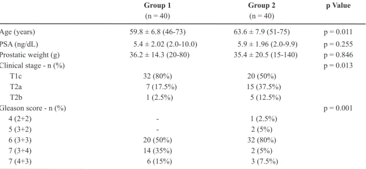
related to the patient’s age, clinical stage and Gleason score. Clinical stage T1c was more common in Group 1 (80%) while cT2 was prevalent in Group 2 (50%). On the other hand, the Gleason score 7 was more prevalent in Group 1 (50% x 12.5%).
The intraoperative data are described in Table-2. Overall surgical time (175 min x 267.6 min; p < 0.001) and estimated blood loss (177.5 mL x 292.4
mL; p < 0.001) were statistically signiicant better in
the Group 1. Two complications (5%) were observed in Group 1, represented by a bleeding from the DVC
and rectal injury. The irst one was controlled after
conversion to the open approach and the last one was treated with intracorporeal suture. Four complica-tions (10%) occurred in Group 2, represented by two cases of bleeding (5%), one bladder (2.5%) and one Table 1 – Preoperative results.
Group 1 Group 2 p Value
(n = 40) (n = 40)
Age (years) 59.8 ± 6.8 (46-73) 63.6 ± 7.9 (51-75) p = 0.011
PSA (ng/dL) 5.4 ± 2.02 (2.0-10.0) 5.9 ± 1.96 (2.0-9.9) p = 0.255 Prostatic weight (g) 36.2 ± 14.3 (20-80) 35.4 ± 20.5 (15-140) p = 0.846
Clinical stage - n (%) p = 0.013
T1c 32 (80%) 20 (50%)
T2a 7 (17.5%) 15 (37.5%)
T2b 1 (2.5%) 5 (12.5%)
Gleason score - n (%) p = 0.001
4 (2+2) - 1 (2.5%)
5 (3+2) - 2 (5%)
6 (3+3) 20 (50%) 32 (80%)
7 (3+4) 14 (35%) 2 (5%)
7 (4+3) 6 (15%) 3 (7.5%)
PSA = prostate-speciic antigen.
Table 2 – Comparison of intraoperative data.
Group 1 Group 2 p Value
(n = 40) (n = 40)
Overall operative time (min) 175.0 ± 48.4 (110-360)
267.6 ± 70.57 (160-540)
p < 0.001
Blood loss (mL) 177.5 ± 148.5
(50-1000)
292.4 ± 173.7 (10-900)
p < 0.001
Open conversion - n (%) 1 (2.5%) 2 (5%) p = 1.000
Complications - n (%)
Overall 2 (5%) 4 (10%) p = 0.675
Complication type - n (%)
Bleeding 1 (2.5%) 2 (5%)
Rectal injury 1 (2.5%) 1 (2.5%)
rectal injury (2.5%). Both bleedings came from the
DVC. The irst one was controlled after conversion
to the open approach and the last one was treated with intracorporeal suture. The bladder injury was recognized and treated by intracorporeal suture. On the other hand, the rectal injury was unrecognized in the intraoperative time and evolved with bloody anal discharge on postoperative day one, which led to an open colostomy.
Postoperative data are described in Table-3. Median time to discharge, early urinary continence and follow-up time were statistically better for Group 2. No statistical difference was observed on early post-operative sexual function, evaluated by the vaginal
penetration rate whether or not using sildenail 100
mg.
The Table-4 shows no statistical difference in overall postoperative complications (52.5% x 35%; p = 0.365). Nonetheless, sub-stratifying the complica-tions, a statistical difference was observed by compar-ing the minor complications durcompar-ing the early and late postoperative time for each group.
The main complications observed in Group 1 were one case each of urinary sepsis, fecal and urinary peritonitis. The sepsis occurred on postoperative day (POD) 8 by Klebsiella pneumoniae despite preopera-tive negapreopera-tive urine culture and trans-operapreopera-tive use of parental ceftriaxone. The patient was readmitted and had an uneventful recovery after appropriate paren-teral antibiotic therapy. The fecal peritonitis occurred on POD 4 due to fecal leakage by the rectal suture line performed intraoperatively. The patient evolved with peritonitis, sepsis and died on POD 35 even after colostomy and parenteral antibiotic therapy. The uri-nary peritonitis occurred due to uriuri-nary leakage from
the posterior aspect of the UV anastomosis, leading to a 1500 cc urine peritoneal collection. After open laparotomy and peritoneal drainage, the patient had an uneventful recovery.
In the Group 2, seven urinary leakages origi-nating from the UV anastomosis occurred and were treated by prolonged bladder catheterization. Of those, six evolved with urinary strictures (bladder neck- 03; bulbar urethra- 02; and meatal urethra- 01), and had an uneventful recovery after appropriate treatment. A further urinary leakage due to the UV anastomosis
evolved with a large retroperitoneal iniltration and
was treated with open drainage, positioning of a tu-bular drain and prolonged bladder catheterization.
Lastly, the Table-5 shows the inal oncological
data. Comparing the results between groups 1 and 2, statistical difference was observed in the biochemi-cal recurrence rate (5% x 20%; p = 0.043), overall incidence of PSM (10.3% x 32.5%; p = 0.016) and pathological stages (pT2: 94.8% x 70% and pT3: 5.2% x 30%; p = 0.005). Nonetheless, no difference was observed when the incidence of PSM was cor-related with the pathological stages. The majority of PSM in Group 1 occurred in pT2c (75%), while this observation was more prevalent in pT3a in Group 2 (61.5%). For pT3b, 100% of PSM occurred in both groups.
COMMENTS
According van Velthoven et al. (4) and Gill et al. (13), about 92% of uro-laparoscopic centers that currently use the extraperitoneal approach, started their laparoscopic programs using the transperitoneal Table 3 – Comparison of postoperative data.
Group 1 Group 2 p Value
(n = 40) (n = 40)
Time to discharge - (days)* 3.0 (3-35) 3.0 (2-17) p = 0.042 Early urinary continence - n (%) 27 (69.2%) 34 (85%) p = 0.033
Sexual intercourse - n (%) 18 (45%) 17 (42.5%) p = 0.368
Follow-up (months) 9.3 ± 7.7 (1-36) 32.9 ± 12.6 (8-50) p < 0.001
route. In general, this observation can cause a bias on results when these accesses are compared in the same series. Such discrepancy in results can even be greater during the initial phase of a LRP program.
Perhaps the best way to overcome the LC in LRP is the incorporation of robotics in clinical practice (14). However, even in robotic LRP, a LC does exist and so far, controversies remain about the choice of the approach to use is these cases. Moreover, the high costs associated with this technique, make it a distant reality for developing countries. Therefore, continuous improvements in LRP technique are
man-datory and identiication of factors that can improve
and shorten the LC is imperative to achieve better results.
The main goal of this retrospective study was to compare the perioperative complication rates of two distinct groups of patients operated each by
the transperitoneal and extraperitoneal approaches during the initial phases of a LRP program. For this, each group was operated by only one urologist, having each a wide experience in retropubic radical pros-tatectomies and in more than 250 uro-laparoscopic surgeries. This study model, despite some points of
criticism, was adopted to analyze the inluence of the
LC over the incidence of complications and to identify factors that could improve the results in this phase. Only Machado et al. (15) performed a similar study and observed better results with the extraperitoneal approach when compared with the transperitoneal route.
Observing the intraoperative data, the patients in Group 1 reached better surgical time and bled
less than the ones in the Group 2 and these indings
can be associated with the better working space and luminosity achieved with the transperitoneal access. Table 4 – Comparison of postoperative complications and reoperations.
Group 1 Group 2 p Value
(n = 40) (n = 40)
Overall complications (before 30th POD): 21 (52.5%) 14 (35%) p = 0.365 Early complications (before 30th POD):
Minor - n (%) p < 0.001
Perineal pain 4 (10%)
-Abdominal wall hematoma 2 (5%)
-Urinary leakage - 7 (17.5%)
Major - n (%) p = 0.241
Fecal peritonitis (death) 1 (2.5%)
-Urinary peritonitis 1 (2.5%)
-Urinary sepsis 1 (2.5%)
-Retroperitoneal urinary iniltration - 1 (2.5%)
Late complications (after 30th POD)
Minor - n (%) p = 0.004
UTI 9 (22.5%)
-Bladder neck stricture 2 (5%) 3 (7.5%)
Urethral stricture 1 (2.5%) 2 (5%)
Urethral meatus stricture - 1 (2.5%)
Major - n (%) 0 0 p = 1.000
Reoperations: n (%) 2 (5%) 2 (5%) p = 1.000
Death 1 (2.5%)
Nonetheless, two major complications were observed in Group 1, causing a urinary and fecal peritonitis, leading to the death of one patient. These results are
the real relex of the LC effect over each LRP program
without any previous experience with LRP.
Urinary leakage can occur up to 28% in LRP during the LC (16-19). Of note, all seven early minor complications observed in the Group 2 were represented by urinary leakages, while only one was observed in the Group 1, which was considered a ma-jor complication. The mama-jority of these cases evolved to urinary strictures and needed surgical treatment. In general, urinary leakages occur due to non-well aligned suture lines, surgery in prostates > 60 grams, use of interrupted sutures and when the extraperito-neal access is chosen (18,19). In general, tension over the UV anastomosis is considered higher when the extraperitoneal access is used instead of the transperi-toneal route, because the bladder remains stacked on the abdominal wall by the urachus (20). In fact, based in these observations, the authors recommend the use of the UV running suture since the initial phases of
the LC. Likewise, to rule out urinary leakage, illing the bladder in with 200 cc of saline after inishing the
UV anastomosis is recommended.
Each group had one major complication on early postoperative time related to urinary leakage, as well as one rectal injury. In Group 1, both com-plications evolved to peritonitis needing reoperation, culminating in one death. On the other hand, these
indings in the Group 2 evolved with less severity
and went well after reoperation.
The incidence of rectal injury occurs in 1.8%-6% (8,21) and is more common during the LC (8,21). According Touijer et al. (22) and Martinez-Piñeiro et al. (19), the majority of injuries occur during the apical dissection. Although the rectal injury had been recognized and sutured during the intraoperative time in one patient in Group 1, the injury presented a fecal leakage on POD 4, leading to peritonitis and death. This fact was attributed to the use of the harmonic shears to dissect the posterior aspect of the prostate, near the apex. Probably, an invisible thermal injury occurred in the rectal wall during the surgery and a later wall necrosis developed, leading to the fecal leakage (19,22).
Important to notice that rectal injury can occur whatever the approach, but this complication tends to have a worse outcome when the transperitoneal route is adopted. The authors strongly recommend the Table 5 – Postoperative pathological data.
Group 1 Group 2 p Value
(n = 39) (n = 40)
Biochemical recurrence 2 (5%) 8 (20%) p = 0.043
Gleason score - n (%) p = 0.365
6 (3+3) 14 (35%) 21 (52.5%)
7
7 (3+4) 7 (4+3)
24 (60%) 19 (47.5%)
5 (12.5%)
16 (40%) 12 (30%) 4 (10%)
8 (3+5) 1 (2.5%) 2 (5%)
8 (4+4) - 1 (2.5%)
BPH 1 (2.5%)
-Positive surgical margins - n (%) 4 (10.3%) 13 (32.5%) p = 0.016
Pathological stage - n (%) p = 0.005
T2 37 (94.8%) 28 (70%)
T3 2 (5.2%) 12 (30%)
use of cold shears instead of the use of any kind of thermal shears to dissect this area to avoid this major complication, no matter which laparoscopic approach chosen.
Bladder injury is considered a rare event and is more common during the LC, reaching 8% (17,23). It can occur with both approaches and usually the injury is recognized and sutured during the surgery. In general, all injuries have an uneventful recovery after appropriate treatment.
Perineal pain is a rare event and was observed in four patients in Group 1 (10%). This was attributed to hyper abduction of legs in order to place the laparo-scopic rack in between. No further cases of this type of complication were observed after discontinuation of this practice.
Epigastric artery injury occurs in about 2% - 6.2% of cases and generally is associated with trocar insertion during transperitoneal surgeries (17,23). This injury rarely occurs during ELRP, because the vessels are easily seen after the extraperitoneal space has been created. The authors suggest puncturing
before the site of trocar placement with a ine needle
in order to verify the route, avoiding this injury. Also is recommended to have a Carter-Thomason device readily to use if necessary.
Finally, urinary tract infection occurs in 1.4% - 2.8% in all cases of LRP, despite of antibiotic prophylaxis (2,24). Generally, these infections are
caused by prolonged indwelling catheter use and/or
inappropriate antibiotic prescription. Currently, the authors suggest the use of quinolones for 14 days after the hospital discharge and the urethral catheter removal as soon as possible, around the postoperative day 7.
The LRP is considered the most challenging laparoscopic surgery in urology. The greatest draw-back of this surgery is its steep LC and consequently the possibility of major complications to occur and weak functional results during this time. Moreover, the initiation of a LRP program demands great caution in order to not overcome the main objective of this surgery: the cure. Therefore, continuous improve-ments and training are mandatory to achieve better outcomes. Based in our results, there was no differ-ence in the inciddiffer-ence of perioperative complications whatever the approach used to operate both groups
during the LC. The incidence and severity of major complications were higher when the transperitoneal approach was adopted.
CONCLUSIONS
The overall complication rate was similar in both approaches. Minor complications occurred in both groups and tended to complete resolution after appropriate treatment. The higher incidence of urinary leakage in Group 2 was directed associated with the interrupted UV anastomosis and indirectly linked with the extraperitoneal route. The transperitoneal ap-proach presented more serious complications during the early postoperative time and this fact is attributed to the potential chance of intraperitoneal peritonitis not observed with the extraperitoneal approach.
CONFLICT OF INTEREST
None declared.
REFERENCES
1. Guillonneau B, Cathelineau X, Barret E, Rozet F,
Vallancien G: Laparoscopic radical prostatectomy. Preliminary evaluation after 28 interventions. Presse Med. 1998; 27: 1570-4.
2. Bollens R, Vanden Bossche M, Roumeguere T, Da-moun A, Ekane S, Hoffmann P, et al.: Extraperitoneal laparoscopic radical prostatectomy. Results after 50 cases. Eur Urol. 2001; 40: 65-9.
3. Porpiglia F, Terrone C, Tarabuzzi R, Billia M, Grande S, Musso F, et al.: Transperitoneal versus extraperito-neal laparoscopic radical prostatectomy: experience of a single center. Urology. 2006; 68: 376-80. 4. van Velthoven RF: Laparoscopic radical
prostatec-tomy: transperitoneal versus retroperitoneal approach: is there an advantage for the patient? Curr Opin Urol. 2005; 15: 83-8.
5. Eden CG, King D, Kooiman GG, Adams TH, Sulli-van ME, Vass JA: Transperitoneal or extraperitoneal laparoscopic radical prostatectomy: does the approach matter? J Urol. 2004; 172: 2218-23.
technique and perioperative morbidity associated with extraperitoneal versus transperitoneal laparo-scopic radical prostatectomy. Urology. 2003; 61: 617-22.
7. Poulakis V, Dillenburg W, Moeckel M, de Vries R, Witzsch U, Zumbé J, et al.: Laparoscopic radical prostatectomy: prospective evaluation of the learning curve. Eur Urol. 2005; 47: 167-75.
8. Ghavamian R, Schenk G, Hoenig DM, Williot P, Melman A: Overcoming the steep learning curve of laparoscopic radical prostatectomy: single-surgeon experience. J Endourol. 2004; 18: 567-71.
9. Fabrizio MD, Tuerk I, Schellhammer PF: Laparoscopic radical prostatectomy: decreasing the learning curve using a mentor initiated approach. J Urol. 2003; 169: 2063-5.
10. Guillonneau B, Vallancien G: Laparoscopic radical prostatectomy: the Montsouris technique. J Urol. 2000; 163: 1643-9.
11. Stolzenburg JU, Neuhaus J, Horn LC, et al.: Inter- and Intrafascial dissection technique of nerve-sparing radical prostatectomy. In: Stolzenburg JU, Gettman MT, Liatsikos EN (ed.), Endoscopic extraperitoneal radical prostatectomy. Laparoscopy and robot-assisted surgery. Berlin, Springer. 2007; pp. 20-23.
12. Van Velthoven RF, Ahlering TE, Peltier A, Skarecky DW, Clayman RV: Technique for laparoscopic running urethrovesical anastomosis:the single knot method. Urology. 2003; 61: 699-702.
13. Gill IS, Clayman RV, Albala DM, Aso Y, Chiu AW, Das S, et al.: Retroperitoneal and pelvic extraperitoneal laparoscopy: an international perspective. Urology. 1998; 52: 566-71.
14. Mavrich Villavicencio H, Esquena S, Palou Redorta J, Gómez Ruíz JJ: Robotic radical prostatectomy: overview of our learning curve. Actas Urol Esp. 2007; 31: 587-92.
15. Machado MT, Juliano RV, Tristão RA, Watanabe M, Forseto Jr PH, Wroclawski ER: Laparoscopic prosta-tectomy: a comparative study between transperitoneal and extraperitoneal approaches during the learning curve. Einstein. 2007; 5: 203-8.
16. Ghavamian R, Knoll A, Boczko J, Melman A: Com-parison of operative and functional outcomes of lapa-roscopic radical prostatectomy and radical retropubic prostatectomy: single surgeon experience. Urology. 2006; 67: 1241-6.
17. Amón Sesmero JH, Estébanez Zarranz J, Conde Re-dondo C, Rodríguez Toves A, Robles Samaniego A, Valle del González N, et al.: Intraoperative complica-tions and morbidity of laparoscopic radical
prostatec-tomy (LRP) during the learning curve. Arch Esp Urol. 2004; 57: 417-24.
18. Mochtar CA, Kauer PC, Laguna MP, de la Rosette JJ: Urinary leakage after laparoscopic radical prosta-tectomy: a systematic review. J Endourol. 2007; 21: 1371-9.
19. Martinez-Piñero L, Pérez-Chrzanowska H, Gonzaléz JS, de La Peña JJ. Handling complications in laparo-scopic radical prostatectomy. In: La Rossete JJMCH, Gill IS (ed.), Laparoscopic urologic surgery in malig-nancies. 1st ed. Berlin: Springer, 2005: p. 185-200. 20. Brown JA, Rodin D, Lee B, Dahl DM: Transperitoneal
versus extraperitoneal approach to laparoscopic radical prostatectomy: an assessment of 156 cases. Urology. 2005; 65: 320-4.
21. Abbou CC, Salomon L, Hoznek A, Antiphon P, Cicco A, Saint F, et al.: Laparoscopic radical prostatectomy: preliminary results. Urology. 2000; 55: 630-4. 22. Touijer K, Trabulsi E, Hassen W, Guillonneau B:
Lapa-roscopic radical prostatectomy: The transperitoneal antegrade approach. In: de La Rossete JJ. MCH, Gill IS (ed.), Laparoscopic urologic surgery in malignan-cies. 1st ed. Berlin, Springer. 2005; pp. 141-8. 23. Martorana G, Manferrari F, Bertaccini A, Malizia M,
Palmieri F, Severini E, et al.: Laparoscopic radical prostatectomy: oncological evaluation in the early phase of the learning curve comparing to retropubic approach. Arch Ital Urol Androl. 2004; 76: 1-5. 24. Gregori A, Simonato A, Lissiani A, Bozzola A, Galli
S, Gaboardi F: Laparoscopic radical prostatectomy: perioperative complications in an initial and con-secutive series of 80 cases. Eur Urol. 2003; 44: 190-4; discussion 194.
Accepted after revision: December 20, 2009
Correspondence address:
Dr. Tibério M. Siqueira, Jr
Av. Agamenon Magalhães, 4775 / 201
Recife, Pernambuco, 50070-160, Brazil Fax: + 55 81 2125-7402