ent
-Kaurane diterpenes from the stem bark of
Annona vepretorum
(Annonaceae) and cytotoxic evaluation
Lívia M. Dutra
a, Larissa M. Bomfim
b, Suellen L. A. Rocha
b, Angelita Nepel
c, Milena B. P. Soares
b,d,
Andersson Barison
c, Emmanoel V. Costa
e, Daniel P. Bezerra
b,⇑
aDepartment of Chemistry, Federal University of Sergipe, São Cristóvão, Sergipe, Brazil
bGonçalo Moniz Research Center, Oswaldo Cruz Foundation (Fiocruz), Rua Waldemar Falcão, 121, Candeal, 40296-710 Salvador, Bahia, Brazil cNMR Center, Department of Chemistry, Federal University of Paraná, Curitiba, Paraná, Brazil
dCenter of Biotechnology and Cell Therapy, Hospital São Rafael, Salvador, Bahia, Brazil eDepartment of Chemistry, Federal University of Sergipe, Itabaiana, Sergipe, Brazil
a r t i c l e
i n f o
Article history:
Received 8 April 2014 Revised 29 May 2014 Accepted 2 June 2014 Available online 11 June 2014
Keywords: Annona vepretorum
Annonaceae
ent-Kaurane diterpenes Cytotoxicity
a b s t r a c t
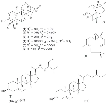
This work describes a novelent-kaurane diterpene,ent-3b-hydroxy-kaur-16-en-19-al along with five knownent-kaurane diterpenes,ent-3b,19-dihydroxy-kaur-16-eno, ent-3b-hydroxy-kaur-16-eno,
ent-3b-acetoxy-kaur-16-eno,ent-3b-hydroxy-kaurenoic acid and kaurenoic acid, as well as caryophyllene oxide, humulene epoxide II, b-sitosterol, stigmasterol and campesterol from the stem bark of
Annona vepretorum Mart. (Annonaceae). Cytotoxic activities towards tumor B16-F10, HepG2, K562 and HL60 and non-tumor PBMC cell lines were evaluated forent-kaurane diterpenes. Among them, ent-3b-hydroxy-kaur-16-en-19-al was the most active compound with higher cytotoxic effect over K562 cell line (IC50of 2.49
l
g/mL) and lower over B16-F10 cell line (IC50of 21.02l
g/mL).Ó2014 Elsevier Ltd. All rights reserved.
Annona vepretorum
Mart. (Annonaceae), popularly known as
‘bruteira’, is a shrub or tree of 2.5–10 m tall native from the
Brazilian biome Caatinga. Its fruits are consumed raw or in juice
form as a nutritional source.
1When softened, its roots present
pop-ular medicinal indication to bite of bees and snakes, inflammatory
conditions and pains in the heart, while the leaves (decoction) are
used in bath to allergies, skin diseases, yeast and bacteria infection
(oral communications received from the local population). Previous
phytochemical and pharmacological investigations on this species
described the chemical composition of essential oil from the
leaves that showed trypanocidal, antifungal and antioxidant properties,
and revealed mainly the presence of bicyclogermacrene,
spathule-nol and
a-phellandrene.
2Moreover, Diniz et al.
3described that
ethanolic extract from the leaves has sedative effect. In our
contin-uous research for bioactive compounds from Annonaceous plants a
novel e
nt
-kaurane diterpene, ent-3
b
-hydroxy-kaur-16-en-19-al
(
1
), along with five known
ent
-kaurane diterpenes,
ent
-3
b
,19-dihy-droxy-kaur-16-eno (
2
),
ent
-3
b
-hydroxy-kaur-16-eno (
3
),
ent
-3
b
-acetoxy-kaur-16-eno (
4
),
ent
-3
b
-hydroxy-kaurenoic acid (
5
) and
kaurenoic acid (
6
), as well as caryophyllene oxide (
7
), humulene
epoxide II (
8
),
b
-sitosterol (
9
), stigmasterol (
10
) and campesterol
(
11
) were found in the stem bark of
A. vepretorum
(
Fig. 1
).
4Cytotoxic activities towards tumor and non-tumor cells lines were
investigated for compounds
1
–
6
. This is the first phytochemical
and biological investigation of the stem bark of
A. vepretorum
.
Compound
1
was obtained as an white amorphous powder with
the molecular formula, C
20H
30O
2, as determined by HR-ESIMS
(observed
m
/
z
325.2140 [M+Na]
+) and NMR data.
5,6LR-MS tandem
analysis showed a fragment at
m
/
z
285 indicating a lost of H
2O
[M+H H
2O]
+. Its infrared (IR) spectrum showed absorptions bands
at 3267, 2726 and 1712 cm
1typical of hydroxyl and aldehyde
groups. The low frequency of the carbonyl group of the aldehyde,
as well as hydroxyl group is due to the hydrogen bonding. The
1
H NMR spectrum showed signals for two tertiary methyl groups
at
d
0.94 and 1.27 (3H each), that are typical of axial C-20 and
equatorial C-18 methyl groups of
ent
-kaurane diterpenes with a
axial C-19 aldehyde group (
Fig. 2
). The signal for this aldehyde
group was observed at
d
9.76 (1H). Additionally, two signals were
observed at
d
4.75 and 4.81 (1H each) typical of hydrogens from an
exocyclic double bond, as well as a signal to a carbinolic hydrogen
at
d
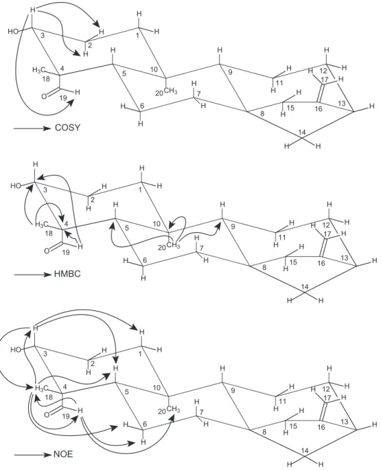
3.16 (1H). The
1H–
1H COSY NMR experiment revealed that the
aldehyde hydrogen was coupling with H-3 at
d
3.16 (1H), probably
due to a ‘W’ orientation (
Fig. 2
), supporting the equatorial C-18
(methyl group) and axial C-19 (aldehyde group) configurations in
the structure of
1
.
7The
13C{
1H} and DEPT135 NMR spectra, as well
as one-bond and long-range
1H–
13C correlation maps from HSQC
and HMBC NMR experiments indicated a total of 20 carbons
http://dx.doi.org/10.1016/j.bmcl.2014.06.005
0960-894X/Ó2014 Elsevier Ltd. All rights reserved.
⇑
Corresponding author. Tel./fax: +55 71 3176 2272.E-mail address:[email protected](D.P. Bezerra).
Contents lists available at
ScienceDirect
Bioorganic & Medicinal Chemistry Letters
(
Table 1
). These carbons comprised two methyl, nine methylenes,
four methines, four quaternary carbons, and one aldehyde at
d
208.1. The hydrogen at
d
3.16 and shown one-bond
1H–
13C
corre-lation with the carbon at
d
77.4 (C-3) and long-range
1H–
13C
corre-lation with the carbons at
d
52.6 (C-4) and
d
208.1 (C-19),
supporting the presence of a hydroxyl group at C-3 and the
alde-hyde at C-19 in the structure of
1
(
Fig. 2
). Moreover, the hydrogen
at
d
3.16 (H-3) showed only two additional
1H–
1H correlation from
COSY NMR experiment, with hydrogens at
d
1.86 and
d
1.89 (H-2),
supporting the substitution at C-3. The aldehyde group at C-19 was
supported on the basis of HMBC NMR experiment, since the
aldehyde hydrogen at
d
9.76 (H-19) shown long-range
1H–
13C
correlation with the carbons at
d
52.6 (C-4) and
d
77.4 (C-3)
(
Fig. 2
). The
b
-orientation of the hydroxyl group at C-3 was
estab-lished by comparing its NMR data with those described in the
lit-erature for 3-hydroxy-kauranoic acids.
7,8Hydrogens at C-3 in
a
(axial OH) and
b
(equatorial OH) isomers are described as having
1
H NMR chemical shifts at
d
4.11 and
d
3.14, respectively.
There-fore, the
1H NMR chemical shifts found in this work to H-3 at
d
3.16 are in accordance with an
b
orientation. This fact was also
supported by 1D NOE NMR selective experiments. In these, the
selective irradiation of the resonance frequency of H-3 at
d
3.16
caused a NOE enhancement in the signals at
d
1.27 (H-18), 1.03
(H-5) and 0.95 (H-1ax) (
Fig. 2
). Moreover, the selective irradiation
of the resonance frequency of the methyl hydrogens H-18 at
d
1.27
showed a NOE intensification of the signals at
d
9.76 (HCO),
d
3.16
(H-3),
d
1.88 (H-6 eq), and
d
1.03 (H-5), although any enhancement
of the signal of H-20 at
d
0.94 (
Fig. 2
). On the other hand, the
selec-tive irradiation of the resonance frequency of the hydrogen H-19 at
d
9.76 showed a NOE intensification of the signals at
d
1.27 (H-18),
d
0.94 (H-20) and
d
1.59 (H-6ax), although no enhancement on the
signal of H-5 at
d
1.03 (
Fig. 2
). The overall analysis of 1D and 2D
NMR experiments enabled us to fully establish the structure and
to completely assign the
1H and
13C NMR chemical shifts of
1
(
Table 1
).
9Therefore, compound
1
was identified as a new
ent
-kau-rane diterpene named as
ent
-3
b
-hydroxy-kaur-16-en-19-al.
Compounds
2
–
11
were identified by comparing their
spectro-metric data
6with those reported in the literature which were in
accordance with
ent
-3
b
,19-dihydroxy-kaur-16-ene (
2
),
ent
-3
b
-hydroxy-kaur-16-ene (
3
),
ent
-3
b
-acetoxy-kaur-16-ene (
4
),
ent
-3
b
-hydroxy-kaur-16-en-19-oic acid (
5
),
ent
-kaur-16-en-19-oic acid
(
6
), mixture of caryophyllene oxide (
7
) and humulene epoxide II
(
8
), and a mixture of
b
-sitosterol (
9
), stigmasterol (
10
) and
campesterol (
11
).
7,10–18Nevertheless, compounds
2
–
4
have been
described a long time ago and its NMR data are incomplete as well
as have some ambiguities. Therefore, the complete and
unequivo-cal NMR data for these diterpenes were reviewed according to 1D
and 2D NMR experiments (
Table 1
).
The absolute configurations of the diterpenoids
ent
and normal
series can be established on the basis of its negative and positive
specific rotation ([
a
]
D), since they can be correlated to similar
kauranoid diterpenes with defined absolute configurations.
Therefore, those that divert the light polarized to the left ( )
belong to the
ent
series, while those that divert to the right (+)
belong to the normal series.
8In this work, all diterpenes diverted
the light polarized to the left ( ) according to
ent
series.
6The
ent
-kaurane diterpenes are common in Annonaceae plants,
particularly in species of
Annona
and
Xylopia
.
19–22In
Annona
, this
class of compound is well represented and considered as
chemo-taxonomic markers. Among them, compound
6
is the most
repre-sentative within the family Annonaceae, mainly in
Annona
such
as
Annona cherimolia
,
Annona glabra
,
Annona senegalensis
and
Annona squamosa
, and
Xylopia
such as
Xylopia frutescens
,
Xylopia
laevigata
,
Xylopia sericeae
.
7,19–23Compound
5
has been described
in
Xylopia laevigata
(Annonaceae), although it was described in
species of the family Asteraceae.
7,24,25Therefore, the presence of
ent
-kaurane diterpenes in
A. vepretoru
m supports that this is a
typical species of the family Annonaceae. Compound
2
was found
(
7
)
O
H
H
12
16 17
R
1H
R
2H
1 2 3
4 5 6 7 8 9
10
11 13 14
15
19 18 20
(
8
)
(
1
) R
1
= OH; R
2
= CHO
(
2
) R
1
= OH; R
2
= CH2OH
(
3
) R
1
= OH; R
2
= CH3
(
4
) R
1
= OOCCH3 (or OAc); R
2
= CH
3(
5
) R
1
= OH; R
2
= COOH
(
6
) R
1
= H; R
2
= COOH
O
22
HO
H
H
H
23
(
9
)
(
10
)
22(23)
HO
H
H
H
(
11
)
in
Stachys lanata
(Labiatae) and
Cacalia pilgeriana
(Asteraceae).
10,26Compound
3
was obtained from
Laetia thamnia
(Flacourtiaceae),
Guarea kunthiana
(Meliaceae) and
Phyllanthus flexuosus
(Phyllanth-aceae).
27–29Compound
4
was observed in
Phyllanthus flexuosus
(Phyllanthaceae).
27The other compounds (
7
–
11
) are commonly
found in species of Annonaceae.
7,30–32Compounds
1
–
6
were evaluated for their cytotoxicity on
B16-F10 (mouse melanoma), HepG2 (human hepatocellular
carcinoma), K562 (human chronic myelocytic leukemia) and
HL-60 (human promyelocytic leukemia) tumor cell lines.
33–36The
compounds
1
,
3
,
5
and
6
showed cytotoxic activity, while
2
and
4
were not cytotoxic at the experimental concentration used
(IC
50>25
lg/mL), in any tumor cell lines tested (
Table 2
). The
cytotoxicity of
2
,
3
and
6
was previously assessed,
28,37,38whereas
the cytotoxic activity of the compounds
1
,
4
and
5
were evaluated
for first time in this work.
Regarding structure-cytotoxicity relationship of
ent
-kaurane
diterpenes, related compounds with the
a-methylene
cyclopenta-none moiety and/or
a
,
b
-unsaturated ketone moiety had been
reported to have cytotoxic activity.
39,40On the other hand, the
ent
-kaurane diterpenes investigated in this work do not present
these features and are able to inhibit cell proliferation. Cavalcanti
et al.
37found that the exocyclic double bond (
D
16(17)) is a
fundamental pharmacophoric group for the cytotoxic activity of
ent
-kaurane diterpenes. All
ent
-kaurane diterpenes evaluated in
this work has exocyclic double bond, although compound
4
was
inactive. This fact can be related to the presence of an acetate
group at C-3. In this work, compounds
1
,
3
and
6
were higher
cyto-toxic than the compounds
2
,
4
and
5
, suggesting that acetoxy
(OOCCH
3) group on C-3 or hydroxymethyl (CH
2OH) group on C-4
decrease the cytotoxic activity of
ent
-kaurane diterpenes. These
results may help in the identification of novel
ent
-kaurane
diterpene-like structures with optimized cytotoxicity to be tested
for cancer treatment.
Compound
1
–
6
were also cytotoxic to non-tumor PBMC cells,
presenting low selectivity (
Table 2
). Compound
1
shown a
selec-tivity index (SI) of 2.9 for leukemia (K562), while doxorubicin
showed a SI of 7.5 for the same tumor cell line. Compound
6
HMBC
NOE
CH3 H H H H H H H H H H H OH3C H
H H HO H H H H H H H H 20 19 18 10 9 8 7 6 5 4 17 16 15 14 13 12 11 3 2 1 H CH3 H H H H H H H H H H H O
H3C H
H H HO H H H H H H H H 20 19 18 10 9 8 7 6 5 4 17 16 15 14 13 12 11 3 2 1 H CH3 H H H H H H H H H H H O
H3C H
H H HO H H H H H H H H 20 19 18 10 9 8 7 6 5 4 17 16 15 14 13 12 11 3 2 1 H
COSY
has been previously reported as non-selective cytotoxic
compound.
37Therefore, the stem bark of
A. vepretorum
is an important source
for cytotoxic
ent
-kaurane diterpenes.
Acknowledgements
The authors are grateful to the Brazilian agencies CAPES, CNPq,
FINEP, FAPITEC/SE, FAPESB and UFPR for financial support and
fellowships as well as to Marlene da S. Cerqueira for technical
assistance and to Prof. Dr. Norberto P. Lopes and José C. Tomaz
for HR-ESIMS analysis.
Supplementary data
Supplementary data (spectrometric data, including NMR, MS
and IR for the new
ent
-kaurane diterpenoid
1
) associated with this
article can be found, in the online version, at
http://dx.doi.org/
10.1016/j.bmcl.2014.06.005
.
References and notes
1. Souza, V. C.; Lorenzi, H. Botânica Sistemática: Guia ilustrado das famílias de Fanerógamas nativas e exóticas no Brasil, baseado em APG I. Instituto Plantarum, 2005, 640.
2. Costa, E. V.; Dutra, L. M.; Nogueira, P. C.; Moraes, V. R.; Salvador, M. J.; Ribeiro, L. H.; Gadelha, F. R.Nat. Prod. Commun.2012,7, 265.
Table 1
1H and13C NMR data (CDCl
3, 400 MHz) forent-kaurane diterpenes1–4
Position 1 2 3 4
dC dHmult. (Jin Hz) dC dHmult. (Jin Hz) dC dHmult. (Jin Hz) dC dHmult. (Jin Hz)
1 38.6 ax 0.95m 38.4 ax 0.90m 38.7 ax 0.90ddd(13.2, 12.4 and 5.2) 38.4 ax 0.98ddd(13.4, 12.8 and 4.7) eq 1.92m eq 1.87m eq 1.85ddd(13.2. 3.7 and 3.4) eq 1.85ddd(13.4, 3.8 and 3.5)
2 28.2 ax 1.86m 27.7 ax 1.71m 27.4 ax 1.60m 23.7 ax 1.65m
eq 1.89m eq 1.84m eq 1.63m eq 1.67m
3 77.4 3.16m 80.9 3.42m 79.1 3.19dd(10.9 and 5.6) 81.1 4.47dd(11.1 and 5.5)
4 52.6 42.9 38.9 37.9
5 56.3 1.03dd(12.6 and 2.3) 55.8 0.87m 55.2 0.76dd(11.8 and 1.9) 55.4 0.85m
6 20.2 ax 1.59m 20.1 ax 1.30m 20.0 ax 1.40m 19.9 ax 1.36m
eq 1.88m eq 1.75m eq 1.55m eq 1.53m
7 41.1 ax 1.51m 41.3 ax 1.48m 41.2 ax 1.48m 41.1 ax 1.51m
eq 1.61m eq 1.51m eq 1.52m eq 1.53m
8 43.8 43.9 44.0 44.1
9 54.5 1.05m 55.9 1.03m 55.9 1.03m 55.9 1.06m
10 39.1 38.7 39.1 39.0
11 18.5 ax 1.55m 18.4 ax 1.53m 18.3 ax 1.53m 18.3 ax 1.54m
eq 1.65m eq 1.63m eq 1.62m eq 1.64m
12 39.8 ax 1.15dddd
(11.5, 5.2, 1.8 and 1.5) 39.6 ax 1.09
dddd
(11.4, 5.0, 1.8 and 1.5) 39.8 ax 1.11
dddd
(11.4, 5.1, 1.8 and 1.6) 39.8 ax 1.11
dddd
(11.4, 5.0, 1.8 and 1.6) eq 1.93dm(11.5) eq 1.92dm(11.4) eq 1.98dm(11.4) eq 1.97dm(11.4)
13 43.7 2.66m 43.8 2.64m 43.97 2.64m 44.0 2.64m
14 32.9 pax 1.49m 33.1 pax 1.48m 33.2 pax 1.48m 33.0 pax 1.48m
peq 1.59m peq 1.63m peq 1.62m peq 1.64m
15 48.8 pax 2.05m 48.9 pax 2.05m 49.0 pax 2.05m 49.0 pax 2.06m
peq 2.06m peq 2.06m peq 2.06m peq 2.07m
16 155.2 155.6 155.8 155.7
17 103.5 4.75m 103.2 4.74m 103.0 4.74m 103.1 4.74m
4.81m 4.80m 4.80m 4.79m
18 19.2 1.27s 22.7 1.23s 28.4 0.98s 28.4 0.86s
19 208.2 9.76s 64.3 3.32d(11.2) 15.5 0.78s 16.6 0.85s
4.20d(11.2)
20 16.5 0.94s 18.2 0.98s 17.6 1.02s 17.7 1.05s
CH3COO-3 171.0
CH3COO-3 21.3 2.04s
Table 2
Cytotoxic activity (IC50values)afor compounds1–6
Cell lines IC50in
l
g/mL (l
M)/compounds1 2 3 4 5 6 Doxorubicinb
Tumor cellsc
B16-F10 21.02 (69.55) >25 (82.17) 19.12 (66.33) >25 (75.70) >25 (78.56) 16.56 (54.79) 2.30 (4.23) HepG2 15.50 (51.29) >25 (82.17) 19.38 (67.23) >25 (75.70) >25 (78.56) 15.33 (50.72) 0.23 (0.42) HL-60 9.92 (32.82) >25 (82.17) 9.86 (34.21) >25 (75.70) 24.21 (76.08) 13.33 (44.11) 0.83 (1.53) K562 2.49 (8.24) >25 (82.17) 2.94 (10.20) >25 (75.70) 20.21 (63.51) 21.92 (72.53) 0.68 (1.25)
Non-tumor cellsd
PBMC 7.20 (23.82) 8.93 (29.35) 6.49 (22.52) >25 (75.70) >25 (78.56) 24.41 (80.77) 5.09 (9.36)
aData are presented as IC
50values in
l
g/mL (l
M) obtained by nonlinear regression from three independent experiments performed in duplicate, measured by Alamar blueassay after 72 h incubation.
b Doxorubicin was used as positive control.
c Tumor cells: B16-F10 (mouse melanoma), HepG2 (human hepatocellular carcinoma), HL-60 (human promyelocytic leukemia) and K562 (human chronic myelocytic
leukemia).
3. Diniz, T. C.; Araújo, C. S.; Silva, J. C.; Oliveira-Júnior, R. G.; Lima-Saraiva, S. R. G.; Quintans-Júnior, L. J.; Nunes, X. P.; Almeida, J. R. G. S.J. Med. Plants Res.2013,7, 2729.
4. Botanical material:The stem bark ofA. vepretorumwas collected in ‘Serra da Guia’, in the city of Poço Redondo [coordinates: 09°5705400S, 37°5104600W],
Sergipe State, Brazil, in April 2010. The identity of the plant was confirmed by Dr. A. P. do N. Prata, a plant taxonomist of Department of Biology from Federal University of Sergipe (UFS), Brazil and a voucher specimen (#15441) has been deposited in the Herbarium of UFS. The authors have authorization from the Chico Mendes Institute for Biodiversity Conservation from Brazilian Ministry of the Environment for plant collection (#25637-1). This work was performed according to the special authorization for access to genetic resources in Brazil # 010240/2013-6, issued by CNPq/MCTI.
5. Extraction and isolation:The dried and powdered stem bark ofA. vepretorum
(653 g) was successively extracted with petroleum ether followed by MeOH, to yield petroleum ether (39.03 g) and MeOH (85.00 g) extracts. The petroleum ether extract (5.0 g) was initially subjected to silica gel column chromatography (CC) eluted with increasing concentrations of CH2Cl2 in
n-hexane (100:0 to 10:90, v/v), followed by EtOAc in CH2Cl2(100:0 at 30:70, v/
v), and MeOH in EtOAc (100:0 to 70:30, v/v), affording 201 fractions (30 mL each). The eluted fractions were evaluated and pooled according to TLC analysis, to afford 19 groups (GF1 to GF19). Group GF3 (88.3 mg) fromn -hexane–CH2Cl2(90:10 and 80:20, v/v) was submitted to preparative TLC eluted
withn-hexane–EtOAc (90:10, v/v, two elution), affording a mixture of7and8 (38.8 mg). Group GF10 (69.0 mg) fromn-hexane–CH2Cl2(40:60 and 30:70) was
submitted to preparative TLC eluted withn-hexane–EtOAc (95:05, v/v, three elution) yielding 6 (38.6 mg) and also a mixture of 7 and 8(15.1 mg), respectively. GF12 (648.9 mg) from n-hexane–CH2Cl2 (10:90 v/v), CH2Cl2
(100%,v)), and CH2Cl2–EtOAc (95:05, v/v) was also subjected to a preparative
TLC eluted withn-hexane–EtOAc (90:10, v/v, two elution) giving4(15.2 mg) and6(346.0 mg), respectively. GF13 (2457.8 mg) from CH2Cl2–EtOAc (90:10
and 80:20, v/v) was submitted to a new silica gel CC eluted with the same methodology as describe for initial CC (petroleum ether extract), affording 90 subfractions (30 mL each) that were subsequently pooled into 21 groups (GF13.1 to GF13.21), according to TLC analysis. Group GF13.2 (157.6 mg) was also submitted to preparative TLC eluted withn-hexane–EtOAc (90:10, v/v, two elution) again affording6(111.6 mg). Group GF13.3 (119.6 mg) was submitted to the same conditions as for GF13.2 also resulting in6(49.2 mg). GF13.4 (321.9 mg) was submitted to a new silica gel CC eluted with increasing concentrations of CH2Cl2inn-hexane (100:0 to 30:70, v/v), followed by EtOAc
in CH2Cl2(100:0 to 50:50, v/v), affording 21 subfractions (30 mL each), that
were evaluated and pooled according to TLC analysis, to afford 10 groups (GF13.4.1–GF13.4.10). The groups GF13.4.6 and GF13.4.7 were pooled (270.7 mg) and also submitted to TLC preparative eluted withn-hexane–EtOAc (80:20, v/v, three elution), yielding3(154.6 mg). Group GF13.5 (183.4 mg) was also submitted to preparative TLC eluted withn-hexane–EtOAc (90:10, v/v, three elution) again affording 3 (99.0 mg). Group GF13.8 (85.7 mg) was submitted to preparative TLC eluted withn-hexane–EtOAc (80:20, v/v, two elution), again yielding 3 (40.4 mg). Group GF13.9 (325.0 mg) was also subjected to a preparative TLC eluted with n-hexane–EtOAc (80:20, v/v, two elution) giving3(68.2 mg) and1(10.9 mg), respectively. Current fractions of the preparative of GF13.9 were contained and submitted successive CC and preparative TLC eluted in the same conditions that GF13, which resulted in the isolation of the mixture of9,10and11(15.5 mg). GF13.10 (145.9 mg) was submitted to a new silica gel CC eluted with increasing concentrations of CH2Cl2inn-hexane (100:0 to 20:80, v/v) and EtOAc in CH2Cl2(100:0 to 80:20,
v/v) giving 28 subfractions (30 mL each) that were pooled into 8 groups (GF13.10.1–GF13.10.8), according to TLC analysis. The group GF13.10.1 (47.8 mg) was subjected to a preparative TLC eluted withn-hexane–EtOAc (80:20, v/v, two elution), yielding3(23.4 mg). Group 13.10.2 (57.1 mg) was submitted to the same conditions as for GF13.10.1 resulting in1(32.1 mg). Group 13.11 was also subjected to a preparative TLC eluted withn-hexane– EtOAc (80:20, v/v, three elution), again affording1(82.7 mg). The group GF16 (238.4 mg) from CH2Cl2–EtOAc (70:30, 60:40 and 50:50, v/v) was submitted to
a new silica gel CC eluted with the same methodology as describe at the initial CC (petroleum ether extract), affording 69 subfractions (30 mL each) that were subsequently pooled into 7 groups (GF16.1–GF16.7), according to TLC analysis. Group GF16.5 was subjected to a preparative TLC eluted withn-hexane–EtOAc (70:30, v/v, four elution) giving2(100.4 mg). The group GF17 (76.7 mg) from CH2Cl2–EtOAc (40:60 and 30:70, v/v) and EtOAc–MeOH (100:0 and 95:05 v/v)
was also submitted to a preparative TLC eluted with n-hexane–EtOAc (60:40, v/v, three elution) yielding2(6.3 mg). Group GF18 (115.9 mg) from EtOAc–MeOH (90:10 v/v) was submitted to preparative TLC eluted with
n-hexane–EtOAc (60:40, v/v, two elution), affording5(13.0 mg).
6. General experimental procedures: Melting points (mp) were measured on a Microquímica MQAPF 301 apparatus. IR spectra were acquired in KBr pellets on a Shimadzu IR Prestige-21 spectrophotometer. Optical rotations were recorded in CHCl3on a Jasco P-2000 polarimeter. GC-MS analyses were performed on a
Shimadzu QP5050A GC-MS system equipped with an AOC-20i auto-injector. The separation of the compounds was achieved employing on RTxÒ
-5SilMS fused capillary chromatography column (30 m0.25 mm0.25
l
m film thickness) coated with 5%-diphenyl-95%-dimethylpolysiloxane. The column temperature program was 200°C/5 min, a rate of 10°C/min to 320°C, and then320°C/10 min (27 min total time analysis); carrier gas, He (99.999%; 1.2 mL/min);
split ratio, 1:20; injection volume, 0.5 mL of the compound in CH2Cl2(5.0 mg/mL).
MS were taken at 70 eV with a scan interval of 0.5 s and fragments from
40–500 Da. Low Resolution Mass Spectra (LRMS) were determined using an ultra-high performance chromatography–mass spectrometry system (Acquity UHPLC-TQD–Waters) with an ESI and APCI source in the positive and negative ion mode. High Resolution Mass Spectra (HR-ESIMS) measurements were performed on a Bruker UltrOTOF-Q MS spectrometer featuring a quadrupole time-of-flight mass analyzer equipped with an electrospray source. 1D and 2D NMR data were recorded at 293 K in CDCl3on a Bruker
Avance III 400 NMR spectrometer, operating at 9.4 Tesla, observing1H and13C
at 400.13 and 100.61 MHz, respectively. The spectrometer was equipped with either, a 5-mm multinuclear direct detection probe (1D NMR experiments) or a 5-mm multinuclear inverse detection probe (1D NOE and 2D NMR experiments) both with z-gradient. One-bond and long-range 1H–13C
correlation from HSQC and HMBC NMR experiments were optimized for an average coupling constant1J
(C,H)andLRJ(C,H)of 140 and 8 Hz, respectively. All1H
and13C NMR chemical shifts (d) are given in ppm related to the TMS signal at
0.00 ppm as an internal reference, and the coupling constants (J) in Hz. Silica gel 60 (70–230 mesh) was used for column chromatography, while silica gel 60 F254 was used for analytical (0.25 mm), and preparative (1.00 mm) TLC.
Compounds were visualized by exposure under UV254/365light and spraying
ofp-anisaldehyde reagent followed by heating on a hot plate.
7. Silva, D. M.; Costa, E. V.; Nogueira, P. C. L.; Moraes, V. R. S.; Cavalcanti, S. C. H.; Salvador, M. J.; Ribeiro, L. H. G.; Gadelha, F. R.; Barison, A.; Ferreira, A. G.Quím. Nova2012,35, 1570.
8. Velandia, J. R.; Carvalho, M. G.; Braz-Filho, R.Quim. Nova1998,21, 397. 9. ent-3b-Hydroxy-kaur-16-en-19-al (1):White solid (purity 98.5%); mp 117.0–
118.1°C; [
a
]D20 93.1°(c0.4, CHCl3); IR(KBr)m
max3267 (OH), 3078, 2924,2854, 2726 (HCO), 1712 (C@O), 1642 (C@C), 1479, 1445, 1402, 1350, 1101, 1058, 1007, 869/cm;1H and13C NMR data, seeTable 1; EI-MSm/z302 [M]+;
LR-APCIMS [M H2O]+ m/z 285.15; HR-ESIMS m/z 325.2140 (calcd for
C20H30O2+Na+, 325.2143).
10. Piozzi, F.; Savona, G.; Hanson, J. R.Phytochemistry1980,19, 1237.
11. Facundo, V. A.; Polli, A. R.; Rodrigues, R. V.; Militão, J. S. L. T.; Stabelli, R. G.; Cardoso, C. T.Acta Amazonica2008,38, 733.
12. ent-3b,19-Dihydroxy-kaur-16-ene (2):Yellow crystals (n-hexane–EtOAc 7:3) (purity 98.7%); mp 180.9–182.2°C (lit. 190°C);10[
a
]D20 26.4°(c1.5, CHCl3);IR(KBr)
m
max3341 (OH), 3068, 2966, 2923, 2846, 1653 (C@C), 1465, 1431, 1362, 1064, 1004, 859/cm;1H and13C NMR data, seeTable 1; EI-MSm/z304 [M]+.LR-APCIMS [M H2O]+m/z287.17.
13. ent-3b-Hydroxy-kaur-16-ene (3): White solid (n-hexane–EtOAc 8:2) (purity 98.7%); mp 161–163.0°C (lit. 177–178°C);27[
a
]D20 149.2°(c0.45, CHCl3);IR(KBr)
m
max3345 (OH), 3070, 2924, 2854, 1651 (C@C), 1487, 1427, 1101, 1041, 989, 869/cm;1H and13C NMR data, seeTable 1; EI-MSm/z288 [M]+.LR-APCIMS [M H2O]+m/z271.24.
14. ent-3b-Acetoxy-kaur-16-ene (4):Yellowish amorphous solid (purity 92.7%); mp 160.3–162.5°C (lit. 162–164°C);27[
a
]D20 28.56°(c0.5, CHCl3); IR(KBr)m
max3078, 2924, 2846, 1651 (C@C), 1720 (C@O), 1454, 1367, 1238, 1015, 869, 757/ cm;1H and 13C NMR data, seeTable 1; EI-MS m/z 330 [M]+. LR-APCIMS
[M OCOCH3]+m/z271.24.
15. ent-3b-Hydroxy-kau-16-en-19-oic acid (5): White crystalline powder (n-hexane–EtOAc 6:4) (purity 100.0%); mp 204.4–205.0°C (lit. 218–219°C);7
[
a
]D20 107.19°(c0.7, CHCl3); IR(KBr)m
max3482 (OH), 3371, 3070, 2941, 2854,2623, 1737 (C@O), 1668 (C@C), 1454, 1350, 1256, 1195, 989, 860, 757, 636, 542/cm;1H and13C NMR data in agreement with those from the literature;7 EI-MSm/z318 [M]+; LR-ESIMS [M H] m/z317.27.
16. ent-Kaur-16-en-19-oic acid (6):White needles (n-hexane–EtOAc 9:1) (purity 96.8%); mp 160.5–162.2°C (lit. 160–162°C);7[
a
]D20 93.56°(c1.0, CHCl3);IR(KBr)
m
max3448 (OH), 3379, 3070, 2924, 2846, 2588, 2356, 1695 (C@O), 1642 (C@C), 1462, 1402, 1264, 1170, 955, 869, 782, 636/cm;1H and13C NMR data in agreement with those from the literature;23EI-MSm/z302 [M]+. LR-ESIMS[M H] m/z301.28.
17. Mixture of caryophyllene oxide (7) (rate 75.3%) and humulene epoxide II (8) (rate 24.7%):Colorless oil;1H and13C NMR data in agreement with those from the
literature; EI-MSm/z220 and 218 [M]+.
18. Mixture of b-sitosterol (9) (rate 50,1%), stigmasterol (10) (rate 20.9%) and campesterol (11) (rate 19.8%):White needles (n-hexane–EtOAc 8:2); mp 135.3– 136.7°C (lit. 138–140°C);311H and13C NMR data in agreement with those
from the literature;7,11EI-MSm/z414, 412 and 400 [M]+.
19. Leboeuf, M.; Cavé, A.; Bhaumik, P. K.; Mukherjee, B.; Mukherjee, R.
Phytochemistry1982,21, 2783.
20. Takahashi, J. Á.; Boaventura, M. A. D.; Bayma, J. C.; Oliveira, A. B.Phytochemistry
1995,40, 607.
21. Takahashi, J. A.; Vieira, H. S.; Boaventura, M. A. D.; Hanson, J. R.; Hitchcock, P. B.; Oliveira, A. B.Quim. Nova2001,24, 616.
22. Chen, C. Y.; Chang, F. R.; Wu, Y. C.J. Chin. Chem. Soc.1997,44, 313. 23. Vieira, H. S.; Takahashi, J. A.; Boaventura, M. A.J. Agric. Food Chem.2002,50,
3704.
24. Barrero, A. F.; Oltra, J. E.; Cabrera, E.; Reyes, F.; Álvarez, M.Phytochemistry1999,
50, 1133.
25. Rezende, M. C.; Urzua, A.; Bortoluzzi, A. J.; Vásquez, L.J. Ethnopharmacol.2000,
72, 459.
26. Li, E. W.; Gao, K.; Jia, Z. J.J. Asian Nat. Prod. Res.2007,9, 191. 27. Tanaka, R.; Matsunaga, S.Phytochemistry1988,27, 2273.
28. Henry, G. E.; Adams, L. S.; Rosales, J. C.; Jacobs, H.; Heber, D.; Seenam, N. P.
Cancer Lett.2006,244, 190.
30. Da Silva, F. M. A.; Koolen, H. H. F.; Barisson, A.; De Souza, A. D. L.; Pinheiro, M. L. B.J. Pharm. Sci.2012,4, 522.
31. Costa, E. V.; Marques, F. A.; Pinheiro, M. L. B.; Braga, R. M.; Delarmelina, C.; Duarte, M. C. T.; Ruiz, A. L. T. G.; Carvalho, J. E.; Maia, B. H. L. N. S.J. Braz. Chem. Soc.2011,22, 1111.
32. Trigo, J. R.; Oliveira, J.; Andrade, E. H. A.; Zoghbi, M. G. B.Rev. Bras. Plants Med.
2007,9, 113.
33. Cell lines: Cytotoxicity was evaluated to tumor cells lines B16-F10 (mouse melanoma), HepG2 (human hepatocellular carcinoma), K562 (human chronic myelocytic leukemia) and HL-60 (human promyelocytic leukemia). All cell lines were donated by Hospital A.C. Camargo, São Paulo, SP, Brazil. Cells were maintained in Roswell Park Memorial Institute-1640 (RPMI-1640) medium supplemented with 10% fetal bovine serum, 2 mML-glutamine and 50
l
g/mL gentamycin. Adherent cells were harvested by treatment with 0.25% trypsin EDTA solution. All cell lines were cultured in cell culture flasks at 37°C in 5%CO2 and sub-cultured every 3–4 days to maintain exponential growth.
Cytotoxicity experiments were conducted with cells in exponential growth phase. All cell lines were tested for mycoplasma with a LookoutÒ
Mycoplasma qPCR detection kit and found to be free from contamination.
34. Primary culture of human lymphoblast:In order to investigate the selectivity of the compounds toward a non-tumor proliferating cell, human lymphoblast cells were obtained by primary culture. Heparinized blood (from healthy, 20–35 years old, non-smoker donors who had not taken any drug at least 15 days prior to sampling) was collected and peripheral blood mononuclear cells (PBMC) were isolated by a standard protocol using Ficoll density gradient in a GE Ficoll-Paque Plus. PBMC were washed and resuspended at a concentration of 0.3106cells/mL in RPMI 1640 medium supplemented with 20% fetal bovine serum, 2 mM glutamine, 50
l
g/mL gentamycin at 37°Cwith 5% CO2. In addition, concanavalin A was used as a mitogen to trigger cell
division in T-lymphocytes. ConA (10
l
g/mL) was added at the beginning of culture and, after 24 h, cells were treated with the test drugs. The Research Ethics Committee of the Oswaldo Cruz Foundation (Salvador, Bahia, Brazil) approved the experimental protocol (#031019/2013). All participants signed their written informed consent to participate in the study. For all experiments, cell viability was performed by Trypan blue exclusion (TBE) assay. Over 90% of the cells were viable at the beginning of the culture.35. Cell proliferation assay:Cell growth was quantified by alamar blue assay, as previously described. For all experiments, cells were seeded in 96-well plates (0.7105cells/mL for adherent cells or 0.3106cells/mL for suspended cells in 100
l
L of medium). After 24 h, the compounds (0.39–25l
g/mL) dissolved in DMSO were added to each well and incubated for 72 h. Doxorubicin (purityP95.0%, doxorubicin hydrochloride) was used as positive control(0.08–5
l
g/mL). Negative control received the vehicle used for diluting the tested (0.5% DMSO). Four (for cell lines) or 24 (for PBMC) hours before the end of the incubation, 20l
L of stock solution (0.312 mg/mL) of the alamar blue were added to each well. The absorbance was measured using a SpectraMax 190 multiplate reader and the drug effect was quantified as the percentage of control absorbance at 570 nm and 600 nm. The absorbance of alamar blue in culture medium was measured at a higher wavelength and a lower wavelength. The absorbance of the medium was also measured at the higher and lower wavelengths. The absorbance of the medium alone was subtracted from the absorbance of medium plus alamar blue at the higher wavelength. This value was called AOHW. The absorbance of the medium alone wassubtracted from the absorbance of medium plus alamar blue at the lower wavelength. This value was called AOLW. A correction factorR0was calculated
from AOHW and AOLW, where R0= AOLW/AOHW. The percent alamar blue
reduced then was expressed as follows: % reduced =ALW (AHWR0)100. 36. Statistical analysis:Data are presented as half maximal inhibitory concentration
(IC50) values obtained by nonlinear regression from three independent
experiments performed in duplicate. All analyses were carried out using the GRAPHPAD software. SI was determined as IC50[PBMC]/IC50[K562].
37. Cavalcanti, B. C.; Bezerra, D. P.; Magalhães, H. I.; Moraes, M. O.; Lima, M. A.; Silveira, E. R.; Câmara, C. A.; Rao, V. S.; Pessoa, C.; Costa-Lotufo, L. V.J. Appl. Toxicol.2009,29, 560.
38. Fatope, M. O.; Audu, O. T.; Takeda, Y.; Zeng, L.; Shi, G.; Shimada, H.; McLaughlin, J. L.J. Nat. Prod.1996,59, 301.
39. Fujita, E.; Nagao, Y.; Node, M.; Kaaneko, K.; Nakazawa, S.; Kuroda, H.
Experientia1976,15, 203.