Printed in Brazil - ©2007 Sociedade Brasileira de Química 0103 - 5053 $6.00+0.00
Article
*e-mail: [email protected]
Natural and semi-Synthetic Clerodanes of
Croton cajucara
and their Cytotoxic Effects
against Ehrlich Carcinoma and Human K562 Leukemia Cells
Maria Aparecida M. Maciel,a Jenilce R. Martins,b Angelo C. Pinto,b Carlos R. Kaiser,b
Andressa Esteves-Souzac and Aurea Echevarria*,c
a
Departamento de Química,Universidade Federal do Rio Grande do Norte, Campus Universitário, 59072-970 Natal - RN, Brazil
b
Instituto de Química, Universidade Federal do Rio de Janeiro, Centro de Tecnologia, 21945-970 Rio de Janeiro - RJ, Brazil
c
Departamento de Química, Instituto de Ciências Exatas,Universidade Federal Rural do Rio de Janeiro, 23851-970 Seropédica - RJ, Brazil
O diterpeno clerodano trans-desidrocrotonina (1), constituinte majoritário de Croton cajucara, tem sua ocorrência correlacionada com o uso dessa planta na medicina popular. Investigações fitoquímicas levaram ao isolamento dos metabólitos 1, cajucarinolida (6), isocajucarinolida(7), trans-crotonina (2), trans-cajucarina B (3), cis-cajucarina B (4), trans -cajucarina A (5), N-metiltirosina, ácido vanílico e ácido 4-hidróxi-benzóico. 6 e 7 foram sintetizados em bons rendimentos através da oxidação de 1 com oxigênio-singlete. Todos os clerodanos foram ensaiados frente a células da leucemia humana K562 e do carcinoma de Ehrlich. Os efeitos inibitórios do crescimento celular foram dependentes da dose para os ensaios com o carcinoma de Ehrlich com IC50 = 166 µM (1), 164 µM (2), 65 µM (6) e 10 µM (7). Além disso, atividade citotóxica moderada foi observada contra as células da leucemia K562 com IC50 = 38 µM (3), 33 µM (5), 36 µM (6) e 43 µM (7). Os 6 e 7 semi-sintéticos mostraram resultados semelhantes quando comparados com os correspondentes clerodanos naturais.
The clerodane-type diterpene, trans-dehydrocrotonin(1) the major component of Croton cajucara has shown striking correlation with its therapeutic use in traditional folk medicine. Phytochemical investigations led to the isolation of the metabolites 1, cajucarinolide (6), isocajucarinolide(7), trans-crotonin (2), trans-cajucarin B (3), cis-cajucarin B (4), trans-cajucarin A (5), N-methyltyrosine, vanillic acid and 4-hydroxy-benzoic acid. 6 and 7 were synthesized in good yield by regiospecific oxidation of 1 using singlet-oxygen. All clerodanes were studied for their cytotoxic effects against human K562 leukemia and Ehrlich carcinoma cells. Ehrlich carcinoma assays with IC50 = 166 µM (1), 164 µM (2), 65 µM (6) and 10 µM (7) related to cell growth inhibitory effects were dose dependent. Furthermore, moderate cytotoxic activity against K562 leukemia cells was observed with IC50 = 38 µM (3), 33 µM (5), 36 µM (6) and 43 µM (7). The semi-synthetic 2, 6 and 7 showed similar results when compared to the corresponding natural clerodanes.
Keywords: Croton cajucara,trans-dehydrocrotonin, cajucarinolide, isocajucarinolide, cytotoxic activity
Introduction
According to traditional folk medicine, clerodane
trans-dehydrocrotonin (1) possesses striking correlation
with therapeutic use of Croton cajucara and included
among its biologic properties are antiinflammatory and
antinociceptive,1-3 antioestrogenic,4 antigenotoxic5,6 and
antiulcerogenic1,7 activities. In addition, hypoglycemic,8
antiatherogenic and hypolipidaemic effects9 were also
recorded. Clerodanes trans-crotonin (2), trans-cajucarin
B (3), cis-cajucarin B (4), trans-cajucarin A (5),
cajucarinolide (6) and isocajucarinolide (7) were
proved to possess antiinflammatory activity11 and 2 showed
antiinflammatory, antinociceptive and antiulcerogenic effects.1,2
Through generations, the use of Croton species in the
Brazilian Amazon culture is believed to provide health
benefits to its users.1,12,13 However, during the 1990s,
several cases of toxic hepatitis were reported by public hospitals of Belém city (capital of Pará state) located in the Amazon region. This disease resulted from the abusive use, e.g., extended treatment and high doses of this plant. In spite of the warning about the toxicity effects of its leaves towards hepatitis, many people still use them to reduce weight, catching this disease as a result. In the early years of this new century, a Brazilian TV program called ‘Globo Reporter’, warned about the toxicity of
Croton cajucara, emphasizing that it could cause hepatitis. This folk medicine observation may be correlated to the extended use required for losing weight and slimming, for C. cajucara toxicological effects were not observed
by Farias et al.14 They have found an absence of acute
toxicity of a hydroalcoholic extract obtained from its
leaves. This observation does not agree with Kubo et al.15
who had found cytotoxic effects of the methanol extract
of the stem bark of C. cajucara in hepatocytes of mice in
vitro. Reinforcing the C. cajucara traditional use of the
major constituent of its stem bark, 1, was not genotoxic
to mice.5 The folk toxic warning about the use of C.
cajucara combined with our early phytopharmacological results prompted us to examine the possible cytotoxicity
of 1-7 on the growth of ascitic Ehrlich carcinoma cells
O
O H
H O
O
O
O H
H O
O
1 2
O
O H
H O
O O HO
O
O H
H O
O OH O
6 7
H O
O
CO2CH3
H
H O
O
CO2CH3
H
H O
O
CO2CH3
CHO
and human K562 leukemia cells. We also expanded the stem bark chemical investigations, obtaining two phenolic compounds and an amino acid, which have not been reported yet as constituents of this plant. The
semi-synthetic 6 and 7 were synthesized by regiospecific
oxidation of 1, affording a new tool in the program of
chemical transformation of the target molecule 1.
Experimental
Plant material
The stem bark of Croton cajucara was collected in
Jacundá, Pará state (Amazon region-Brazil) and identified by Nelson A. Rosa. A voucher specimen (No. 247) was deposited in the Museu Paraense Emílio Goeldi Herbarium (Belém-Brazil).
General chemical procedures
Melting points were determined with a Kofler (Jasco DIP-370) apparatus and were not corrected. Optical
activities were measured in CHCl3 on a Perkin-Elmer 341
polarimeter. IR spectra (KBr and CHCl3) were taken on a
Perkin-Elmer FT-16PC spectrophotometer and UV spectra (MeOH) on a GBC UV/VIS 911A CG instrument. The
13C(1H)/DEPT135 spectra were recorded on Varian-Gemini
and Bruker-Advance spectrometers (300 MHz for 1H and
75 MHz for 13C). Mass spectra were run on a CG/MS
Finnigan-4000 and VG Auto Spec Q instruments at 70 eV. TLC was carried out on 0.25 mm layers of silica gel PF 254 (Merck). Classical column chromatography was performed with silica gel 60 (70-230 mesh).
Extraction and isolation of the constituents
The dried and powdered stem barks (6 kg) of native mature plants were extracted in a Soxhlet apparatus with hexane and then methanol (MeOH) each for 48 h. The respective extracts were concentrated in a vacuum, at 40 °C, affording residues of 471.8 and 202.0 g, respectively. For chemical isolation, the obtained extracts were submitted to chromatographic fractionation. The hexane extract was chromatographed on a silica gel
column with hexane affording fraction A; CH2Cl2 (fraction
B) and then, MeOH (fraction C). Fraction B was
chromatographed using mixtures of hexane/CH2Cl2
affording the new fractions FB1-6 [hexane/CH2Cl2 (9:3)],
FB7-16 (6:4), FB17-20 (5:5), FB21-24 (1:4) and FB25-28 (0:1) and then with hexane/EtOAc affording fractions FB29,30 (9:1), FB31,32 (7:3), FB33-36 (1:1) and
FB37-40 (0:1). Fractions FB7-36 gave a solid material which
was crystallized from hexane/Me2CO (1:1) affording
37.2 g of 1 [colourless crystals; mp 139-140 °C;
literature15,16 138.5-140.5 °C; [α]
D +10.6° (CHCl3, c 0.6).
The mother liquor residue from FB9-24 was chromatographed on silica gel [hexane/EtOAc (7:3, 1 L) and (1:1, 2 L)] to yield an amorphous solid material and an oily residue. The solid material was crystallized from
hexane/Me2CO (1:1) affording 0.151 g of 2 [colorless
crystals; mp 130-132 °C, literature15 131-132 °C; [α]
D
+1.5° (CHCl3, c 0.8). The oily residue was subjected to
preparative TLC using hexane/EtOAc (8:2) as solvent
(eluted four times; Rf 0.4) to yield 0.308 gof 3 [colourless
oil; [α]D –10.2° (CHCl3, c 1.6)] and 0.064 g of 4 [colourless oil; [α]D –13.1° (CHCl3, c 1.7)].
Fraction C was chromatographed with mixtures of
hexane/EtOAc (8:2 to 0:1) to yield 22.3 gof 1, which was
obtainedfrom fractions FC3 (1:1 of hexane/EtOAc), FC4
(1:3) and FC5 (0:1). The remaining material of these fractions gave a solid material which, after crystallization,
[CHCl3/Me2CO (9:2)] afforded 0.020 gof 6 (colorless
needles; mp202- 204 °C, literature11 202-203 °oC) and
0.006 g of 7 (colorless needles; mp204-205 °C, literature11
205-206 °C).
The MeOH extract was chromatographed with
mixtures of hexane/EtOAc affording fr1 (7:3), fr2 (6:4), fr3
(5:5), fr4 (1:3) and fr5 (0:1) and then with mixtures of
EtOAc/MeOH to obtain two fractions [fr6 (1:1) and fr7
(0:1)]. The fractions fr1-fr7 produced 26.3g of 1. The
remaining material from fractions fr1 and fr2 afforded
0.051g of a phenolic acids mixture (vanillic acid and 4-hydroxy-benzoic acid). This mixture was subjected to preparative TLC using hexane/EtOAc (6:4) as eluting
solvent (it was eluted five times, Rf 0.5) to yield 0.037 g
of vanillic acid (white needles, mp 209-210 °C) and 0.005 g of 4-hydroxy-benzoic acid (white needles, mp
214-215 °C). The remaining material from fractions fr3 to fr5
was chromatographed with mixtures of hexane/EtOAc (7:3
to 0:1) affording 0.062 g of 5, 0.062 g of 6 [crystalline
material which was crystallized from hexane/Me2CO
(1:1)] and 0.143 g of N-methyltyrosine [white powder;
mp 240-241 °C; crystallization from MeOH/H2O/HCl
(8:1.5:0.5); its TLC analysis was performed using n
-BuOH/Me2CO/HOAc/H2O (2.0:3.5:3.5:1.0), detection
performed with Ninhydrin reagent, Rf 0.4].
Oxidation of 1 using singlet oxygen
Using the regioselective oxidation method previously
described for furanic compounds,17 a solution of 1 (0.3 g)
rose bengal catalyst (2 mL) was stirred at –78 °C under oxygen atmosphere and irradiated with a 500 W Tungsten incandescent lamp. Aliquots of the reaction mixture were
periodically analyzed by TLC until reaction appeared to
be completed (6 h). The reaction mixture was allowed to warm to room temperature and filtered through a pad of cotton, and the solvent was evaporated under a vacuum. The synthetic material after evaporation was submitted to chromatography on a silica gel column eluted with a mixture of hexane/EtOAc (1:1) and then MeOH to obtain
the semi-synthetic derivatives 6 and 7. The methanolic
fraction afforded 0.2 g (65%) of colorless needles, mp
204-205 oC corresponding to 7 isomeric with 6 (20% of
colorless needles, mp 202-204 °C, literature11
202-203 °C). The semi-synthetic derivatives 6 and 7 were
purified by similar techniques to those used for the natural cajucarinolides.
Spectroscopic data of the isolated and semi-synthetic compounds
The characterization of the natural 1-7 was previously
performed using spectroscopic methods such as IR, UV,
MS and 1H and 13C NMR.1,10,18 The semi-synthetic
derivatives 6 and 7 were compared with authentic samples
and shown to be identical. Aromatic acids (mixture of
vanillic acid and 4-hydroxy-benzoic acid) and its
derivatives 1 and 2, and also N-methyltyrosine were
characterized by spectroscopic methods such as IR, UV, MS and NMR.
Assays
The assays against human K562 leukemia and murine Ehrlich carcinoma were performed in three independent experiments. The cells were grown on RPMI-1640 medium supplemented with 5 % fetal bovine serum, 2
mmol L-1 glutamine, 100 µg mL-1 streptomycin, and 100
U mL-1 penicillin, and were incubated at 37 oC under 5%
CO2 atmosphere. In these experiments, cells were seeded
in quadruplicate onto 96-wells plates (2×10⁴ cell mL-1 for
K562, and 5×10⁵ cell mL-1 for Ehrlich) with the tested
compounds dissolved in DMSO (final DMSO concentration in culture of 0.2% v/v) at various concentrations. The cultures were incubated for 96 h (for K562) and 48 h (for Ehrlich). Upon incubation, MTT was added and after three hours, formazan was dissolved in 100 mL acidified isopropanol. Tumor cell growths were quantified by the ability of living cells to reduce the yellow
dye MTT to a purple formazan product.19 The absorbance
was measured at 550 nm using an Elisa microplate reader,
and for each tested compound the concentration required
to reduce the absorbance by 50% (IC50) in comparison to
control cells, was determined.
Results and Discussion
Hexane and methanol extracts afforded the major
constituent 19-nor-clerodane 1 (1.4%), and the minor
components 2 (0.002%), 3 (0.005%), 4 (0.001%), 5 (0.001%),
6 (0.0003%) and 7 (0.0001%).1,10,18 In addition, the common
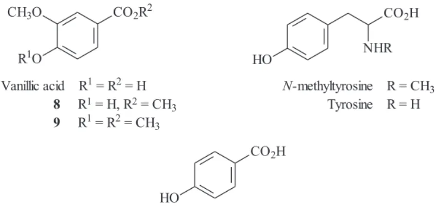
metabolites 4-hydroxy-3-methoxy-benzoic acid (vanillic acid) and 4-hydroxy-benzoic acid [0.005% in the ratio 87:13 (RMN), respectively] and an amino acid
2-methylamino-3-(4-hydroxyphenyl)-propanoic acid (0.014%, N-
methyl-tyrosine) were isolated. The structures of vanillic acid,
4-hydroxy-benzoic acid and N-methyltyrosine were identified
by spectroscopic analysis (including 300 MHz1H and 13C
NMR and MS experiments) and by chemical transformation of the vanillic acid, with diazomethane giving the two
methylated derivative esters (8 and 9). 1H and 13C NMR
(DMSO/DCl) data of N-methyltyrosine were in accordance
with the authentic sample of 2-amino-3-(4-hydroxyphenyl)-propanoic acid (tyrosine) obtained from commercial sources.
The different observed peaks were assigned to the N-Me
group of N-methyltyrosine.
Two of the aromatic acids vanillic and 4-hydroxy-benzoic acid have shown remarkable antioxidant activity
in other species.20,21 Based on such results, C. cajucara
could be expected to possess antioxidant properties. Reinforcing this suggestion, several kaempferol
metabolites have proved to be antioxidant agents22-24 and
C. cajucara leaves also contain two of them, e.g.
kaempferol 3,4’,7-trimethyl ether and 3,7-dimethyl ether.1
Within the research program for the major biological
constituents of 1, we are improving its chemical
transformation, which means preparing derivative
compounds for screening as pharmacological agents.1 In
the present work, using the method of regioselective
oxidation17 the 7-derivative was synthesized in good yields
(67%) by synthetic transformation of the furan moiety of
1, with singlet oxygen generated from molecular oxygen
by irradiating a polymer-bound rose bengal catalyst in
dichloromethane solution at –78 oC (in the presence of a
hindered base, such as diisopropylethylamine). In the
absence of a hindered base, the reaction of 1 with singlet
oxygen yielded an isomeric mixture of 6 and 7, which
was purified by TLC chromatography. The structures of
both 6 and 7-derivatives were determined by spectroscopy,
including 2D-NMR experiments (COSY 45, HSQC, HMBC) and the observed data corresponded to those of
Pharmacological profiling
In order to identify the antiproliferative action of
Croton cajucara constituents we studied the effects of natural clerodanes 1, 3, 4, 5, 6 and 7 and also the
semi-synthetic 6 and 7-derivatives against ascitic Ehrlich
carcinoma and human leukemia K562 cells. The
experiments were performed using the MTT assay,19 with
quercetin and vincristine as positive controls for Ehrlich and leukemia K562, respectively.
In vitro tests using Ehrlich carcinoma cells, natural
6, 7, 1 and 2, showed a significant cytotoxicity with dose
dependent responses over a 48 h culture period. The IC50
values were 166 µM for 1, 164 µM for 2, 65 µM for 6,
and 10 µM for 7, compared to quercetin with an IC50 = 44
µM (Table 1). The semi-synthetic cajucarinolide
derivatives (6 and 7) showed similar antiproliferative
activities compared to natural 6 and 7. The remaining
natural clerodanes 3, 4 and 5 do not show cytotoxic activity
against Ehrlich carcinoma cells.
Natural clerodanes 1-7 and also the semi-synthetic 2,
6 and 7 were also tested for their cytotoxic effects against
human K562 leukemia cell line using the Mossman assay19
and 48 h of cell culture. Among the natural clerodanes
assayed, only 3, 4, 6 and 7 (natural and semi-synthetic)
showed concentration-dependent growth inhibiting
activities on cultured K562 leukemia cells. The IC50 values
were 33 µM (3), 38 µM (5), 36 µM (6) and 43 µM (7). The cajucarinolide-derivatives indicated similar significant antiproliferative activity. Figure 1 shows the comparative cytotoxic effects of the tested clerodanes against both Ehrlich carcinoma and human K562 leukemia cells. The phenolic acids were also assayed against Ehrlich and K562 leukemia cells, but were inactive, under the same experimental conditions.
3 structure has only one α,β-insaturated ketone and
its cytotoxicity on K562 cells was similar to that observed
for 6, which has this moiety in addition to the conjugated
lactone carbonyl in the hydroxybutenolide ring. The
similar cytotoxic effects on K562 cells observed for 3,
5, 6 and 7, suggested no significant contribution of
hydroxybutenolide ring. Meanwhile, 4, the
dias-tereoisomer of 3, lacked efficacy indicating the
importance of the decaline system stereochemistry.
Further, 1 and 2 did not present significant cytotoxic
effect against K562 cultured cells until 50 µM. This result
was in accordance with the lower cytotoxic activity of 1
against promyelocytic HL60 cells with IC50 = 300 µM,
after 24 h, and 180 µM, after 96 h of culture.26
According to Wattenberg25 compounds containing an α,β
-unsaturated carbonyl moiety have been shown to bind to receptors that induce increased activities of phase II enzymes responsible for metabolizing xenobiotic agents. Thus, the
hydroxybutenolide moiety present in 6 and 7, probably led
to increase of activities in the Ehrlich carcinoma assay. Taking
into account the cytotoxic effect of 7 (10 µM), we suggest
that its higher antiproliferative action on growth Ehrlich cells
compared to 6 (65 µM) is related to the stereoposition of the
hydroxyl group in the hydroxybutenolide moiety (7 with OH
at position 16 and its isomeric stereoisomer 6 with OH at
position 15). Further, the clerodanes 3, 5 and 6 showed
comparable effects against human K562 leukemia cells.
HO
CO2H CO2R2
CH3O
R1O HO
CO2H
NHR
Vanillic acid R1= R2= H N-methyltyrosine R = CH3
8 R1= H, R2= CH
3 Tyrosine R = H
9 R1= R2= CH3
4-Hydroxy-benzoic acid
Table 1. IC50 values of metabolites from Croton cajucara against Ehrlich carcinomaand human K562 leukemia cells
Ehrlich carcinoma K562 cells Metabolites IC50/(µM) IC50/(µg mL-1) IC
50/(µM) IC50/(µg mL -1)
1 166 52.2 n.a. n.a.
2 164 51.8 n.a. n.a.
3 n.a. n.a. 33 11.1
4 n.a. n.a. n.a. n.a.
5 n.a. n.a. 38 12.6
6 65 22.4 36 12.6
7 10 3.5 43 14.9
Quercetin 44 13.3 -
-Vicristine - - 0.060 0.049
Conclusions
The further phytopharmacological studies of Croton
cajucara have demonstrated that the therapeutic potential of this plant is undeniable. The tested natural bioactive
clerodanes 1-7 and also the semi-synthetic derivatives
of 1 had their purity confirmed by NMR analysis. The
clerodanes 1, 2, 6 and 7 showed weak to moderate
cytotoxic effect against Ehrlich carcinoma cells and moderate efficacy against human K562 leukemia cells
was observed for the clerodanes 3, 5, 6 and 7. Only the
hydroxybutenolide clerodanes 6 and 7 (natural and
semi-synthetic) showed antiproliferative effect on both Ehrlich and K562 cells.
Synthetic transformation of 1, with satisfactory yields
by simple and low cost methodologies, afforded 6,which
was isolated as minor natural constituents of C. cajucara.
Further new phytochemical investigation yielded two aromatic acids (vanillic acid and 4-hydroxy-benzoic acid)
and an amino acid (N-methyltyrosine).
Acknowledgments
The authors wish to thank financial support from CNPq, CAPES and CAPES-PRODOC Programa CNRNM for the availability of the 600 MHz NMR instrument.
References
1. Maciel, M. A. M.; Pinto, A. C.; Arruda, A. C.; Pamplona, S. G. S. R.; Vanderlinde, F. A.; Lapa, A. J.; Cólus, I. M. S.; Echevarria, A.; Grynberg, N. F.; Farias, R. A. F.; Luna, A. M. C.; Rao, V. S. N.; J. Ethnopharmacol. 2000, 70, 40.
2. Maciel, M. A. M.; Pinto, A. C.; Veiga Jr., V. F.; Martins, J. R.; Grynberg, N. F.; Echevarria, A.; Lapa, A. J.; Vanderlinde, F.
A.; Phytochem. Pharmacol. II Series Recent Prog. Med. Plants
2002, 8, 502.
3. Carvalho, J. C. T.; Silva, M. F. C.; Maciel, M. A. M.; Pinto, A. C.; Nunes, D. S.; Lima, R. M. L.; Bastos, J. K.; Sarti, S. J.;
Planta Med. 1996, 62, 402.
4. Luna, A. M. C.; Silva, J. C. R.; Campos, A. R.; Rao, V. S. N.; Maciel, M. A. M.; Pinto, A. C.; Phytother. Res. 1999, 13, 689. 5. Agner, A. R.; Maciel, M. A. M.; Pinto, A. C.; Pamplona, S. G. R. S.; Cólus, I. M. S.; Teratog. Carcinog. Mutagenesis 1999,
19, 377.
6. Agner, A. R.; Maciel, M. A. M.; Pinto, A. C.; Cólus, I. M. S.;
Planta Med. 2001, 67, 815.
7. Hiruma-Lima, C. A.; Spadari-Bratfisch, R. C.; Grassi-Kassisse, D. M.; Brito, A. R.; Planta Med. 1999, 65, 325.
8. Farias, R. A. F.; Rao, V. S. N.; Viana, G. S. B.; Silveira, E. R.; Maciel, M. A. M.; Pinto, A. C.; Planta Med. 1997, 66, 558. 9. Silva, R. M.; Santos, F. A.; Maciel, M. A. M.; Pinto, A. C.;
Rao, V. S.; Planta Med. 2001, 67, 763.
10. Maciel, M. A. M.; Pinto, A. C.; Brabo, S. N.; Silva, M. N.;
Phytochemistry 1998, 49, 823.
11. Ichihara, Y.; Takeya, K.; Hitotsuyanagi, Y.; Morita, H.; Okuyama, S.; Suganuma, M.; Fujiki, H.; Motidome, M.; Itokawa, H.; Planta Med. 1992, 58, 549.
12. Di Stasi, L. C.; Hiruma, C. A.; Guimarães, E. M.; Santos, C. M.; Fitoterapia 1994, 65, 529.
13. Van den Berg, M. E.; Plantas Medicinais na Amazônia, Gráfica Falangola: Belém, 1993, pp.124-125, 168.
14. Farias, R. A. F.; Neto, M. F. O.; Viana, G. S. B.; Rao, V. S. N.;
Phytother. Res. 1996, 10, 697.
15. Kubo, I.; Asaka, Y.; Shibata, K.; Phytochemistry 1991, 30, 2545. 16. Itokawa, H.; Ichihara, Y.; Shimizu, M.; Takeya, K.; Motidome,
M.; Chem. Pharm. Bull. 1990, 38, 701.
17. Kernan, R. M.; Faulkner, D. J.; J. Org. Chem. 1988, 53, 2773. 18. Maciel, M. A. M.; Pinto, A. C.; Kaiser, C. R.; Magn. Reson.
Chem. 2003, 41, 278.
19. Mossmann, T. J.; J. Immunol. Methods 1983, 65, 55. 20. Hung, C.; Yen, G.; J. Agric. Food Chem. 2002, 50, 2993. 21. Ohsugi, M.; Fan, W.; Hase, K.; Xiong, Q.; Tezuka, Y.; Komatsu,
K.; Namba, T.; Saitoh, T.; Tazawa, K.; Kadota, S.; J. Ethnopharmacol. 1999, 67, 111.
22. Marfak, A.; Trouillas, P.; Allais, D. P.; Champavier, Y.; Calliste, C. A.; Duroux, J. L.; J.Agric. Food Chem. 2003, 51, 1270. 23. Jonson, E. L.; Schmidt, W. F.; Emche, S. D.; Biochem. Syst.
Ecol. 2003, 31, 59.
24. Bonina, F.; Puglia, C.; Ventura, D.; J. Cosmet. Sci. 2002, 53, 321. 25. Wattenberg, L. J.; J. Cell. Biochem. 1995, 22, 162.
26. Anazetti, M. C.; Melo, O. S.; Duran, N.; Haun, M.; Toxicology
2003, 188, 261.
Received: September 27, 2005
Web Release Date: March 30, 2007