Article
J. Braz. Chem. Soc., Vol. 26, No. 6, 1152-1159, 2015. Printed in Brazil - ©2015 Sociedade Brasileira de Química 0103 - 5053 $6.00+0.00
A
*e-mail: [email protected]
New Alkaloids from
Margaritopsis carrascoana
(Rubiaceae)
Raimundo R. G. Nascimento,a Antônia T. A. Pimenta,a Pedro de Lima Neto,b José R. C. Junior,b Letícia V. Costa-Lotufo,c Elthon G. Ferreira,c Luzineide W. Tinoco,d
Raimundo Braz-Filho,e Edilberto R. Silveiraa and Mary A. S. Lima*,a
aDepartamento de Química Orgânica e Inorgânica and bDepartamento de Química Analítica e Físico-Química,
Centro de Ciências, Universidade Federal do Ceará, CP 12.200, 60021-940 Fortaleza-CE, Brazil
cDepartamento de Farmacologia e Fisiologia, Centro de Ciências da Saúde, Universidade Federal
do Ceará, CP 12.200, 60430-270 Fortaleza-CE, Brazil
dNúcleo de Pesquisas de Produtos Naturais, Centro de Ciências da Saúde, Universidade Federal do
Rio de Janeiro, 21941-902 Rio de Janeiro-RJ, Brazil
eFAPERJ, Laboratório de Ciências Químicas, Universidade Estadual do Norte Fluminense, Instituto
de Química, Universidade Federal Rural do Rio de Janeiro, 28013-602 Rio de Janeiro-RJ, Brazil
Two new alkaloids containing Nb-methyl-triptamine units linked to bis-quinoline moieties, N-8”-formyl-calycosidine and N-8”-methyl-N-1’-demethyl-iso-calycosidine, were isolated from the aerial part of Margaritopsis carrascoana, along with known calycosidine and hodgkinsine. The cytotoxicity of the isolated alkaloids has been evaluated against cancer cell lines. The first two compounds showed only weak activity against HL-60 cell line, while hodgkinsine was the most active compound against HCT-116, OVCAR-8, HL-60 and SF-295 cancer cell lines. The structures of the isolated compounds were established by spectroscopic and molecular mechanical methods.
Keywords: Margaritopsis carrascoana, Rubiaceae alkaloids, N-8”-methyl-N -1’-demethyl-iso-calycosidine, N-8”-formyl-calycosidine, calycosidine, hodgkinsine
Introduction
The Rubiaceae family is known as a prolific source of alkaloids with great structural diversity and pharmacological properties. Among these, the pyrrolidinoindoline polymers, resulting from the condensation of several Nb -methyl-triptamine units, and those containing Nb-methyl-triptamine units linked to a bis-quinoline part, seem to be specific to the Psychotrieae tribe, particularly to the genera
Calycodendron, Psychotria and Hodgkinsonia.1-3
Thegenus Margaritopsis comprises about 50 species with pantropical occurrence, 27 of which are recognized in America and 15 are found in Brazil. It consists mainly of species that have been removed from the genus Psychotria
L. (Rubiaceae) based on molecular data and geographic distribution.4,5 The chemical studies of this genus is restricted to M. cymuligera, that revealed the presence of pyrrolidinoindoline alkaloids, largely found in species of
Psychotria.6 Margaritopsis carrascona is an endemic shrub
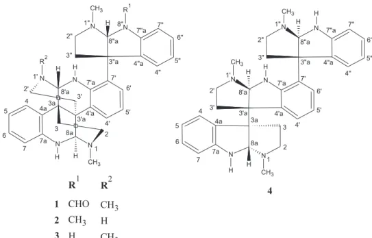
from northeastern Brazil flora growing in the sandy soils of the region of Ibiapaba and Araripe plateaus in Ceará State. Previous chemical studies of its roots revealed glucosylated flavonoids with antioxidant activity.7 In this work, we report the isolation and structural characterization of two new alkaloids containing a Nb-methyl-triptamine unit linked to a bis-quinoline moiety, N-8”-formyl-calycosidine (1) and N-8”-methyl-N-1’-demethyl-iso-calycosidine (2), in addition to the known alkaloids calycosidine (3)1 and hodgkinsine (4)2 (Figure 1). The cytotoxicity of the isolated alkaloids have been evaluated against colorectal adenocarcinoma (HCT-116), ovarian carcinoma (OVCAR-8), human promyelocytic leukemia (HL-60) and glioma (SF-295) cancer cell lines.
Experimental
General experimental procedures
equipped with an inverse detection probehead and z-gradient accessory working at 500.13 (1H) and at 125.77 MHz (13C). All pulse sequences are standard in the Bruker XWIN-NMR software, and all experiments were conducted at room temperature. The samples, in 5 mm tube, were dissolved in CD3OD (0.6 mL). Infrared (IR) spectra were recorded on a PerkinElmer FT-IR 1000 spectrometer (Waltham, USA). High resolution mass spectra were recorded on a QTOF mass spectrometer (Xevo-Waters, Milford, USA) by positive ionization mode of the electrospray ionization (ESI) source. Optical rotations were obtained on a PerkinElmer Q-2000 polarimeter (Waltham, USA) at 589 nm and 25 °C. High performance liquid chromatography (HPLC) analyses were performed on a Shimadzu chromatographer equipped with a ternary pump (Shimadzu LC-20AT) and UV detector (Shimadzu SPD-M20A, Kyoto, Japan), using Phenomenex RP-18 column (analytical: 250 × 4.6 mm, 5 µm; semi-preparative: 250 × 10 mm, 10 µm). HPLC grade solvents were purchased from Tedia Co. (São Paulo, Brazil) and the HPLC grade water was obtained by a Milli-Q purification system (Millipore, Bedford, USA). Thin layer chromatography (TLC) was performed on precoated silica gel aluminum sheets Merck (Kenilworth, USA) and the compounds were visualized by UV detection and spraying with Dragendorff reagent or vanillin/perchloric acid/EtOH solution, followed by heating.
Plant material
Aerial parts of Margaritopsis carrascoana (Delprete & E. B. Souza) C. M. Taylor & E. B. Souza were
collected at the Moreilândia County, Araripe Plateau (Pernambuco State, Northeast of Brazil). A voucher specimen (No. 22075) has been deposited at the Herbário Prisco Bezerra (EAC) and identified by Prof Edson Paula Nunes, Departamento de Biologia, Universidade Federal do Ceará, Brazil.
Extraction and isolation
Dried aerial parts of M. carrascoana (7.6 kg) were exhaustively extracted with EtOH (3 × 9.0 L) at room temperature, followed by solvent evaporation under vacuum, yielding 295.5 g of extract. A portion of the extract (33.0 g) was treated with 10% aqueous HCl and the acid solution was extracted with EtOAc (3 × 150 mL). The aqueous phase was basified with NH4OH to pH 9-10 and extracted with EtOAc (3 × 150 mL). The organic fractions were combined, and the solvent was evaporated to yield an alkaloid fraction (1.1 g), which was then chromatographed on a C18 cartridge [(H2O/trifluoroacetic acid (TFA) 0.1%)/ MeOH 1:1; (H2O/TFA 0.1%)/MeOH 1:2; (H2O/TFA 0.1%)/ MeOH 1:3, and MeOH] yielding four fractions: F-1 (80 mL, 231.9 mg), F-2 (60 mL, 563.4 mg), F-3 (40 mL, 46.1 mg) and F-4 (100 mL, 84.8 mg). Fraction F-2 (563.4 mg) was submitted to semi-preparative RP-18 HPLC, using (H2O/TFA 0.1%)/MeOH solutions with a gradient of 25% to 75% MeOH, to give five sub-fractions: F-2 (1) (15.1 mg, tR = 5.5 min), F-2 (2) (95.6 mg, tR = 6.2 min), F-2 (3) (90.0 mg, tR = 8.9 min), F-2 (4) (59.2 mg, tR = 12.1 min), and F-2 (5) (163.8 mg). Sub-fraction F-2 (1) (15.1 mg) was resubmitted to semi-preparative RP-18 HPLC, using
CH3 CH3
R2 H H
N N
H R1
N N
H H
N N
6"
5" 7"
4" 4"a
7"a
3"a 8"a
3" 2"
1"
6'
5' 7'
4' 4'a 7'a
3'a 3' 2'
8'a 3a 1'
3
2 4a
4 5
6
7 7a
8a
1 8"
R1 R2
CHO CH3
H CH3
H CH3
1
2
3
4 CH3
CH3
CH3 H H
H
H
H
H
N N
N N
N N
1 2 3 3a 4a
4
5
6
7 7a
8a 4'a 3'a
3' 2'
1'
8'a
4' 5' 6' 7' 7'a
4"a 3"a 3"
2" 1"
8"a
4" 5" 6" 7" 7"a
(H2O/TFA 0.1%)/MeOH 3:1 as eluent, to afford 1 (4.2 mg) and 2 (5.6 mg). Sub-fractions F-2 (2) (95.6 mg) and F-2 (4) (15.5 mg) were resubmitted to the former HPLC conditions to yield calycosidine 3 (50.6 mg) and hodgkinsine 4
(30.2 mg), respectively.
Molecular mechanics calculation
The lowest energy structures were obtained by a scanning method through the rotation of the dihedral angles of 60 degrees each. The corresponding structures were relaxed using the Gaussian 09 program from Gaussian INC, through de density functional theory (DFT) method B3LYP with the base-set 6-31+G (d,p). The continuous solvatation method polarizable continuum model (PCM) was applied to simulate the medium of the NMR experiment. Methanol was used as solvent. The semiempirical Austin model 1 (AM1) method yielded seven distinct and sterically allowed conformations for compound 1, and two conformations for compound 2. The lowest energy conformations were relaxed with the DFT method.
Cytotoxic assays
Tumor cell lines colorectal adenocarcinoma (HCT-116), ovarian carcinoma (OVCAR-8), human promyelocytic leukemia (HL-60) and glioma (SF-295), provided by the National Cancer Institute (Bethesda, MD, USA), were used in this work. Cells were maintained in RPMI 1640 medium supplemented with 10% fetal bovine serum, 2 mmol L−1 glutamine, 100 U mL−1 penicillin, and 100 ug mL−1 streptomycin at 37 °C with 5% CO
2. The cytotoxicity of all compounds was tested against tumor cell lines using the 3-(4,5-dimethyl-2-thiazolyl-2,5-diphenyl-2H-tetrazolium bromide) (MTT) (Sigma Aldrich Co., St. Louis, MO, USA) reduction assay.8 For all experiments, cells were plated in 96-well plates (105 cells per well for adherent cells or 3 × 10⁵ cells per well for suspended cells in 100 µL of medium). The tested compounds (0.05-25 µg mL−1) dissolved in dimethyl sulfoxide (DMSO) were added to each well, using the high-throughput screening-biomek (HTS) 3000-Beckman Coulter (Inc. Fullerton, California, USA), and incubated for 72 h. Doxorubicin was used as positive control. Control groups received the same amount of DMSO. After 69 h of incubation, the supernatant was replaced by fresh medium containing MTT (0.5 mg mL−1). Three hours later, the MTT formazan product was dissolved in 150 µL of DMSO, and absorbance was measured at 595 nm (DTX 880 Multimode Detector, Beckman Coulter, Inc. Fullerton, CA, USA).
N-8”-Formyl-calycosidine (1)
Brown resin; [α]D
25 = −4.6 (c 0.1, MeOH); IR (KBr)
νmax / cm−1 3346, 2950, 2927, 2843, 1678, 1487, 1463,
1206, 1136, 841, 801, 756, 724; 1H NMR (500 MHz,
CD3OD) and 13C NMR (125 MHz, CD3OD), Table 1; HRESIMS (positive mode) m/z 547.3199 [M + H]+ (calcd. for C34H39N6O, 547.3185).
N-8”-Methyl-N-1’-demethyl-iso-calycosidine (2)
Brown resin; [α]D
25 = −3.3 (c 0.05, MeOH); IR (KBr)
νmax / cm−1 3347, 2924, 2843, 1679, 1457, 1205, 1139, 845,
801, 724; 1H NMR (500 MHz, CD
3OD) and
13C NMR
(125 MHz, CD3OD), see Table 1; HRESIMS (positive
mode) m/z 519.3236 [M + H]+ (calcd. for C
33H39N6, 519.3246).
Results and Discussion
Compound 1 was isolated as a brown resin,
[α]D
25 = −4.6° (c 0.1, MeOH). The IR spectrum showed the presence of N−H (3346 and 2950 cm−1), two absorptions related to aldehyde carbonyl (2843 and 1678 cm−1) and benzene ring (1678, 1487 and 1463 cm−1) functionalities. The molecular formula of compound 1 was established by the positive mode HRESIMS peak at m/z 547.3199 [M + H]+. Comparison of its mass spectrometry data with those obtained for the alkaloids hodgkinsine and calycosidine, isolated from other Psychotria species, suggested the same trimeric-type structure for compound 1. The principal features were the presence of important ions at m/z 490.2646 and 433.2057, due to subsequent loss of C3H7N, typical fragments of the analogous calycosidine skeleton formed by one pyrrolidinoindoline and one calycanthine subunits attached at C-7’and C-3”a (Figure 2).9
The 1H NMR spectrum of 1 corroborated the suggestion of the trimeric structure from the signals corresponding to six diastereotopic methylenes at dH3.12 (m, H-2a), 2.74 (m, H-2b);dH2.60 (m, H-2’a) and 2.18 (m, H-2’b); dH 3.05 (m, H-2”a) and 2.98 (t, J 12.7 Hz, H-2”b), dH2.06 (m, H-3a) and 1.73 (d, J 14.7 Hz, H-3b), dH2.18 (m, H-3’a) and 1.62 (d,
J 12.2 Hz, H-3’b); dH 2.57 (m, H-3”a) and 2.42 (m, H-3”b), in addition to three N-methyl groups at dΗ 2.81 (s, CH3-1), 2.41 (s, CH3-1’) and 2.66 (s, CH3-1”), three methines at
dH 7.38 (H-4’) ↔ 7.15 (H-5’) ↔ 7.68 (H-6’) (subsystem II) and dH 7.64 (H-4”) ↔ 7.32 (H-5”) ↔ 7.43 (H-6”) ↔ 7.56 (H-7”) (subsystem III). In addition, the following vicinal and geminal correlations for three pairs of diastereotopic methylenes were observed: dH 3.12 (m, H-2a), 2.74 (m, H-2b) and dH 2.06 (m, H-3a), 1.73 (d,
J 14.7 Hz, H-3b); dH 2.60 (m, H-2’a), 2.18 (m, H-2’b) and
dH 2.18 (m, H-3’a), 1.62 (d, J 12.2 Hz, H-3’b); and dH 3.05 (m, H-2”a), 2.98 (t, J 12.7 Hz, H-2”b) and dH 2.57 (m, H-3”a), 2.42 (m, H-3”b). The unambiguous assignments of all carbons and hydrogens were possible by the HMQC spectrum analysis (Table 1). From these data it was possible to deduce the presence of the pyrrolidinoindole unit by the typical signal related to the methine carbon at dC 85.6 (C-8”a). The other methines at dC 76.2 and 73.9 accounted for carbons C-8’a and C-8a of the calycanthine unit.9 The HMBC spectrum showed long-range correlations in the pyrrolidinoindoline moiety between the methine proton at dH 6.58 (H-8”a) with the carbons at dC 135.5 (C-4”a),
140.2 (C-7”a) and 35.1 (C-3”), besides the correlations of the N-methyl group at N-1” (dH 2.66) with carbon at dC 46.3 (C-2”). The aromatic protons at dH 7.56 (H-7”) and 7.32 (H-5”) showed correlations with the carbon at dC 135.5 (C-4”a), whereas H-6” (dΗ 7.43) and H-4” (dΗ 7.64)
correlated with the carbon at dC 140.2 (C-7”a).
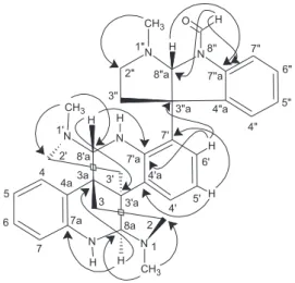
The position of the formyl group at N-8” was deduced on the basis of the correlations of the hydrogen at dH 9.18 (CHO) with carbons at dC 140.2 (7”a) and 85.6 (C-8”a) (Figure 3). The HMBC correlations for the calycanthine-type moiety were observed through the methine at
dH 5.48 (H-8a) with carbons at dC 143.2 (C-7a), 36.4 (C-3a) and 47.9 (C-2), whereas the aromatic protons at
dH 7.04 (H-5) and 6.95 (H-7) correlated with the carbon at dC 120.1 (C-4a), and the protons at dH 7.37 (H-4) and 7.29 (H-6) showed correlations with the carbon at
dC 143.2 (C-7a). The other methine at dH 5.49 (H-8’a) showed correlations with carbons at dC 38.5 (C-3’a) and 146.5 (C-7’a), the aromatic proton at dH 7.15 (H-5’)
CH3 CH3 CH3 H H N N H N N H H N N
O+ H H 6" 5" 7" 4" 4"a 7"a 3"a 8"a 3" 2" 1" 6' 5' 7' 4' 4'a 7'a 3'a 3' 2' 8'a 3a 1' 3 2 4a 4 5 6 7 7a 8a 1 8" C
H2 CHNHCH3
O+ H
H H CH3 N N H CH3 N N N H 6" 5" 7" 4" 4"a 7"a 3"a 8"a 3" 2" 1" 6' 5' 7' 4' 4'a 7'a 3'a 3' 2' 8'a 3a 1' 4a 4 5 6 7 7a 8a 8" C
H2 CHNHCH3
O+ H
N H CH3 N N N H 6" 5" 7" 4" 4"a 7"a 3"a 8"a 3" 2" 1" 6' 5' 7' 4' 4'a 7'a 3'a 3a 4a 4 5 6 7 7a 8a 8" CH3 CH3 H H N N H CH3 N N+ H H N N H H 6" 5" 7" 4" 4"a 7"a 3"a 8"a 3" 2" 1" 6' 5' 7' 4' 4'a 7'a 3'a 3' 2' 8'a 3a 1' 3 2 4a 4 5 6 7 7a 8a 1 8" C
H2 CHNHCH3
CH3 H H N N H CH3 N N+ N H H 6" 5" 7" 4" 4"a 7"a 3"a 8"a 3" 2" 1" 6' 5' 7' 4' 4'a 7'a 3'a 3' 2' 8'a 3a 1' 4a 4 5 6 7 7a 8a 8" C H2 CHNH2
CH3 N H CH3 N N+ N H 6" 5" 7" 4" 4"a 7"a 3"a 8"a 3" 2" 1" 6' 5' 7' 4' 4'a 7'a 3'a 3a 4a 4 5 6 7 7a 8" m/z547.3199
(calcm/z547.3212) m/z490.2646
m/z433.2057
m/z519.3246 (calcm/z519.3236)
m/z462.2684
m/z419.5402
C34H39N6O C
31H32N5O
C28H25N4O
C
33H39N6 C30H32N5
C28H27N4 Compound2
Compound1
correlated with carbons at dC 128.8 (C-4’a) and 116.7 (C-7’), whereas H-6’(dH 7.68) and H-4’ (dH 7.38) showed correlations with the carbon at dC 146.5 (C-7’a). The N-1 methyl group at dH 2.81 showed correlation with the carbons at dC 47.9 (C-2) and 73.9 (C-8a), while N-1’-Me
(dH 2.41) correlated with the carbons at dC 47.8 (C-2’) and 76.2 (C-8’a).
Molecular mechanics methods generated the most stable conformation for 1, that was consistent with the observed transanular nOe interactions in the NOESY
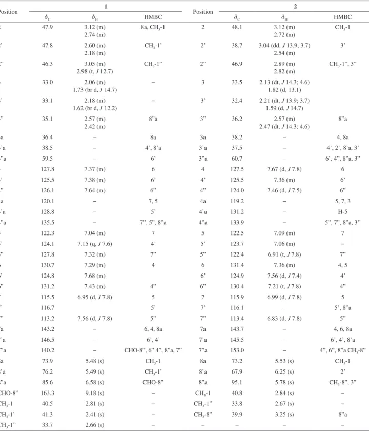
Table 1. 1H NMR and 13C NMR (d in ppm and J in Hz) data and HMBC correlations of compounds 1 and 2
Position 1 Position 2
dC dH HMBC dC dH HMBC
2 47.9 3.12 (m)
2.74 (m)
8a, CH3-1 2 48.1 3.12 (m)
2.72 (m)
CH3-1
2’ 47.8 2.60 (m)
2.18 (m)
CH3-1’ 2’ 38.7 3.04 (dd, J 13.9; 3.7) 2.54 (m)
3’
2” 46.3 3.05 (m)
2.98 (t, J 12.7)
CH3-1” 2” 46.9 2.89 (m)
2.82 (m)
CH3-1”, 3”
3 33.0 2.06 (m)
1.73 (br d, J 14.7)
− 3 33.5 2.13 (dt, J 14.3; 4.6)
1.82 (d, 13.1)
3’ 33.1 2.18 (m)
1.62 (br d, J 12.2)
− 3’ 32.4 2.21 (dt, J 13.9; 3.7)
1.59 (d, J 14.7)
3” 35.1 2.57 (m)
2.42 (m)
8”a 3” 36.2 2.57 (m)
2.47 (dt, J 14.3; 4.6)
8”a
3a 36.4 − 8a 3a 38.2 − 4, 8a
3’a 38.5 − 4’, 8’a 3’a 37.5 − 4’, 2’, 8’a, 3’
3”a 59.5 − 6’ 3”a 60.7 − 6’, 4”, 8”a, 3”
4 127.8 7.37 (m) 6 4 127.5 7.67 (d, J 7.8) 6
4’ 125.5 7.38 (m) 6’ 4’ 125.5 7.36 (m) 6’
4” 126.1 7.64 (m) 6” 4” 124.0 7.46 (d, J 7.5) 6”
4a 120.1 − 7, 5 4a 119.2 − 5, 7, 3
4’a 128.8 − 5’ 4’a 131.2 − H-5
4”a 135.5 − 7”, 5”, 8”a 4”a 133.9 − 5”, 7”, 8”a, 3’’
5 122.3 7.04 (m) 7 5 122.5 7.09 (m) 7
5’ 124.1 7.15 (q, J 7.6) 4’ 5’ 123.7 7.06 (m) −
5” 127.8 7.32 (m) 7” 5” 122.4 6.91 (t, J 7.8) 7”
6 130.7 7.29 (m) 4 6 131.4 7.36 (m) 4, 5
6’ 124.8 7.68 (m) 6’ 124.9 7.56 (d, J 7.4) 4’
6” 131.2 7.43 (m) 4” 6” 130.4 7.21 (t, J 7.8) 4”
7 115.5 6.95 (d, J 7.8) 5 7 115.9 6.99 (d, J 7.8) 5
7’ 116.7 5’ 7’ 116.1 − 5’, 8”a
7” 113.2 7.56 (d, J 7.8) 5” 7” 113.4 6.83 (d, J 7.8) 5”
7a 143.2 − 6, 4, 8a 7a 143.7 − 4, 6, 8a
7’a 146.5 − 6’, 4’ 7’a 145.5 − 6’, 4’, 8’a
7”a 140.2 − CHO-8”, 6” 4”, 8”a, 7” 7”a 153.0 − 4”, 6”, 8”a CH3-8”
8a 73.9 5.48 (s) CH3-1 8a 73.2 5.53 (s) CH3-1
8’a 76.2 5.49 (s) CH3-1’ 8’a 67.9 6.25 (s) 2’
8”a 85.6 6.58 (s) CHO-8” 8”a 95.1 5.78 (s) CH3-8”, 3”
CHO-8” 163.3 9.18 (s) − CH3-1 40.8 2.84 (s) −
CH3-1 40.5 2.81 (s) − CH3-1” 33.8 2.67 (s) −
CH3-1’ 41.3 2.41 (s) − CH3-8” 39.9 3.25 (s) 8”a
spectrum (Figure 4). The key nOe cross-peaks observed between the pairs dH 1.62 (H-3’b) ↔ 5.48 (H-8a) ↔ 7.38 (H-4’); dH 2.06 (H-3a) ↔ 5.49 (H-8a’) ↔ 7.37 (H-4); and dH 6.58 (H-8a”) ↔ 9.18 (CHO) ↔ 7.56 (H-7”) indicated that they were oriented in the same direction, and definitively determined the relative stereochemistry of 1, asameso-calycanthine central subunit linked at C-7’ by one pirrolidineindole unit.
Compound 2 was isolated as a brown resin,
[α]D
25 = −3.3° (c 0.05, MeOH). The IR spectrum showed bands relative to the presence of N−H (3346 and 2924 cm−1) and aromatic ring (1678, 1457 and 1205 cm−1) functionalities. The comparative analysis of its IR data with those observed for compound 1, revealed that formyl band was missing. The molecular formula of compound 2 was
established by the positive mode HRESIMS peak at m/z
[M + H]+ 519.3246, which undergo further fragmentations similar to those of 1, to form the ion peaks at m/z 462.2684 and 419.5402, relative to subsequent loss of C3H7N and C2H5N (Figure 2).
The support for a trimeric alkaloid calycosidine-type for compound 2 came from the comparative analysis of its NMR data with those observed for compounds 1 and 3, revealing them to be very similar. In the 1H NMR spectrum of 2, the same signals related to the meso-calycanthine central subunit linked at C-7’ by one pirrolidineindole unit were observed (Table 1). However, the signal relative to the hydrogen of the formyl group at N-8’’ at d 9.18 observed for
1 was absent in 2, as well as the absence of carbonyl group at the 13C NMR spectrum. The long-range correlations in the HMBC spectrum between the typical methine of the pirrolidineindole moiety at dΗ 5.78 (H-8”a) with the carbons
at dC 46.9 (C-2”), 36.2 (C-3”) and 133.9 (C-4”a), and
between both protons at dΗ 2.67 (CH3-1”) and 3.25 (CH3-8”) with the methine carbon at dC 95.1 (C-8”a) determined the
position of the N-methyl groups at N-1” and N-8”. Thus, compound 2 was determined as a structural analogue of calycosidine (3), where the N-group has migrated from N-1’ to N-8’’ (Figure 4). This suggestion was corroborated by the protonated molecule at m/z 519.3246 in the HRESIMS, that undergo further fragmentations to form the ion peaks at m/z 462.2684 and 419.5402, relative to subsequent loss of C3H7N and C2H5N (Figure 2).
The most stable conformation of 2 was generated by molecular mechanics methods, and supported the spatial interactions observed on the NOESY spectrum (Figure 5).
CH3
CH3 CH3
H H N N
H N N
H H
N N
O H
H
H 4"a 7"a
2 8"a
5' 7'
4'a 7'a
3'a 8'a 3a
7a 8a
6' 2"
3"
3"a
4" 5" 6" 7"
4' 3'
2' 4
5
6
7 4a 1'
3
1
1" 8"
Figure 3. HMBC correlations (H → C) of 1.
The nOe cross peaks observed in the NOESY spectrum were in agreement with those proposed by the diagnostic correlations observed for the proton at dH 5.53 (H-8a) with those at dH 1.59 (H-3’b), 7.36 (H-4’) and 2.84 CH3-1; the proton at dH 6.25 (H-8’a) with those at dH 1.82 (H-3b) and 7.67 (H-4), in addition to the correlations of the proton at dH 5.78 (H-8”a) with those at dH 2.57 (H-3”a), 3.25 (CH3-8”) and 2.67 (CH3-1”).
The alkaloids calycosidine (3) and hodgkinsine (4) are common in Psychotria species, and were isolated as the major constituents from the aerial parts of M. carrascoana. Calycosidine (3) was also obtained by transformation of hodgkinsine (4) under mild acidic conditions,9 and has been suggested as an artifact resulting from acid-base reaction during extraction the and isolation processes. Hodgkinsine is cited as anti-microbial, antinociceptive and as potent cytotoxic agent against HTC cell line.10,11
The cytotoxicity of pyrrolidinoindoline alkaloids has been examined for potential applications as anti-cancer agents, and the statement of relationship between some structural patterns and the biological effects has been investigated.12 Studies using hepatoma (HTC) cells
in vitro revealed that the β-β’ bond in these compounds is important for the activity.13 The cytotoxic activity of 1-4 was assessed against human cancer cell lines HCT-116, OVCAR-8, HL-60 and SF-295. Based on data collected from three independent experiments, results showed that only hodgkinsine (4) exhibited effective action against HCT-116, SF-295 and OVCAR-8 cell lines with IC50 values of 1.67, 1.0 and 1.41 µg mL−1, respectively. Compound 2 exhibited a moderate effect against the HL-60 cell line with an IC50 value of 14.19 µg mL−
1, whereas 1 and 3 showed IC50 > 50 µg mL−1. IC50 values for doxorubicin were 0.12 (HCT-116), 0.02 (HL-60), 0.24 (SF-295) and 0.26 µg mL−1 (OVCAR-8).
CH3 CH3 H H H
N N
H CH3 N N
H H
N N
H
H 4"a
7"a
3"a 8"a
5' 7'
4'a 7'a
3'a 8'a 3a
4a 7a
8a
6' 2"
3"
1" 8"
2
7"
6"
5"
4" 1'
2'
3'
3 4'
4
5
6
7 1
Figure 5. HMBC correlations (H → C) of 2.
Conclusions
The isolation of alkaloids N-8’’-formyl-calycosidine (1) and N-8’’-methyl-N-1’-demethyl-iso-calycosidine (2) containing Nb-methyl-triptamine units from M. carrascoana reinforced the close relationship of the genera Margaritopsis
and Psychotria, since both calycosidine (3) and hodgkinsine (4) have been found in P. rostrata. However, this is the first report of the isolation of structural analogues of calycosidine.
The cytotoxic activity of all compounds was evaluated against HCT-116, OVCAR-8, HL-60 and SF-295 cancer cell lines, where compounds 1 and 2 showed weak activity against HL-60, while hodgkinsine 4 was the most active compound against HCT-116, SF-295 and OVCAR-8. The current results suggest that, as already observed for
Psychotria, the genus Margaritopsis is a prolific source of structurally interesting and biologically active alkaloids.
Supplementary Information
Supplementary data associated with this paper are available free of charge at http://jbcs.sbq.org.br as PDF file.
Acknowledgements
The authors are grateful to CNPq, CAPES, PADCT, PRONEX, FUNCAP and FINEP for the fellowships and financial support. We also thank to CENAUREMN and LEMANOR of Universidade Federal do Ceará, for NMR and high resolution mass spectra, respectively.
References
1. Libot, F.; Miet, C.; Kunesch, N.; Poisson, J.; Pusset, J.; Sévenet, T.; J. Nat. Prod. 1987, 50, 468.
2. Verrotta, L.; Pilati, T.; Tatò, M.; Elisabetsky, E.; Amador, T. A.; Nunes, D. S.; J. Nat. Prod.1998, 61, 392.
3. Parry, K. P.; Smith, G. F.; J. Chem. Soc., Perkin Trans. 1 1978, 1671.
4. Andersson, L.; Syst. Geogr. Pl. 2001, 71, 73. 5. Taylor, C. M.; Syst. Geogr. Pl. 2005, 75, 161.
6. Brand, G.; Henriques, A. T.; Passos, C. S.; Baldoqui, D. C.; Santin, S. M. O.; Costa, W. F.; Saragiotto, M. H.; Biochem. Syst. Ecol. 2012, 45, 155.
7. Nascimento, R. R. G.; Monteiro, J. A.; Pimenta, A. T. A.; Trevisan, M. T. S.; Braz-Filho, R.; Souza, E. B.; Silveira, E. R.; Lima, M. A. S.; Quim. Nova2014, 38, 60.
8. Mosmann, T. J.; J. Immunol. Methods 1983, 65, 55.
9. Libot, F.; Kunesch, N.; Poisson, J.; Kaiser, M.; Duddeck, H.;
10. Hassan-Elready, A. S.; El-Sharkawy, S. H.; Shier, W. T.; Planta Med. 1995, 61, 313.
11. Amador, T. A.; Verotta, L.; Nunes, D. S.; Elisabetsky, E.; Planta Med. 2000, 66, 770.
12. Roth, A.; Kuballa, B.; Bounthanh, C.; Cabalion, P.; Sévenet, T.; Beck, J. P.; Anton, R.; Planta Med. 1986, 52, 450.
13. Adjibadé, Y.; Saad, H.; Kuballa, B.; Beck, J. P.; Sévenet, T.; Cabalion, P.; Anton, R.; J. Ethnopharmacol. 1990, 29, 127.
Submitted: December 12, 2014