Short communication
Antiparasitic effect of
Dinoponera quadriceps
giant ant venom
Danya Bandeira Lima
a, Paloma Le
ao Sousa
~
a, Alba Fabíola Costa Torres
a,
Klinger Antonio da França Rodrigues
b, Clarissa Perdig
ao Mello
~
a,
Ramon R
oseo Paula Pessoa Bezerra de Menezes
c, Louise Donadello Tessarolo
a,
Yves Patric Quinet
d, M
arcia Rosa de Oliveira
e, Alice Maria Costa Martins
a,*aDepartment of Clinical and Toxicological Analysis, Faculty of Pharmacy, Federal University of Ceara, Fortaleza, Ceara, Brazil
bPost-graduate Program in Natural Products and Synthetic Bioactives, Health Sciences Center, Federal University of Paraíba, Joao Pessoa, Paraíba, Brazil cDepartment of Physiology and Pharmacology, Faculty of Medicine, Federal University of Ceara, Fortaleza, Ceara, Brazil
dInstitute of Biomedical Sciences, State University of Ceara, Fortaleza, Ceara, Brazil
eDepartment of Molecular Biology, Center of Exact Sciences and of the Nature, Federal University of Paraiba, Joao Pessoa, Paraíba, Brazil~
a r t i c l e
i n f o
Article history:
Received 23 June 2016 Received in revised form 10 August 2016 Accepted 11 August 2016 Available online 13 August 2016
Keywords:
Dinoponera quadriceps Trypanosoma cruzi Leshmania amazonensis
Venom
a b s t r a c t
Neglected tropical diseases (NTD) are treated with toxic therapy of limited efficacy. Previously, we studied the antimicrobial effect ofDinoponera quadricepsvenom (DqV) against bacteria. To continue the study, we report in this short communication the antimicrobial effect of DqV againstLeishmania ama-zonensisandTrypanosoma cruzi. DqV inhibits the promastigote forms ofL.amazonensisand allT.cruzi developmental forms, with low toxicity in host cells. DqV causes cell death inT.cruzithrough necrotic and apoptotic mechanisms observed by staining the cells with annexin V-FITC (AX) and propidium io-dide (PI), loss of mitochondrial membrane potential byflow cytometry analyses and confocal microscopy and morphological alterations, such as loss of membrane integrity and cell shrinkage by scanning electron microscopy (SEM). In conclusion, we suggest there is an antimicrobial effect also on parasites. ©2016 Elsevier Ltd. All rights reserved.
1. Introduction
The World Health Organization (WHO) estimates that the Neglected Tropical Diseases (NTDs) affect proximally 37% of the world's poorest individuals (WHO, 2010b; Berkowitz et al., 2015). The NTDs cause significant disfigurement, morbidity and mortality, accounting for 1% of the global burden of disability-adjusted life years lost in 2010 (Hotez et al., 2014).
Chagas disease and Leishmaniosis are thefirst and third most prevalent NTDs in the world, respectively. It is estimated that 7 to 8 million people are infected withT.cruziand 1.3 million are infected with Leishmania (WHO, 2010b; WHO, 2013). The treatment of parasitic diseases is of limited effectiveness and has severe collat-eral effects (WHO, 2010a; Murcia et al., 2012; Silva et al., 2014). Therefore, several studies have sought to discover novel antipara-sitic substances from natural sources (Passero et al., 2007; Adade et al., 2011, 2013; 2014).
Recently, our group produced aDinoponera quadricepsvenom
gland cDNA library to investigate the toxin repertoire present in the venom used in this study. This venom has 30% of dinoponeratoxins in its composition and has high homology with ponericins, anti-microbial peptides (Torres et al., 2014). Antimicrobial peptides from venoms of many organisms have shown antiparasitic effect on Leishmania and Trypanosoma genera (Mcgwire and Kulkarni, 2010). Recently, we demonstrated the antimicrobial action of D.
quadricepsvenom against bacteria (Lima et al., 2014). To continue the study of the antimicrobial effect ofD.quadricepsvenom (DqV), we report in this short communication its action onTrypanosoma cruziandLeishmania amazonensis.
2. Material and methods
2.1. Venom
D.quadricepsvenom (DqV) was obtained as described bySousa et al. (2012). For the experimental assays,final concentrations of 200, 100, 50, 25, 12.5, 6.25, 3.12, 1.56 and 0.78
m
g/mL were utilized, with Sterile PBS being used as negative control (pH 7.4).*Corresponding author.
E-mail address:[email protected](A.M.C. Martins).
Contents lists available atScienceDirect
Toxicon
j o u r n a l h o m e p a g e : w w w . e l s e v i e r . c o m / l o c a t e / t o x i c o n
http://dx.doi.org/10.1016/j.toxicon.2016.08.008
2.2. Effect of D. quadriceps venom on promastigote forms of L. amazonensis
Promastigote forms of L. amazonensis (IFLA/BR/67/PH8) were cultivated in NNN/Schneider medium (SigmaeAldrich™, St. Louis,
USA) supplemented with 20% of fetal bovine serum (FBS) and an-tibiotics with different concentrations of DqV and amphotericin B (Cristalia, S~ao Paulo, Brazil) at 26C for 72 h. Parasite growth
in-hibition was quantified in a Neubauer chamber (Torres et al., 2010).
2.3. Effect of D. quadriceps venom on epimastigote forms of T. cruzi
Epimastigote forms of T. cruzi strain Y were plated in Liver Infusion Tryptose medium supplemented with antibiotics and 10% of FBS with different concentrations of DqV and Benznidazole (Bz), incubated at 28C for 24 and 48 h. Parasite growth inhibition was
quantified in a Neubauer chamber (Abe et al., 2002; Gonçalves et al., 2002).
2.4. Effect of D. quadriceps venom on trypomastigote forms of T. cruzi
The trypomastigote forms of T. cruzi obtained by infecting LLCMK2 cells, were incubated at 37C in an atmosphere with 5%
CO2in DMEM medium (Vitrocell, S~ao Paulo, Brazil) supplemented
with antibiotics and 2% of FBS (Aparicio et al., 2004). Cells were incubated with different concentrations of DqV and Bz for 24 h. Parasite growth inhibition was quantified in a Neubauer chamber (Abe et al., 2002; Gonçalves et al., 2002).
2.5. Cytotoxicity to mammalian cells (MTT assay)
Cell viability was also measured using a standard MTT assay (Mosmann, 1983). LLC-MK2 cells were plated in the DMEM me-dium, treated with different concentrations of DqV and incubated at 37C for 24 h. MTT (Amresco, Ohio, USA; 5 mg/mL) was added
and the cells were incubated for 4 h, when 10% Sodium dodecyl sulphate (SDS; Vetec, S~ao Paulo, Brazil) was added to solubilize the formazan product. Cell viability measurements were performed at 570 nm on a microplate reader (Biochrom®
Asys Expert Plus). Selectivity index (SI) was calculated by the ratio of cytotoxic/try-panocidal activity (Galle et al., 2013).
2.6. Effect of D. quadriceps venom on amastigote forms of T. cruzi
LLC-MK2 cells were seeded in 24-well plates containing glass coverslips (13-mm diameter) cultivated in DMEM supplemented with 10% FCS, and maintained at 37C in a 5% CO
2atmosphere for
24 h. Cells were infected with trypomastigote forms (parasite: host cell ratio of 20:1) in DMEM medium containing 2% FCS. After 48 h of infection, the non-internalized parasites were removed and the cells were cultivated in 2% FCS DMEM medium with or without DqV (1.98 and 3.96
m
g/mL) and Bz (73.4 and 146.8m
g/mL). The coverslips were collected up to 24 h and 48 h, washed with PBS, fixed in Bouin's solution and stained with Giemsa (Adade et al., 2011). The percentage of infected cells and the number of intracellular amas-tigotes per 100 cells was determined by counting 300 cells in triplicate.2.7. Cytometryflow analysis
Epimastigote forms treated with IC50 of DqV (28.32
m
g/mL) incubated for 24 h were stained with FITC-conjugated to annexinV/ propidium iodide according to the manufacturer's instructions (BD Pharmigen, California, USA). The population of AX-PI viable cellswas evaluated. Mitochondrial transmembrane potential and swelling of reservosomes were also determined. Epimastigote forms were treated with IC50 (28.32
m
g/mL) and 2 x IC50 (56.64m
g/ mL) of DqV for 24 h and stained with Rhodamine 123 (10m
g/mL) and Acridine Orange (5m
g/mL) according to the manufacturer's instructions (SigmaeAldrich™, St. Louis, USA). The results wereestablished by determining the fold change (treated/non-treated cell ratio) of the geometric mean offluorescence. At the end of each incubation period, the cells were washed and submitted to cytometryflow analysis. All data were collected in a FACSCalibur system and analyzed using the Cell Quest software (Becton-Dick-inson, California, USA).
2.8. Confocal microscopy
Epimastigote forms were incubated with IC50 (28.32
m
g/mL) and 2 x IC50 (56.64m
g/mL) of DqV for 24 h at 28C. The parasiteswere washed and labeled with Rhodamine 123 (10
m
g/mL) to observe the mitochondrial transmembrane potential. At the end of the incubation time, the cells were washed and the slides were mounted. The cells were analyzed under a LSM 710 confocal laser scanning microscope (Zeiss, Germany).2.9. Scanning electron microscopy (SEM)
Epimastigotes forms were treated with IC50 (28.32
m
g/mL) and 2 x IC50 (56.64m
g/mL) of DqV for 12 h. After incubation, the par-asites werefixed for 2 h with 2.5% glutaraldehyde (Electron Mi-croscopy Sciences, Hatfield, Pennsylvania) washed, dehydrated, dried with CO2, coated with gold and observed in a FEG Quanta 450 scanning electron microscope (FEI, Oregon, USA). Digital images were acquired and stored in a computer.2.10. Statistical analysis
All tests were performed in triplicate. The statistical analysis was performed using the GraphPad Prism 5 program (GraphPad Software, San Diego, CA, USA). IC50 values were determined by nonlinear regression. Data were analyzed using one-way analysis of variance (ANOVA) followed by Dunnett's post-test. Significance was defined as *p<0.05.
3. Results and discussion
Venom components are of great biotechnological interest, as they are useful to identify new therapeutic targets and establish molecular models for the development of new drugs, with improved efficacy and less toxicity (Harvey, 2014).
Benznidazole and nifurtimox are the only drugs currently being used to treat Chagas' disease; both are highly toxic and rarely beneficial during the chronic phase, with a cure rate of approxi-mately 20% (Urbina and Docampo, 2003). Meglumine antimoniate, sodium stibogluconate, amphotericin B, pentamidine, and milte-fosine are the current therapeutic options used to treat Leish-maniasis, which are also highly toxic (D'Annessa et al., 2015).
Leishmanicidal activity by animal venom has been demon-strated (Passero et al., 2007; Pinto et al., 2014). In this study, the growth of promastigote forms ofL.amazonensiswas inhibited by DqV with IC50 of 63.76±20
m
g/mL whereas IC50 of amphotericin B was 0.18±0.2m
g/mL after 72 h of incubation.in dose-dependent growth inhibition, with an IC50 of DqV of 1.98±0.23
m
g/mL whereas the IC50 of BZ was 73.40±20m
g/mL. Considering these results, we continued the experiments withT.cruzi, the most sensitive parasite.
Venoms from hymenopteran insects have also been reported as showing trypanocidal activity (Adade et al., 2012, 2013). In the present study, D. quadriceps venom was able to kill all T. cruzi
developmental forms and the toxicity evaluated in LLC-MK2 cells showed IC50 of 222±30
m
g/mL (Fig. 1A), resulting in an SI of 112.12 for trypomastigotes forms. When the SI is>50, it means that the venom meets the criteria required by the WHO/TDR forT.cruzito be considered a hit (Nwaka and Hudson, 2006).The effect of the DqV on the amastigote forms showed reduction in the percentage of infected cells and number of amastigotes/ 100 cells with 1.98 and 3.96
m
g/mL of DqV at 24 and 48 h, as well as Bz with 73.4 and 146.8m
g/mL (Fig. 1B; 1C). Similar results were observed with Apis mellifera venom (Adade et al., 2012) and melittin (Adade et al., 2013), showing a reduction in intracellular amastigotes in both periods.Recently, we demonstrated that DqV showed antimicrobial properties causing membrane damage in bacteria (Lima et al., 2014). To identify the cell death mechanism induced by DqV treatment inT.cruzi,flow cytometry and confocal microscopy were performed. In this study, DqV treatment caused an increase in the number of AX-/PIþcells, indicating rupture of plasma membrane,
AXþ/PI- cells indicating phosphatidylserine exposure and AXþ/
PI þ cells (Fig. 1D), suggesting involvement of necrotic and
apoptotic pathways.
These results show the involvement of both types of cell death in the DqV-treated epimastigote forms. Adade et al. (2012)
demonstrated that autophagy and apoptosis are the most frequently observed pathways with A. mellifera venom. In the present study, acridine orange staining was used to analyze swelling of reservosomes, a characteristic of autophagic cell death, but no evidence of autophagy was observed.
Alterations in mitochondrial membrane potential can be observed in both types of cell death, necrosis and apoptosis (Krysko et al., 2008). In this study, loss of mitochondrial membrane po-tential was observed withflow cytometry and confocal microscopy in IC50 and 2 x IC50 of DqV, being more expressive with the higher concentration in both methods (Fig. 1EeH).
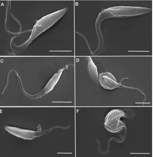
Epimastigote forms treated with DqV showed morphological alterations that were observed by SEM. The parasites showed loss of membrane integrity with the presence of pores (Fig. 2C; 2E) and cell shrinkage (Fig. 2D; 2F), also showing characteristics of apoptosis and necrosis at both concentrations.
The most importantfindings of the present study were the ac-tivity of DqV against the intracellular amastigote form and the high selective index. Amastigotes develop and maintain the infections by T. cruzi and significantly lower DqV concentrations, when compared to those required to cause damage to host cells, inhibit amastigote proliferation.
In conclusion,D.quadricepsvenom was be able to inhibit the promastigote form of L. amazonensis and the epimastigote,
Fig. 2.Scanning electron microscopy of control epimastigotes (AeB) and treated epimastigotes with IC50 of DqV (CeD) and 2 x IC50 of DqV (EeF). Morphological alterations were
trypomastigote and amastigote forms ofT.cruzi, strain Y, with the involvement of necrotic and apoptotic mechanisms. Therefore, DqV and its components can offer potential tools for the development of new drug candidates against neglected diseases.
Acknowledgements
We thank Analytic Centre - UFC/CT - INFRA/Proe Equipment
CAPES. We thank National Council of Technological and Scientific Development (CNPq), Cearense Foundation for the Support of Sci-entific and Technological Development (Funcap) and Coordination for Enhancement of Higher Education Personnel (CAPES) for their
financial support.
Transparency document
Transparency document related to this article can be found online athttp://dx.doi.org/10.1016/j.toxicon.2016.08.008.
References
Abe, F., Nagafuji, S., Yamauchi, T., Okabe, H., Maki, J., Higo, H., et al., 2002. Trypa-nocidal constituents in plants 1. Evaluation of some mexican plants for their trypanocidal activity and active constituients in guaco, roots of Aristolochia taliscana. Biol. Pharm. Bull. 25, 1188e1191.
Adade, C.M., Carvalho, A.L., Tomaz, M.A., Costa, T.F., Godinho, J.L., Melo, P.A., et al., 2014. Crovirin, a snake venom cysteine-rich secretory protein (CRISP) with promising activity against Trypanosomes and Leishmania. PLoS Negl. Trop. Dis. 8, e3252.
Adade, C.M., Chagas, G.S., Souto-Padron, T., 2012. Apis mellifera venom induces different cell death pathways in Trypanosoma cruzi. Parasitology 139, 1444e1461.
Adade, C.M., Cons, B.L., Melo, P.A., Souto-Padron, T., 2011. Effect of Crotalus viridis viridis snake venom on the ultrastructure and intracellular survival of Trypa-nosoma cruzi. Parasitology 138, 46e58.
Adade, C.M., Oliveira, I.R., Pais, J.A., Souto-Padron, T., 2013. Melittin peptide kills Trypanosoma cruzi parasites by inducing different cell death pathways. Toxicon 69, 227e239.
Aparicio, I.M., Scharfstein, J., Lima, A.P.C.A., 2004. A new cruzipain-mediated pathway of human cell invasion by Trypanosoma cruzi requires trypomasti-gota membranes. Infect. Immun. 72, 5892e5902.
Berkowitz, A.L., Raibagkar, P., Pritt, B.S., Mateen, F.J., 2015. Neurologic manifesta-tions of the neglected tropical diseases. J. Neurol. Sci. 349, 20e32.
D'Annessa, I., Castelli, S., Desideri, A.1, 2015. Topoisomerase 1B as a target against leishmaniasis. Mini Rev. Med. Chem. 5, 203e210.
Galle, J.B., Attioua, B., Kaiser, M., Rusig, A.M., Lobstein, A., Vonthron-Senecheau, C., 2013. Eleganolone, a diterpene from the French marine alga Bifurcaria bifurcata inhibits growth of the human pathogens Trypanosoma brucei and Plasmodium falciparum. Mar. Drugs 11, 599e610.
Gonçalves, A.R., Soares, M.J., de Souza, W., DaMatta, R.A., Alves, E.W., 2002. Ultra-structural alterations and growth inhibition of Trypanosoma cruzi and Leish-mania major induced by Bothrops jararaca venom. Parasitol. Res. 88, 598e602. Harvey, A.L., 2014. Toxins and drug discovery. Toxicon 15, 193e200.
Hotez, P.J., Alvarado, M., Basanez, M., Bolliger, I., Bourne, R., Boussinesq, M., et al., 2014. The global burden of disease study 2010: interpretation and implications for the neglected tropical diseases. PLOS NTD 8, e2865.
Krysko, D.V., Vanden Berghe, T., D'herde, K., Vandenabeele, P., 2008. Apoptosis and necrosis: detection, discrimination and phagocytosis. Methods 44, 205e221. Lima, D.B., Torres, A.F., Mello, C.P., de Menezes, R.R., Sampaio, T.L., Canuto, J.A., et al.,
2014. Antimicrobial effect of Dinoponera quadriceps (Hymenoptera: For-micidae) venom against Staphylococcus aureus strains. J. Appl. Microbiol. 117, 390e396.
Mcgwire, B.S., Kulkarni, M.M., 2010. Interactions of antimicrobial peptides with Leishmania and trypanosomes and their functional role in host parasitism. Exp. Parasitol. 126, 397e405.
Mosmann, T., 1983. Rapid colorimetric assay for cellular growth and survival: application to proliferation and citotoxicity. J. Immunol. Methods 65, 55e63. Murcia, L., Carrilero, B., Albajar Vinas, P., Segovia, M., 2012. Nifurtimox chemo-~
therapy: collateral effects in treated Trypanosoma cruzi infected patients. Rev. Esp. Quimioter. 25, 74e75.
Nwaka, S., Hudson, A., 2006. Innovative lead discovery strategies for tropical dis-eases. Nat. Rev. Drug Discov. 5, 941e955.
Passero, L.F., Tomokane, T.Y., Corbett, C.E., Laurenti, M.D., Toyama, M.H., 2007. Comparative studies of the anti-leishmanial activity of three Crotalus durissus ssp. venoms. Parasitol. Res. 101, 1365e1371.
Pinto, E.G., Antoniazzi, M.M., Jared, C., Tempone, A.G., 2014. Antileishmanial and antitrypanosomal activity of the cutaneous secretion of Siphonops annulatus. J. Venom. Anim. Toxins Incl. Trop. Dis. 24, 20e50.
Sousa, P.L., Quinet, Y., Ponte, E.L., Vale, J.F., Torres, A.F.C., Pereira, M.G., Assreuy, A.M.S., 2012. Venom's antinociceptive property in the primitive ant Dinoponera quadriceps. J. Ethnopharmacol. 144, 213e216.
Silva, G.M.S., Mediano, M.F., Alvarenga Americano do Brasil, P.E., da Costa Chambela, M., da Silva, J.A., de Sousa, A.S., Xavier, S.S., Rodrigues da Costa, A., Magalh~aes Saraiva, R., Hasslocher-Moreno, A.M., 2014. A clinical adverse drug reaction prediction model for patients with chagas disease treated with benz-nidazole. Antimicrob. Agents Chemother. 58, 6371e6377.
Torres, A.F., Huang, C., Chong, C.M., Leung, S.W., Prieto-da-Silva, A.R., Havt, A., Quinet, Y.P., et al., 2014. Transcriptome analysis in venom gland of the predatory giant ant Dinoponera quadriceps: insights into the polypeptide toxin arsenal of hymenopterans. PLoS ONE 9, e87556.
Torres, A.F.C., Dantas, R.T., Toyama, M.H., Diz Filho, E., Zara, F.J., Rodrigues De Queiroz, M.G., et al., 2010. Antibacterial and antiparasitic effects of Bothrops marajoensis venom and its fractions: phospholipase A2 and L-amino acid oxi-dase. Toxicon 55, 795e804.
Urbina, J.A., Docampo, R., 2003. Specific chemotherapy of Chagas disease: contro-versies and advances. Trends. Parasitol. 19, 495e501.
WHO, 2010a. Control of the Leishmaniasis: Report of a Meeting of the WHO Expert Committee on the Control of Leihmaniases (WHO Technical Report Series, Geneva, Switzerland).
WHO, 2010b. Working to Overcome the Impact of Neglected Tropical Diseases. First WHO Report on Neglected Tropical Diseases.