MR IMAGING FOR EVALUATION OF
LESIONS OF THE CRANIAL VAULT
A pictorial essay
Lázaro Amaral
2, Miriam Chiurciu
1, João Ricardo Almeida
1,
Nelson Fortes Ferreira
2, Renato Mendonça
2, Sérgio Santos Lima
3ABSTRACT - Purpose: A variety of diseases affect the calvaria. They may be identified clinically as palpable masses or incidentally in radiologic examinations. There are many diagnostic possibilities, including congenital, neoplastic, inflammatory and traumatic lesions. The purpose of this study is to illustrate the main calvarial lesions through MR imaging, their signal intensity and extension to neighboring sites. Method: A retrospective analysis of 81 cases, from November 1996 to July 2001, was conducted. The examinations were performed on a 1.5 T equipment and each one of the cases was pathologically proven. Results: The results were: dermoid cysts [4 cases (5%)], epidermoid cysts [2 cases (2.5%)], cephalocele [14 cases (17.5%)], sinus pericranii [3 cases (3.7%)], leptomeningeal cysts [4 cases (5%)], Langerhans cell histiocytosis [10 cases (12.5%)], lipoma [4 cases (5%)], fibrous dysplasia [13 cases (16.2%)], osteoma [8 cases (10%)], hemangioma [1 case (1.2%)], meningioma [3 cases (3.7%)], chondrosarcoma [5 cases (6.2%)], hemangiosarcoma [1 case (1.2%)], multiple myeloma [3 cases (3.7%)], sarcomatous transformation of Paget disease [1 case (1.3%)], and metastasis [5 cases (6.2%)]. Conclusion: MRI identifies bone marrow abnormalities and invasion of adjacent tissues at an early stage. Therefore, it is an essential method when it commes to properly evaluating calvarial lesions.
KEY WORDS: calvaria, MRI, tumors, congenital lesions, infammatory lesions.
Avaliação por ressonância magnética das lesões da calota craniana: ensaio ilustrado
RESUMO - Objetivo: A calota craniana é sede de diversas doenças, as quais podem ser identificadas clinicamente como massas palpáveis ou incidentalmente em estudos radiológicos. O diagnóstico diferencial é variado e inclui lesões de natureza congênita, neoplásica, inflamatória e traumática. O objetivo do nosso ensaio é ilustrar as principais lesões da calota craniana através de avalição por imagens de ressonância magnética (RM), expondo suas características de sinal e demonstrando sua extensão para os espaços adjacentes. Método: Foi realizada análise retrospectiva de 81 casos no período compreendido entre novembro de 1996 a julho de 2001. Os exames foram efetuados em aparelho de RM de 1,5T. Resultados: Cisto dermóide [4 casos (5%)], cisto epidermóide [2 casos (2,5%)], cefalocele [14 casos (17,5%)], sinus pericranii [3 casos (3,7%)], cisto leptomeníngeo [4 casos (5%)], histiocitose de células de Langerhans [10 casos (12,5%)], lipoma [4 casos (5%)], displasia fibrosa [13 casos (16,2%)], osteoma [8 casos (10%)], hemangioma [1 caso (1,2%)], meningeoma [3 casos (3,7%)], condrossarcoma [5 casos (6,2%)], hemangiossarcoma [1 caso (1,2%)], mieloma multiplo [3 casos (3,7%)], transformação sarcomatosa da doença de Paget [1 caso (1,3%)], e metastase [5 casos (6,2%)]. Conclusão: A RM detecta precocemente as alterações que envolvem a medula óssea, bem como demonstra acuradamente o envolvimento dos tecidos adjacentes. Este método é fundamental na avaliação detalhada das lesões da calota craniana.
PALAVRAS-CHAVE: calota craniana, ressonância magnética, tumor, lesões congênitas, lesões inflamatórias.
MEDIMAGEM - Hospital Beneficência Portuguesa, São Paulo SP, Brasil: 1Médico estagiário do setor de ressonância magnética; 2Médico neurorradiologista;3Chefe do Departamento de Imagens.
Received 29 November 2002, received in final form 20 February 2003. Accepted 8 March 2003.
Dr. Lázaro Amaral - Rua Luiz Gottschalk 151/111 - 04008-070 São Paulo SP - Brasil. E-mail: [email protected]
There is a wide variety of calvarial lesions that are identified as palpable masses or as incidental fin-dings in radiographic studies. The radiologist has a long list of differential diagnosis which includes
522 Arq Neuropsiquiatr 2003;61(3-A)
for the assessment of bone lesions. However, mag-netic resonance image (MRI) is able to show these lesions at an earlier stage while they are restricted to the bone marrow. It also shows the soft tissues and intracranial involvement when paramagnetic contrast agents are used.
The aim of this essay is to demonstrate the MR findings of vault lesions, their signal intensities and their extension to soft tissues.
METHOD
A retrospective analysis of 81 cases, from November 1996 to July 2001, was conducted. The examinations were performed on a 1.5 T equipment (GE Medical Systems) with sequences SE T1WI pre and post-contrast (Gd-DTPA), FSE T2WI, FLAIR and Gradient echo (T2*WI). All cases were pathologically proven.
Anatomy
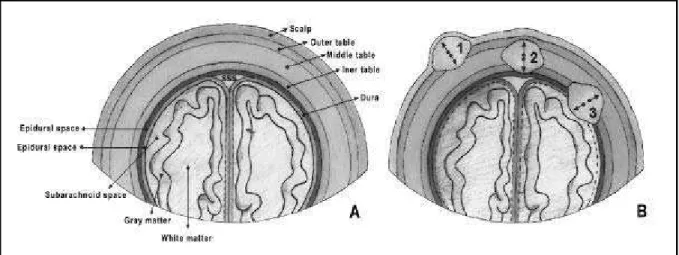
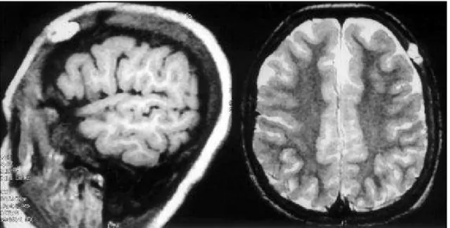
Calvarial lesions may arise from the bone (primary le-sions) or invade it by means of distant metastasis or thro-ugh contiguity when primary to the adjacent tissues (secondary lesions). In order to distinguish them as primary or secondary, it is essential to recognise the three main anatomic regions: the scalp, the vault and the meninges (Fig 1A and B)
The scalp consists of skin, fibrous and fatty tissue, galea aponeurotica and its connective tissue. The skin itself is subdivided into the epiderm, derm, hair follicles, subcuta-neous tissue, superficial blood vessels and nerves. These blood vessels are connected to the ones intracranially, after running through the vault either partially or fully. Lesions found here are called extracranial1.
The calvaria consists of two cortical layers and the mar-row (diploe) between them. There are frontal, parietal, occipital, temporal and sphenoid bones that are separated by sutures.
The outer membrane, dura mater or pachymeninge has an external layer attached to the bone called the peri-osteal membrane, and an internal one called the meningeal membrane. They are held together, being parted only when they circumscribe the dural sinuses, the cerebral falx and the cerebellar tend. The arachnoid membrane is thin and avascular with trabeculae that reaches the pia mater. The pia mater itself is a thin membrane of connective tissue involving the cerebral surface, even penetrating the fissures. The extra or epidural space is outside the periosteal dura. The subdural space is inside the meningeal dura. The cerebrospinal fluid (CSF) flows between the arachnoid membrane and the pia mater. There are extradural, sub-dural and subarachnoid spaces outside the brain paren-chyma, which are thus extra-axial spaces. Lesions in these spaces cause cortical effacement and widening of the suba-rachnoid space1,2.
RESULTS
The results were: dermoid cysts [4 cases (5%)], epidermoid cysts [2 cases (2.5%)], cephalocele [14 cases (17.5%)], sinus pericranii [3 cases (3.7%)], lep-tomeningeal cysts [4 cases (5%)], Langerhans cell histiocytosis [10 cases (12.5%)], lipoma [4 cases (5%)], fibrous dysplasia [13 cases (16.2%)], osteoma [8 cases (10%)], hemangioma [1 case (1.2%)], me-ningioma [3 cases (3.7%)], chondrosarcoma [5 ca-ses (6.2%)], hemangiosarcoma [1 case (1.2%)], mul-tiple myeloma [3 cases (3.7%)], sarcomatous trans-formation of Paget disease [1 case (1.3%)], and me-tastasis [5 cases (6.2%)].
DISCUSSION
The main calvarial lesions were studied using MR imaging. Therefore, their signal intensities and ex-tension to neighboring sites were evaluated. The
most important differential diagnosis are considered in this study, including congenital, traumatic, inflam-matory and neoplastic lesions.
Congenital lesions
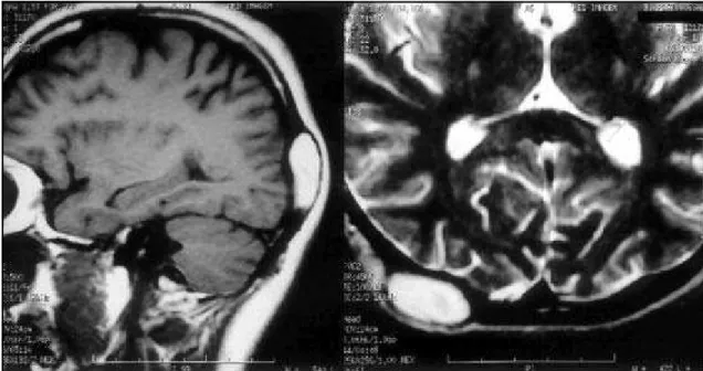
Dermoid and epidermoid cysts - Both dermoid and epidermoid cysts (DC and EC) consist of an ecto-dermal inclusion that differs in complexity. ECs are made up solely of squamous epithelium, while DCs also include hair, sebaceous and sudoriparous glands (Fig 2). They occur inside the orbit, the diploic space of the calvaria and intracranially (posterior and middle fossa). ECs account for most cases3.
ECs are typically laterally located. They may be either extradural or intradiploic in origin, occurring mainly in parietal and temporal bones2.
DCs account for 20% of vault lesions and are lo-cated at sutures. They occur in newborns and chil-dren up to 3 years old. Most of these lesions are solitary masses with the upper edge extending be-yond the limits of the calvaria. Their commonest lo-cation is in the anterior fontanele4.
Epidermoid cysts (Fig 3) that are inside the bone (intradiploic) may show erosion with a sclerotic margin and scalloped appearance. The attenuation of EC on CT and their signal intensity on MR studies are similar to that of water (CSF). Occasionally, their content might be hypodense on CT and a little bri-ghter than water on T1 weighted images (T1WI). So-metimes, DCs have an heterogenous appearence, with varied signal intensities, because of their
com-plex composition. The walls of these lesions are thick, may become calcified and are enhanced by contrast agents. Since DCs can contain fatty tissue, their attenuation and signal intensity may be equivalent to those of fat, accounting for low density on CT and high signal intensity on T1WI3,4.
Cephalocele - Cephalocele is a fault involving the dura and the cranial vault associated with herniation of intracranial components. There are four main ty-pes: meningoencephalocele, when there is herniation of CSF, nerves and meninges; meningocele, which is
Fig 2. MRI - sagital T1WI and axial T2WI: dermoid cyst in the right parietal bone.
524 Arq Neuropsiquiatr 2003;61(3-A)
Fig 4. MRI - sagital T1WI and T2WI: occipital cephalocele.
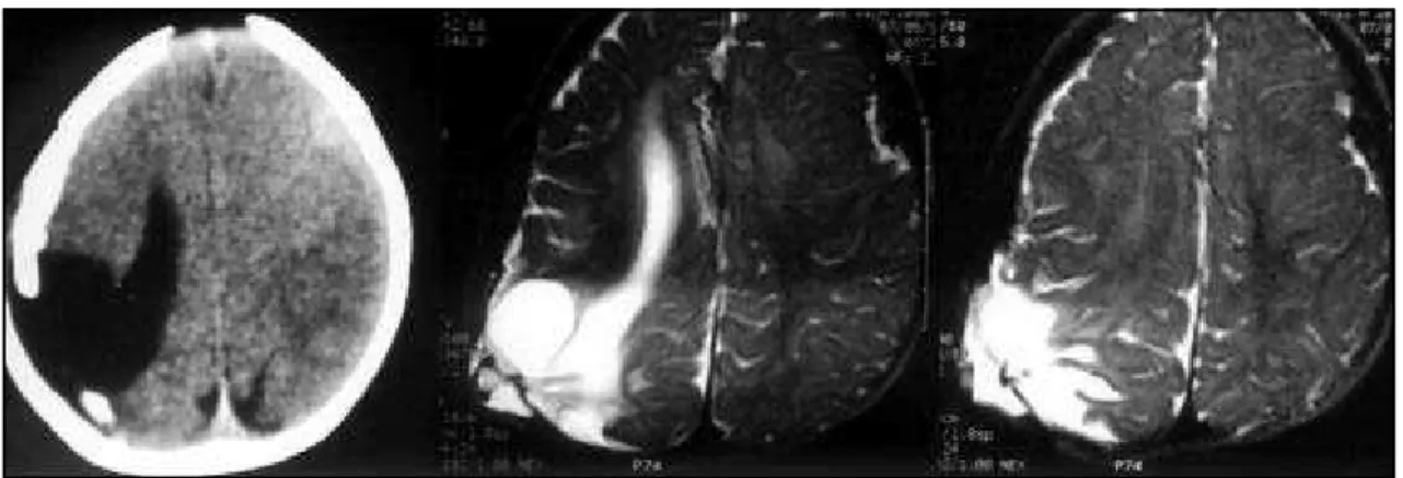
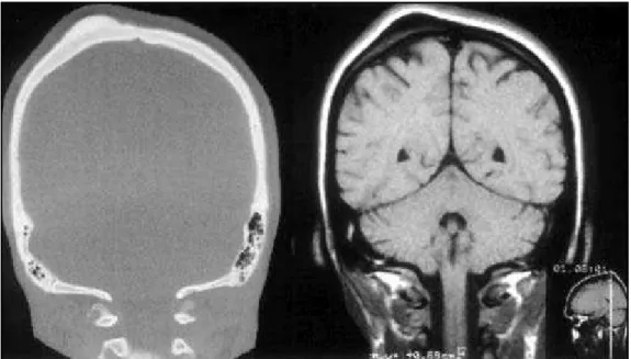
Fig 6. CT axial and MRI axial T2WI: leptomeningeal cyst (previous trauma).
the herniation of the meninges, without brain pa-renchyma or nerves; atresic cephalocele that contains dura mater, degenerated fibrous and nervous tissue; and gliocele, a cyst that is lined by glial cells and filled with CSF. The cause of cephalocele is still essen-tially unknown.
Cephaloceles are classified according to their lo-cation. Therefore, they can be occipitocervical,
occipi-tal, parieoccipi-tal, fronoccipi-tal, temporal, frontoethmoidal, sphenomaxillar and spheno-orbital. Occipital cepha-locele is the most common, accounting for 80% of the cases in Caucasians. The supra and infratentorial structures are often inside the cephalocele. MR de-monstrates the presence of cerebellar cortical dys-plasia, venous sinus anomaly, dorsal interhemispheric cyst, corpus callosum anomalies and Chiari and
Dandy-Walker malformations, all of them being fre-quently associated with cephalocele4(Fig 4)
Sinus pericranii (SP) - SP is a slow growing lesion of the cranial vault, usually occurring in infancy, asso-ciated with an adjacent small bone defect. It is a de-velopmental abnormality, that can also be secondary to trauma. It consists of venous structures which are linked to the intracranial venous system (mainly the superior sagittal sinus) through a small ostium. They may grow in size as a result of Valsalva maneuver, crying, coughing and from jugular vein compression. The most commonly involved bones are the frontal and parietal, followed by the occipital and temporal. Radiographic studies will show the bone defect with regular or irregular margins. A CT scan or MRI may demonstrate dilated vascular structures which are enhanced on both sides of the cranial vault, close to a dural venous sinus or a cortical vein1(Fig 5).
Traumatic lesions
Leptomeningeal cyst - These cysts occur as a late complication in approximately 1% of vault fractures in children under 3 years. When the dura mater is ruptured, the inner table is exposed to the CSF and arachnoid pulsations. So much pressure will cause bone erosion and widening of the fracture, usually weeks after its occurrence. Leptomeningeal cysts are typically elongated, occurring along the fracture line
and enclosing the inner table of the calvaria. Both CT scan and MRI show a bone defect and a cystic lesion4(Fig 6).
Inflammatory lesions
Langerhans cell histiocytosis (LCH) - LCH is a multi-systemic disease of unknown etiology, which consists of abnormal proliferation of histiocytes that may form focal or diffuse clusters. The clinical presenta-tion and evolupresenta-tion vary according to its extension and to the organs that are involved, sometimes re-sembling a true neoplasia. There are three main sub-types: eosinophylic granuloma (EG) accounting for 70% of the cases; Hand-Schüller-Christian (HSC) in 10 to 15% and Letterer-Siwe (LS).
LCH can arise nearly anywhere in the brain and skull. This diagnosis should always be considered when a mass arises from the bones of the face, skull base, or calvaria in children. LCH may result in masses based in the cerebral parenchyma, spinal cord, dura, or choroid plexus.
The most common imaging finding of calvarial LCH is a well-defined mass involving the diploe. On CT, a sharply circumscribed skull lesion is identified with differential involvement of the inner and outer table. MRI shows these bone lesions as sharply defined soft tissue masses, with signal intensity equivalent to that of skeletal muscle and marked enhance after intravenous paramagnetic contrast medium (Fig 7).
Benign neoplastic lesions
Lipoma - Lipomas are rare in the bone marrow, despite the fact that it is rich in fatty tissue. There are 4 main types: 1. soft tissue lipoma which pres-sures the outer table; 2. periosteal lipoma that cau-ses hyperostosis and bone erosion; 3. intraosseus lipoma originating from the bone marrow, with a tendency to expand and distort the bone; 4. liposarcoma.
These lesions affect the metaphysis of the long bones (femur, tibia e fibula) and occasionally the cal-varia. Most patients are assymptomatic but some may complain of pain and edema, symptoms that can persist from weeks to years5.
Radiographic studies show a radiolucent lesion with well circumscribed margins and widening of the medular cavity. There is cortical thinning and pe-riosteal reaction. MRI is useful in the demonstration of typical fatty tissue signal intensity6,7(Fig 8).
526 Arq Neuropsiquiatr 2003;61(3-A)
Fig 8. MRI - sagital T1WI and axial T2WI: left frontal lipoma.
Fig 9. MRI - sagital T1WI and axial T2WI: right frontal bone fibrous dysplasia.
Fig 11. CT - coronal with bone algoritm and MRI coronal T1WI: outer table osteoma in the right parietal bone.
Fibrous dysplasia - Fibrous dysplasia (FD) is a be-nign condition associated with normal bone marrow replacement by proliferative fibro-osseous tissue with varied quantities of stroma and bone. FD may affect
only one (monostotic form) or multiple bones (po-liostotic form).
It is a relatively common disease and is most fre-quent in teenagers and young adults. It occurs in
528 Arq Neuropsiquiatr 2003;61(3-A)
any part of the skeletal system but the most affected bones are: 1) monostotic form - hip, proximal femur, tibia, cranial and facial bones; 2) polyostotic form -femur, tibia, hip and foot.
The most frequent complication is pathological fracture, occuring in 85% of the patients suffering from the polyostotic type. Malign transformation to osteosarcoma, fibrosarcoma and chondrosarcoma occurs very rarely (0.5%)5.
Affected bones will be expanded and strongly enhance after intravenous contrast medium injec-tion. A noncontrasted CT scan with bone algorithm will show the classic “ground glass” appearance of fibrous dysplasia.
MRI shows homogeneous low signal on T1WI, unless there is pathological fracture. On T2WI, the lesion is heterogeneous depending on the amount of fibro-osseous tissue, cellularity, cystic alterations, hemorrhage and cartilaginous tissue. The lesions usually have well defined borders. All the sequences show a hypointense halo that corresponds to scle-rotic margins. MRI is the modality of choice to demonstrate the internal structures of the lesion and evaluate disease extension, especially in cases due for surgical treatment8 (Fig 9).
Osteoma - It is a benign juxtacortical slow-growing tumor, composed by mature dense bone. Little os-teomas are asymptomatic and appear by chance. They may occur in any age, mainly from the third to the fifth decade and mostly in males. Sometimes, mul-tiple lesions are associated with Gardner syndrome.
The paranasal sinuses (frontal and ethmoidal),
cranial vault, jaw and maxilla are among the most affected sites. There are juxtacortical lesions which arise from the periosteal region5. They enclose the
outer table and less frequently the inner table of the cranial vault. Radiographic studies and CT scans show a dense, oval (or round) lesion. On MRI osteomas are homogeneous and markedly hypointense on T1 and T2WI. They do not oftenly enhance after the use of contrast agents (Figs 10 and 11).
Osseous hemangioma - Osseous hemangiomas are benign vascular lesions with capillaries, venous and cavernous vascular channels, histopathologically identical to soft tissue hemangiomas. They are more common in middle aged females, with the peak inci-dence between the fourth and sixth decade.
They are found in dorsal and lumbar vertebral bodies, facial bones and calvaria, mainly frontal and parietal bones. In 15% of the cases they present as multiple lesions. They tend to grow slowly and are not associated with bleedings9.
The outer table is commonly affected, while the inner table is spared. Either radiographic studies or CT scans show a typical lytic lesion, with trabecular matrix, periosteal reaction (“sunbeam”) and sclerotic margins. MRI is the best method to show lesion extension and its relationship to the adjacent neu-rovascular structures. Hemangiomas are typically hyperintense on T1WI and heterogeneously hyperin-tense on T2WI. The calvarial hemangioma isn’t enhanced on CT or MRI9 (Fig 12).
Meningioma - Meningioma is the most frequent extra-axial neoplasia arising from the arachnoid. It
may pressure the adjacent parenchyma causing ede-ma or infiltrate it when ede-maling (meningeal sarcoede-ma). The calvarial convexity is ofently affected, in a para-sagittal position, adjacent to the cerebral falx, in the paraselar or subfrontal regions and close to the sphe-noid bone and clivus.
The meningiomas that occur near the sphenoid bone grow alongside its edge and are thus elongated in shape. They are called “en plaque” meningiomas (Fig 13). The bones react to a greater or lesser degree and respond with severe sclerosis. Sometimes the tumor will invade adjacent structures. Some menin-giomas arise from remnants of arachnoid cells inside the diploe. These are better studied by MRI10.
Malign neoplastic lesions
Chondrosarcoma - Chondrosarcomas are malig-nant cartilaginous tumors which may be primary or secondary to the calvaria. The two main types are classified according to their origin: central or intra-medullar (most cases) and peripheral or juxtacortical.
Fig 14. MRI - coronal T2WI and coronal T1WI post-Gd-DTPA: right fronto-temporal and cavernous sinus chondrosarcoma.
Fig 15. MRI - axial T2WI, coronal T1WI post-Gd-DTPA and axial 3D SPGR post-Gd-DTPA: left fronto-temporal hemangiosarcoma.
530 Arq Neuropsiquiatr 2003;61(3-A)
It may affect any bone, and the calvaria is an unu-sual site. MRI shows a lobulated mass that is isointense or faintly hyperintense compared to the muscle on T1WI and hyperintense on T2WI. However, low grade lesions typically show a homogeneous sig-nal, with thin fibrous septa of low signal intensity on all pulse sequences. The high grade tumors tend to be heterogeneous. The calcified matrix can be pre-sented as very hypointense, with a “dot-like” appea-rance on MRI. Endosteal scalloping is a common finding5 (Fig 14).
Hemangiosarcoma - Hemangiosarcoma is a rare tumor with a high grade intraosseous endothelial cell component. It occurs more oftenly in males from 12 to 74 years old. The most common locations are the femur, tibia and humerus, to an equal extent, accounting for about 50% of the cases. The verte-bral bodies, pelvic bones and hips are less commonly affected. Any bone could, in theory, be affected10.
Radiographic studies show a lytic lesion, with poorly defined margins suggesting malignity. The
bo-ne is usually eroded and the lesion may as well extend to the cortex. Its behavior is aggressive with invasion of the cranial vault and even the dura mater, which has an intermediate intensity on T1 and hyposignal on T2WI, being strongly enhanced by the paramag-netic contrast agent11 (Fig 15).
Multiple myeloma (MM) - MM is a malignant di-sorder of the bone marrow in which there is mono-clonal proliferation of plasma cells. Solitary lesions are known as plasmacytomas. It is the most common pri-mary malignant bone lesion occurring between the fourth and eigth decade, with a slight male predomi-nance. The most common symptom is bone pain5.
The axial skeleton is the predominant site of in-volvement and the most affected bones are the ver-tebrae, ribs, skull, pelvis, and long bones (especially femur), in order of frequency. At the time of diag-nosis, the disease is often widely disseminated throu-ghout the axial skeleton.
On T1WI, MM lesions typically appear hypoin-tense, but may appear of intermediate intensity
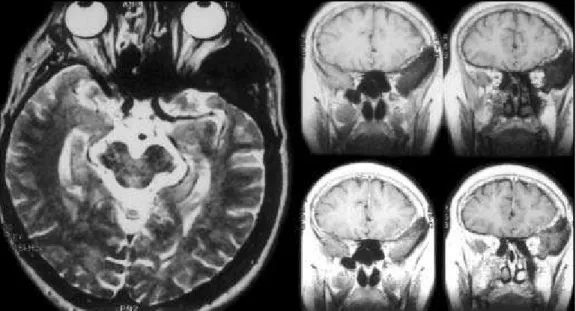
Fig 17. MRI - sagital T1WI and axial T2WI: Paget´s disease with basilar invagination.
Fig 20. MRI - axial T2WI and axial T1WI post-Gd-DTPA: right temporal-occipital-parietal bone metastasis from colonic carcinoma.
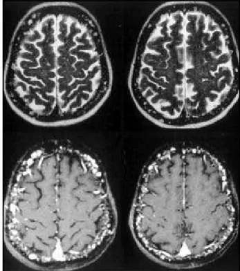
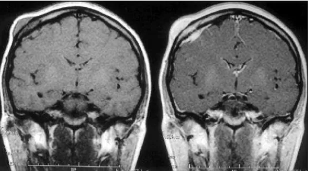
Fig 19. MRI - coronal T1WI pre and post-Gd-DTPA: right parietal bone metastasis from melanoma.
(slightly hyperintense to muscle) and are enhanced by contrast agents6 (Fig 16). On T2WI and STIR images,
the lesions typically exhibit hyperintense signal. Ver-tebral compression fractures may occur. Extension into the soft tissues or epidural space may also be present.
Paget’s sarcoma - Paget’s disease, also known as osteitis deformans, is a disorder of abnormal and excessive bone remodeling which may affect one or more bones. The precise etiology is unknown. Paget’s disease may be divided into three phases: initial (active), middle (intermediate), and late (inactive). Each phase is associated with distinctive pathologic,
radiologic, and clinical features. Paget’s disease is a relativity common disorder affecting about 3% of individuals over the age of 40 and 10% of the popu-lation above 85 years. The vast majority of patients are asymptomatic in all stages. Paget’s disease usually present in the middle phase and most of the symp-toms are related to weakened and thickened bones. Patients frequently complain of pain6. The most
com-mon sites are the pelvis, spine, femur, and skull, follo-wed by the tibia, humerus, scapula, and clavicle.
532 Arq Neuropsiquiatr 2003;61(3-A)
composition, and extent of involvement. In later sta-ges, bone enlargement and cortical thickening are identified. The thickened cortex appears as a peri-pheral rim of signal void on all pulse sequences. Coar-sened trabeculae appear as thick linear areas of signal void. Cyst-like areas of fat filled marrow cavities may also be present. Areas of hemorrhage with variable signal intensity may also be present.
The most feared complication of Paget’s disease is malignant transformation, which occurs in about 1% of all patients and approximately 10% of those with extensive polyostotic disease. The most frequent histologic types of Paget’s sarcomas are osteosarco-ma, malignant fibrous histiocytoosteosarco-ma, fibrosarcoma and chondrosarcoma6,12(Fig 18).
Metastasis - The primary tumors that metastasis to the cranial vault are breast, lung, prostate and kidney carcinomas. They are well circumscribed, fo-cal masses which invade the dura mater and/or cra-nial vault with extension to the cerebral surface, sometimes being diffuse.
The cranial vault metastasis are blastic or lytic le-sions invading both the inner and outer tables (radio-graphy and CT scan). These lesions may be expansive in nature, especially when originating from thyroid and renal cell carcinoma. The MRI shows focal areas of low intensity (isointense to gray matter) on T1WI, that are easily distinguished from the hyperintensity of normal bone marrow. Such lesions may be obs-cured in a post-gadolinium sequence, thus it is essential to perform a previous non-contrasted se-quence13,14(Figs 19 and 20).
CONCLUSION
It may be difficult to render an etiologic diagnosis through imaging studies alone. Many imaging
me-thods can be used to evaluate these lesions and, among them, the CT scan is considered the best exa-mination available to characterize bone alterations. However, MRI identifies earlier abnormalities of the bone marrow, even before the development of cor-tical destruction, which is only shown later on CT. Therefore, MRI is an essential method in defining lesion extension both for the skin and intracranial and extraaxial spaces. It is also important to empha-size that the use of a paramagnetic contrast agent is paramount for such purposes.
REFERENCES
1. Ortiz O, Schochet S, Bastug D. Imaging evaluation and clinicopathologic correlation of mass lesions involving the calvária. Part I: congenital e traumatic lesions. Int J Neuroradiol 1999;5:96-108.
2. Rauschning W. Brain tumors and tumorlike masses: classification and differential diagnosis. In: Osborn AG. (ED) Diagnostic neuroradiology. 1st ed St. Louis: Mosby, 1994:511-519.
3. Smirniotopoulos JG, Chiechi MV. Teratomas, dermoides and epidermoides of head and neck. Radiographics 1995;15:1437-1455. 4. Barkovich AJ. Congenital malformations of the brain and skull. In:
Barkovich AJ. (ED) Pediatric neuroimaging. 3rd ed. Philadelphia:
Lippincott Williams&Wilkins,2000: 280-281.
5. Laorr A, Helms CA. Miscellaneous malignant masses. In: Laorr A, Helms CA. (EDS) MRI of musculoskeletal masses. A practical text and atlas. 1st ed. New York. Tokyo. Igaku-Shoin, 1997:322-324.
6. Ortiz O, Schochet S, Bastug D. Imaging evaluation and clinicopathologic correlation of mass lesions involving the calvária. Part II: tumoral and inflammatory lesions. Int J Neuroradiol 1999;5:151-165.
7. Tomabechi M, Kazuhiro S, Daita G et al. Lipoma involving the skull. J Neurosurg 1992;76:312-314.
8. Jee WH, Choi KH, Choe BY et al. Fibrous dysplasia: MR imaging characteristics with radiopathologic correlation. AJR 1996;167:1523-1527. 9. Bastug D, Ortiz O, Schochet SS. Hemangiomas in the calvaria: imaging
findings. AJR 1995;164:683-687.
10. Atlas SW, Lavi E, Goldberg HI. Extraaxial Brain Tumors. In: Atlas SW. (ED) Magnetic resonance imaging of the brain and spine. 3rd ed.
Philadelphia: Lippincott-Raven, 2002:723-726.
11. Burger PC, Scheithaver BW Atlas of Tumor Pathology - Tumor of the CNS - Third Series, Fascicle 10, Washington, DC: AFIP - 1994. 12. Hacein-Bey L, Pile-Spellman J. Craniofacial pathology. In: Taveras JM.
(ED) Neuroradiology. 3rd ed. New York: Williams&Wilkins, 1996:1164.
13. Ahmadi J, Hinton DR, Segall HD et al. Dural invasion by craniofacial and calvarial neoplasm: MR imaging and histopathologic evaluation. Radiology 1993;188:747-749.