Arq Neuropsiquiatr 2003;61(3-A):533-541
UNUSUAL MANIFESTATIONS OF
NEUROCYSTICERCOSIS IN MR IMAGING
Analysis of 172 cases
Lázaro Amaral
2, Murilo Maschietto
1, Roberta Maschietto
1, Ricardo Cury
1,
Nelson Fortes Ferreira
2, Renato Mendonça
2, Sérgio Santos Lima
3ABSTRACT - Purpose: The typical manifestations of neurocysticercosis are described widely in the literature. The purpose of this study is to demonstrate the uncommon presentations of different forms of neurocysticercosis in MR imaging. Method: A retrospective analysis of 172 cases of neurocysticercosis in MR studies was carried out over a period of 13 years. One hundred and four males and 68 females with a mean age of 32 ± 3.7 years were studied. The studies were performed on 1.5 T GE MR units and T1 was used before and after gadolinium injection, T2 and gradient-echo (T2*) sequences. Results: The authors divided the unusual manifestations of neurocysticercosis into: intraventricular, subarachnoid, spinal, orbital, intraparenchymatous, and reactivation of previously calcified lesions. The results obtained were: intraparenchymatous 95 cases (55.23%); intraventricular 27 cases (15.69%); subarachnoid 20 cases (11.63%); spinal 6 cases (3.49%); orbital 1 case (0.58%); reactivated lesion 1 case (0.58%); association of intraventricular and intraparenchymatous 12 cases (6.98%); association of subarachnoid and intraparenchymatous 6 cases (3.49%); association of subarachnoid and intraventricular, 4 cases (2.32%). Conclusion: MR imaging is a sensitive and specific method in the analysis of different forms of unusual manifestations of neurocysticercosis, which should appear in the differential diagnosis of parenchymal, ventricular, spinal, cisternal, and orbital lesions.
KEY WORDS: cysticercosis, parasites, MRI, subarachnoid space.
Manifestações incomuns na ressonância magnética da neurocisticercose: análise de 172 casos
RESUMO - Objetivo: as manifestações típicas da neurocisticercose já são bem conhecidas. O papel deste estudo foi demonstrar os aspectos incomuns da neurocisticercose na ressonância magnética. Método: foram analisados 172 casos de ressonância magnética de neurocisticercose na Med Imagem num período de 13 (treze) anos em aparelhos GE de 1.5T Signa (Horizon, LX e CVI). Dos casos analisados, foram diversas as formas de apresentação, incluindo intraventricular, intraespinhal, cisternal, orbital, formas atípicas parenquimatosas (simulando tumores), forma miliar e evolução não usual (reativação). Conclusão: A ressonância magnética é método sensível e específico na avaliação das numerosas formas de apresentação atípica da neurocisticercose, as quais devem constar no diagnóstico diferencial de lesões intraventriculares, cisternais, orbitárias e parenquimatosas.
PALAVRAS-CHAVE: cisticercose, parasitas, ressonância magnética, espaço subaracnóide.
MEDIMAGEM - Hospital Beneficência Portuguesa, São Paulo SP, Brasil: 1Médico estagiário do setor de ressonância magnética; 2Médico
neurorradiologista;3Chefe do Departamento de Imagens
Received 29 November 2002, received in final form 20 February 2003. Accepted 8 March 2003.
Dr. Lázaro Amaral - Rua Luiz Gottschalk 151/111 - 04008-070 São Paulo SP - Brasil. E-mail: lazden@terra.com.br The typical manifestations of neurocysticercosis
are widely described in the literature. The purpose of this study is to demonstrate the uncommon aspects of the different forms of neurocysticercosis in magnetic resonance imaging ( MRI ).
METHOD
A retrospective analysis of MRI exams of 172 neuro-cysticercosis patients, performed between 1989 and 2002,
was carried out. There were 104 males and 68 females with a mean age of 32.3 years. The studies were performed on 1.5T GE MRI units with sequences SE T1WI pre- and postcontrast (Gd-DTPA), FSE T2WI, FLAIR and Gradient echo (T2*WI).
RESULTS
in-traparenchymatous. The results obtained were: intra-parenchymatous (95 cases or 55.23%), intraventri-cular (27 cases or 15.69%), subarachnoid (20 cases or 11.63), spinal (6 cases or 3.49%), orbital (1 case or 0.58%), reactivated lesion (1 case or 0.58%), asso-ciation of intraventricular and intraparenchymatous (12 cases or 6.98 %), association of subarachnoid
and intraparenchymatous (6 cases or 3.49%), asso-ciation of subarachnoid and intraventricular (4 ca-ses or 2.33%).
DISCUSSION
Cysticercosis affects 50 million people around the world, with a prevalence of 3 to 6% of the population Table 1. Stages of cysticerci on MRI.
Arq Neuropsiquiatr 2003;61(3-A) 535
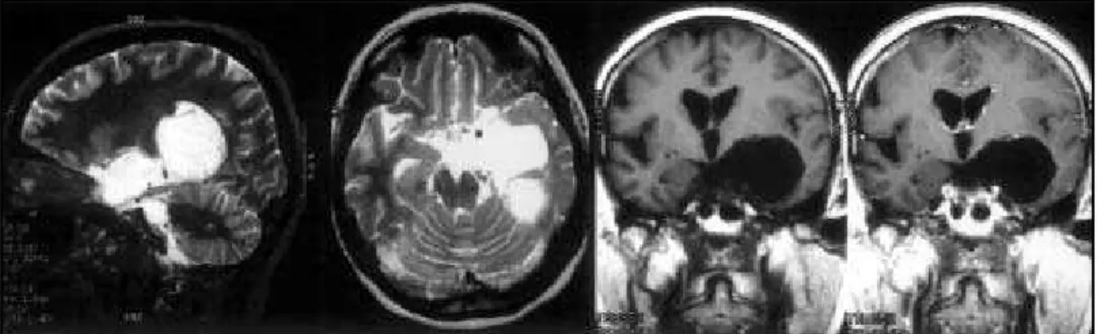
Fig 4. MRI Axial T1WI and CT Cisternography: Racemous form in the basal cisterns with cyst whithin the ventricular atrium. Fig 3. MRI Sagital T1WI and Axial T1WI postcontrast (Gd-DTPA): Racemous form in the cisterna magna with invagination into the IV ventricle.
Fig 2. MRI Sagital T1WI, Axial T1WI and Axial T2WI: Cysticercus in the IV ventricle with high protein content.
in endemic areas such as Central and South America, East Europe, Africa and some regions in Asia1,2.
Cys-ticercosis is the most common parasitic infection of the central nervous system (CNS) and it is caused by
Taenia solium’s invasion in its larval stage. CNS
in-volvement occurs in 60 to 90% of patients with cys-ticercosis3-5. The severity of neurocysticercosis
When invasion of the CNS occurs, the cysticerci develop in four stages identified by MRI (Table 1). With didatic purpose, the unusual forms of neurocys-ticercosis were divided into: intraventricular, suba-rachnoid, spinal, orbital, intraparenchymatous and reactivation of previously calcified lesion.
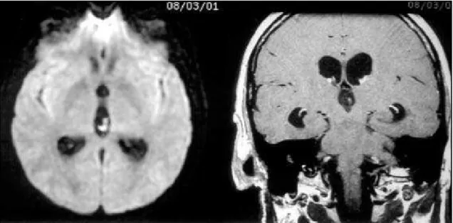
Intraventricular neurocysticercosis
The ventricular system is the second most com-mon site of neurocysticercosis2. It is frequently
cau-sed by Cysticercus cellulosae, however Cysticercus racemosus can also infect the ventricular system. The intraventricular form of the disease is found in more than 54% of patients with intracranial cysticercosis studied by MRI1,2.
It most commonly affects the IV ventricle (54%-64%), followed by the III ventricle (23%-27%), the la-teral ventricles (11% - 14%) and Sylvius aqueduct (9%)7,8. In our study we had 43 intraventricular, being
69% located in the IV ventricle, 12% in the III ven-tricle, 12% in the lateral ventricles and 7% in the Sylvius aqueduct. Computed tomography (CT) does not
fre-Fig 7. MRI Axial T2WI(A), Axial flair(B), Axial T1WI pre- and postcontrast (Gd-DTPA). Bulky cystic lesion in the right CPA cistern and small cyst in the right IV ventricle recess/ Axial Flair(D) after surgery: Removal of the cyst in the right CPA cistern and residual cyst whithin the IV Ventricle.
Fig 5. MRI Axial T1WI postconstrast (Gd-DTPA): Multiple cysts in the basal cisterns with arachnoiditis.
Arq Neuropsiquiatr 2003;61(3-A) 537
Fig 10. MRI Sagital T2WI and Sagital T1WI: Multiple cystic lesions in extra dural situation.
Fig 8. MRI Sagital T2WI, Axial T1WI and Sagital T1WI postcontrast (Gd-DTPA): Multiple cystic lesions in intradural-extramedullary situation leading to spinal cord compression.
Fig 13. MRI T1WI postcontrast (Gd-DTPA) - ring enhanced lesion in the frontal lobe. MR PWI - without high perfusion. Follow-up confirmed cysticercosis.
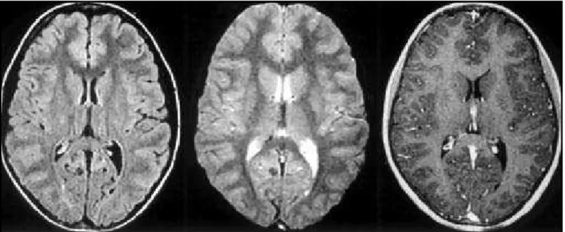
Fig 12. MRI Axial T2WI and T1WI postcontrast (Gd-DTPA):Miliary form.
quently show these lesions, because their density is similar to that of the cerebrospinal fluid (CSF). On MRI they can present hyperintensity on T1WI in compari-son to the CSF due to their protein content. The C. racemosus does not have scolex and multiply by wall proliferation. The C. cellulosae has scolex and one
Arq Neuropsiquiatr 2003;61(3-A) 539
Fig 14. Similar lesion in the left frontal lobe with high perfusion. Follow-up confirmed GBM.
Fig 15. Ring lesion with surrouding edema.
They are frequently associated with aqueductal ste-nosis, which could be secondary to coexistent epen-dimytis, appearing as wall enhancement near the parasite or adhesion by a previous inflamatory process.
Subarachnoid neurocysticercosis
Both the C. cellulosae and the C. racemosus affect the subarachnoid space, being the latter more fre-quent9. The incidence of cysternal involvement is
estimated at 3.5% of all neurocysticercosis cases2 ,
being the third most common site2. In our study, 30
cases presented in this location. They involve basal cisterns, mainly the supra selar, perimesencephalic, magna and Sylvian fissures.
The cystic masses are multiloculated, do not enhance after gadolinium chelates injection and determine cysternal expansion and deformity. They are related to local inflammatory reaction which can cause leptomeningeal thickening, fibrosis and loca-lized calcifications, most probably representing chro-nic meningitis (Fig 5). The inflammatory response could lead to vasculitis, affecting the basal perfora-ting vessels, resulperfora-ting in infarction10.
The major differential diagnosis are: arachnoid cyst, neuroglial cyst and epidermoid tumor (Figs 6 and 7).
Spinal neurocysticercosis
Cysticercosis may involve the spinal space and/or the spinal cord in CSF less than 1% of the cases, being more frequent the involvement of the subarachnoid space than the spinal cord2,9. The forms
observed are: intradural- extramedullary in 54% of the cases, intramedullary in 17% and association of intramedullary and intradural-extramedullary in 17%. Extra dural is very rare with few cases reported.
The intradural-extramedullary involvement occurs predominantly due to larval dissemination from brain to the spinal subarachnoid space11. The cysticercus
in the subarachnoid space leads to inflammatory re-action and colagen proliferation, being the clinical signs of spinal cysticercosis caused by direct com-pression of neurological tissue or due to inflamma-tory reaction. (Ex. arachnoiditis)
cere-Fig 16. Calcified lesion without surrouding edema.
Fig 17. Calcified lesion with late reactivation and perilesional edema.
bral involvement of neurocysticercosis is much more frequent than the spinal one11.
Of the six cases with spinal neurocysticercosis of our casuistic, three were intradural and extrame-dullary (Fig 8), two were intrameextrame-dullary (Fig 9) and one was extradural (Fig 10).
Orbital neurocysticercosis
The cysticercus reaches the orbit through the cho-roid vessels, having primitive sub-retinian location. During its development the cysticercus needs more space and it either stays in the primitive site and leads to retinal detachment or it perforates the reti-na with vitreous invasion. Inside the orbit it can in-duce inflammatory reaction and blindness in 8% of patients12.
The orbital cysticercosis outside the eyeball ge-nerally involves the extra-ocular muscles, leading to myositis which determines motor restriction and squint. The treatment of choice is the surgical remo-val. Occasionally, the cyst can resolve spontaneously.
On T1 weighted non-contrasted images the pa-rasite appears hyperintense, simulating primary cho-roidal melanoma. On T2WI the lesion appears hy-pointense. The differential diagnosis should include retinoblastoma (in children), primary melanoma and metastasis (in older patients)13. In our study we had
identified only one case involving the orbital extrinsic muscles (Fig 11).
Atypical forms of intraparenchymatous neurocysticercosis
There are two atypical forms of intraparenchy-matous presentations: miliary and pseudotumoral.
The miliary form represents massive cysticercus infestation of the CNS and is characterized by multi-ple small cystic formations difusely spread out in the brain parenchyma. It is a rare form of presentation, being observed in only one of our cases (Fig 12).
Arq Neuropsiquiatr 2003;61(3-A) 541
or small, solid or cystic, and can present themselves with wall enhancement or mural nodule, being or not surrounded by edema. The main differential diag-nosis of this kind of lesion should include gliomas, hemangioblastomas, neuronal cell tumors (ganglio-gliomas) and echinococcus. Lesions involving the su-perior cerebellar vermis in children can be indistin-guishable from meduloblastomas or astrocytomas.
In our 113 intraparenchymatous cases, the ma-jority of the non calcified lesions simulated tumors. In one of them, increased MR Perfusion weighted images (PWI) helped us to differentiate cysticercosis from GBM (Figs 13 and 14).
Reactivation of neurocysticercosis
Cerebral calcified lesions in patients with previous neurocysticercosis represent cysticercu’s death (imunologic inactivity). In recent studies some theo-ries try to explain the peri-lesional edema in previo-usly calcified lesions14-16. A plausible explanation
pro-posed is that calcified lesions contain dead cysticer-cus antigens in insoluble and inaccessible forms and for some reason not yet clarified it could be recog-nized by the host triggering inflammatory reaction.
It is not well known, why only some calcified le-sions trigger inflammatory response.
In our study we had only one case of reactivation of a previously calcified lesion (Figs 15, 16 and 17).
CONCLUSION
MRI is a sensitive and generally specific method in the analysis of different forms of unusual
mani-festations of neurocysticercosis, which should appear in the differential diagnosis of parenchymal, intra-ventricular, spinal, cysternal and orbital lesions.
REFERENCES
1. Creasy JL, Alarcon JJ. Magnetic resonance imaging of neurocysticer-cosis. Top Magn Reson Imaging 1994;6:59-68.
2. Shandera WX, White AC Jr, Chen JC, Diaz P, Armstrong R. Neurocysticosis in Houston, Texas: a report of 112 cases. Medicine 1994;73:37-52.
3. Latovitzki N, Abrams G, Clark C, Mayeus R, Ascherl Jr G, and Sciarra D. Cerebral cysticercosis. Neurology, 1978;28:838-842.
4. Schultz TS, and Ascherl J Cerebral cysticersosis: ocurrence in the immigrant population. Neurosurgery 1982;3:164-169.
5. Zee C, Segall HD, Miller C, et al. Unusual neuroradiological features of intracranial cysticercosis. Radiology 1980;137:397-407.
6. Veronesi R, Spina-França A, Focaccia R. Neurocysticercosis. In: Veronesi R (ed). Doenças infecciosas e parasitárias. 8. Ed. Rio de Janeiro: Guanabara Koogan, 1991:820-826.
7. CDC. Editorial note. JAMA 1992;2S67:1183-1184.
8. Thornton CA, Houston S, Latif AS. Neurocysticercosis and human immunodeficiency virus infection: a possible association. Arch Neurol 1992;49:963-965.
9. Byrd SE, Locke GE, Biggers SK, et al. The computed tomographic appearance of cerebral cysticercosis in adults and children. Radiology 1982;144:819-823.
10. Levy AS, Lillehei KO, Rubisttein D, Steres JC. Subaracnoid neurocys-ticercosis with occlusion of the major intracranial arteries: case report. Neurosurgery 1995;36:183-188.
11. Leite CC, Jinkins JR, Escobar BE, et al. MR imaging of intramedullary and intradural: extramedullary spinal cysticercosis. Am J Roent. 1997;169:1713-1717.
12. Silberrt PL, Gubbay SS, Khangure M. MRI findings in a case of neurocysticercosis. Med J Aust 1993;159:185-186.
13. Castillo M, Salgado P, Rojas R, et al. Unusual imaging manifestation of neurocysticercosis. Internat J Neuroradiol 1996;2:168-175.
14. Nash TE, Patronas NJ. Edema associated with calcified in neurocysti-cercosis.Neurology 1999;53:777-785.
15. Shith T, Pilon L, Keystone J, Kercharczyk W. Persistent MR contrast enhancement of calcified neurocysticercosis lesion. Am J Neuroradiol 1998;19:79-82.