Veterinary Immunopathology Laboratory, Paulista University (UNIP), São Paulo SP, Brazil. Received 9 August 2004, received in final form 25 October 2004. Accepted 4 December 2004.
Dr. Maria Anete Lallo - Rua Caconde 125 / 51 - 01425-011 São Paulo SP - Brasil. E-mail: [email protected]
Microsporidia are obligate intracellular protozo-al parasites that infect a variety of cell types in a bro-ad range of vertebrates and invertebrates1. Thir-teen genera of microsporidia have been reported to infect humans2.Encephalitozoon cuniculiis by far the most common spontaneously occurring microspo-ridian parasite of animals2 , 3, including a wide range of hosts, such as rodents, carnivores and primates2 , 3.
ExperimentalE. cuniculiinfections in immuno-competent hosts usually produce only chronic asymp-tomatic brain and kidney lesions4 , 5. In contrast, the inoculation of immunodeficient animals, such as athymic mice, results in the lethal disease4 - 7.
As encephalitozoonosis is an increasingly impor-tant opportunistic protozoan infection in immuno-compromised individuals, such as HIV-positive
pa-EXPERIMENTAL MENINGOENCEPHALOMYELITIS
BY
Encephalitozoon cuniculi
IN
CYCLOPHOSPHAMIDE-IMMUNOSUPPRESSED MICE
Maria Anete Lallo, Eduardo Fernandes Bondan
ABSTRACT - Encephalitozoonosis is an increasingly important opportunistic protozoan infection in immuno-compromised individuals. This study aims to examine the development of an experimentalE n c e p h a l i t o z o o n
cuniculiinfection in the central nervous system of immunosuppressed mice. Adult Balb-C mice were
inoc-ulated intraperitoneally withE. cuniculispores, treated with cyclophosphamide during the experimental period and killed from 15 to 75 days post-inoculation. Tissue samples were collected and processed for light and transmission electron microscopy investigation. Multifocal granulomas were seen in all organs. A lym-phocytic, diffuse, non-suppurative menigoencephalomyelitis was observed, with neuronal degeneration and necrosis, macrophagic infiltration and reactive astrocytosis. E. cuniculi spores were seen in the micro-granulomas or occurred unassociated with inflammatory reaction. The parasites were rarely seen in Hematoxylin-Eosin stained sections, but were Gram-Chromotrope positive. Proliferative forms and spores were found in parasitophorous vacuoles within neural cells and macrophages. Experimental encephalito-zoonosis in immunosuppressed mice provides an useful model for the study of brain lesions associated with these protozoans in man.
KEY WORDS: cyclophosphamide, Encephalitozoon cuniculi,experimental infection, imunosuppression, mice, meningoencephalomyelitis.
Meningoencefalomielite experimental por Encephalitozoon cuniculi em camundongos imunos-suprimidos com ciclofosfamida
RESUMO - A encefalitozoonose constitui protozoose emergente em indivíduos imunocomprometidos. Este estudo visa examinar o desenvolvimento de infecção experimental porEncephalitozoon cuniculino sis-tema nervoso central de camundongos imunossuprimidos. Camundongos Balb-C adultos foram inocula-dos intraperitonealmente com esporos de E. cuniculi,tratados com ciclofosfamida durante o período experimental e sacrificados dos 15 aos 75 dias pós-inoculação. Fragmentos teciduais foram coletados e proces-sados para estudos de microscopia de luz e eletrônica de transmissão. Granulomas multifocais foram vis-tos em todos os órgãos. Foi observada meningoencefalomielite linfocítica, difusa, não-supurativa, com dege-neração e necrose neuronal, infiltração macrofágica e astrocitose reativa. Esporos de E. cuniculi foram vis-tos nos microgranulomas ou ocorreram sem associação com reação inflamatória. Os parasitas raramente foram notados em cortes corados com Hematoxilina-Eosina, mas eram Gram-chromotrope-positivos. Esporos e formas proliferativas foram encontrados em vacúolos parasitóforos dentro de células neurais e macrófagos. A encefalitozoonose experimental em camundongos imunossuprimidos fornece um modelo adequado para o estudo de lesões cerebrais associadas com tais protozoários no homem.
t i e n t s6, this study aims to examine the development of an experimentalE. cuniculiinfection in the cen-tral nervous system (CNS) of mice immunosuppres-sed with cyclophosphamide (CY) as a suitable ani-mal model to study the disease in humans.
METHOD
Adult Balb-C mice were divided into 4 groups - group I (n=50), including immunosuppressed mice inoculated with E. cuniculi; group II (n=50) - immunocompetent mice inoculated with E. cuniculi; group III (n=10) - immunosup-pressed, but non-infected, mice; group IV (n=5), non-in-fected and non-immunosuppressed animals taken as histologic controls. Mice from groups I and II were inoc-ulated with 2x10⁸E. cuniculispores by intraperitoneal route. These spores were grown in Madin Darby Canine Kidney (MDCK) cells and purified by centrifugation over 50% Percoll (Pharmacia) as described by Didier et al.8. Immunosuppression was induced by cyclophosphamide treatment (50mg/kg, twice a week) by intraperitoneal route during the experimental period. The animals were killed at 15, 30, 45, 60 and 75 days post-inoculation and tissue samples, including brain, spinal cord, liver, kidneys, gut and lungs, were collected and processed for light and transmission electron microscopy investigation. He-matoxylin-Eosin (HE) and Gram-Chromotrope (accord-ing to Moura et al.9) staining techniques were perfor-med, as well as immunohistochemical staining (avidinbiotin method) to Glial Fibrillary Acidic Protein (GFAP -Rabbit anti-cow GFA, Code number Z0334, Dako, 1:1000) and Vimentin (VIM, Mouse anti-swine Vimentin, Code number M0725, Dako, 1:200).
For ultrastructural study, thin slices of the cerebrum, brainstem, cerebellum and spinal cord were collected, fixed in a mixture of 2% glutaraldehyde in 0.2M phospha-te buffer (pH 7.2), post-fixed in 1% osmium phospha-tetroxide, dehydrated through a graded ethanol series and embed-ded in Epon, following transitional stages in ethanol. Thick sections were stained with 0.25% alkaline tolui-dine blue. Selected areas were trimmed and thin sections were stained with 2% uranyl acetate and lead citrate and examined using a Zeiss EM-109 transmission elec-tron microscope.
Morphometric analysis was performed by observing hepatic and pontine HE stained sections from mice be-longing to groups I and II. Three sections per mouse from the liver and 3 from the pons were scanned and the num-ber of lesions per mm2 was obtained using Optimas imaging analysis program (Optimas Corporation of Edmonds, Washington, USA). Student’s t-test was used to measure the significance of differences between group means.
This experimental study was approved by the Scien-tific and Ethics Commission of the Paulista University.
RESULTS
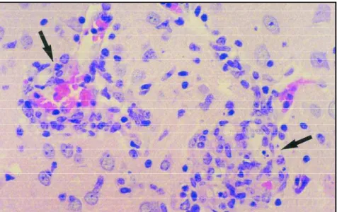
Hepatomegaly and splenomegaly were seen from the 15t hto the 75t hday post-inoculation in ani-mals from group I (immunosuppressed mice), al-though in mice from group II (immunocompetent) only hepatomegaly was evident. All mice inoculat-ed withE. cuniculipresented weight loss, but no mortality was recorded during the experimental period in both groups. Macroscopical lesions in the CNS were not found at necropsy. Microscopi-cally, infected mice presented multifocal granulo-mas, predominantly in the liver, but also occurring in the spleen, kidneys, lungs, guts, cerebrum, ce-rebellum, brainstem (mesencephalon, pons and medulla oblongata) and spinal cord (cervical, tho-racic and lumbar). Table shows the number of le-sions per mm2in the liver and pons from groups I and II. The proportion of such granulomas in both sites was higher in group I comparing to group II. A lymphocytic, diffuse, non-suppurative menigo-encephalomyelitis was observed, with neuronal degeneration and necrosis, secundary demyelina-tion, macrophagic infiltration and reactive astro-cytosis. These granulomas appeared as nests of 20 to 40 pleomorphic cells, presenting acidophilic cy-toplasm and central nuclei (Fig 1). E. cuniculi spo-res were seen in the microgranulomas or occurred unassociated with a tissue reaction throughout the neuropil. Increased astrocytic GFAP
immunos-Table. Mean number of lesions in hepatic and pontine sections of mice from groups I and II.
Group Mean number of lesions Mean number of lesions in the hepatic tissue/mm2 in the pons/mm2
(mean ± s.e.m.) (mean ± s.e.m.)
I* 70.57 ± 55.45a 10.52 ± 4.82a
II 45.70 ± 13.87 3.36 ± 4.73
*Group I immunosuppressed mice inoculated with E. cuniculi; group II immunocompetent mice
Fig 1. Microgranulomas (arrows) in the brainstem 75 days after E. cuniculi inoculation. H-E, Group I, 650x.
Fig 2. Reactive astrocytes showing increased Glial Fibrillary Acidic Protein immunostaining (A) and Vimentin reexpression (B) in mice from group I. A) GFAP, Electron micrograph, 650x; B) VIM, Electron micrograph, 650x.
A
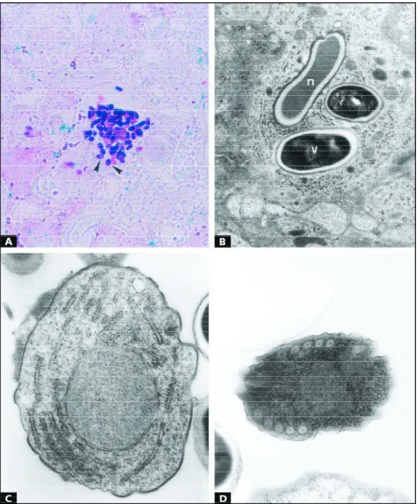
taining was noted (Fig 2A) and reexpression of Vimentin (Fig 2B) was found in reactive astrocytes surrounding the granulomas. The parasites were observed with difficulty in Hematoxylin-Eosin stain-ed sections, but were easily identifistain-ed using Gram-Chromotrope technique as ovoid strutures (Fig 3A) measuring from 2.4 to 3.2 x 1.7 micrometres and presenting a prominent equatorial belt-like
stripe. Some proliferative forms (meronts, sporon-ts, sporoblasts) and spores were found in parasi-tophorous vacuoles within neural cells and macro-phages (Fig 3B). Mature spores were usually so dense that internal organellae were difficult to ob-serve although a coiled filament (polar tubule) and sometimes a single nucleus were seen. The po-lar tubule was arranged into 5 or 6 coils in the
terior region of the spore. The wall of the mature spore was thickened. The spores were surround-ed by a thin, electron-dense exospore that appe-ared to be slightly wrinkled; a thick electron-lucent endospore; and a thin cell membrane that encased the spore contents. Spores with extruded polar tubules were not noted. Meronts were seen as unicellular structures, irregularly round, with one single nucleus, numerous free ribosomes and endo-plasmic reticulum cisternae dispersed in the cyto-plasm. They were delimited by a plasma mem-brane and adhered to the parasitophorous vacuole membrane. Meronts with 2 nuclei and cytoplasmic prolongaments in a characteristic pattern of bina-ry fission were also seen. Sporonts were vebina-ry sim-ilar to meronts, but they showed a thicker and a more electron-dense and ruffled membrane (Fig 3C). Sporoblasts presented a single nucleus, a dou-ble-membrane wall, the most external being cov-ered by an electron-dense material (Fig 3D). Their cytoplasm contained many free ribosomes uni-formly distributed within the cell and a rudimen-tary coiled filament was already seen.
Some degenerating spores within the cyto-plasm of macrophages retained an intact wall, which was depicted as a narrow, corrugated shell (exospore) circumscribing a discrete electron-lucent halo (endospore). The distinctly preserved spore wall usually surrounded a central, homogeneous mass of destroyed organelles. This clear space appears to coincide with the electron-lucent space described above for the mature viable spore. No recognizable organelles, including the cytoplasmic membrane, remained in non-viable spores, all hav-ing been digested to a moderately electron-dense homogeneous mass.
DISCUSSION
Encephalitozoonosis falls into 2 basic patterns4 , 1 0. The first form appears as an acute and clinically de-tectable disease, frequently resulting in death. It is easily produced in neonates as well as in immuno-logically compromised animals4-7,10,11. On the oth-er hand, the second pattoth-ern appears as a chronic and silent infection4 , 1 2. Possible mechanisms of per-sistence of infection may include the sugestion that such protozoans block the fusion of lysosomes to the parasitophorous vacuoles or the supposition that parasites can evade from immune defenses of the host1 3 , 1 4. Much of what is known about mamma-lian microscoporidioses has been understood from experimental models, which provide useful
ap-poaches for the study of humans infected with these protozoans1 5. Human and experimental ani-mal microsporidiosis are both characterized by wi-de dissemination of the organisms, necrosis of in-fected cells, presence of lymphocytic inflammato-ry infiltrates and parasites, free in tissues or in intracellular parasitophorous vacuoles16.
Microsporidia are ubiquitous and occur worldwi-de as obligate intracellular protozoans in most m a-jor groups of the animal kingdom1. Interest in the microsporidia is growing as these organisms are rec-ognized in immunocompromised patients or imm u-nosuppressed recipients of organ transplants5 - 8, as well as in normal individuals, including children and travelers17. Furthermore, concern exists about the potential of zoonotic and waterborne transmission of microsporidia to humans1,17.
Our observations show that E. cuniculi behave in a manner similar to other parasites such as To-xoplasma gondii1 4. Parasites were capable of infect-ing and residinfect-ing within macrophages.E. cuniculi spores with intact walls were found inside parasito-phorous vacuoles, revealing that spores were pha-gocytosed despite the fact that the animals were submitted to an immunosuppressive treatment with CY. Some spores were observed without any sign of internal damage suggesting that they were able to survive in a compartment which failed to fuse with lysosomes. Both merogony and sporo-gony were found in infected macrophages, thus indicating that parasites were able to complete their life cycle. Few endocytosed parasites pre-sented morphological evidences of intracellular hydrolisis such as wall irregularities and cytoplas-mic electron-luscence, and phagosome-lysosome fusion was occasionally seen. As CY treatment in-terferes with immune activation of phagocytic cells, it can be assumed that macrophages from im-munosuppressed mice failed to kill the protozoans and could serve as a vehicule for parasite propa-gation in the host. It remains obscure if E. cuniculi enter macrophages by phagocytosis or by inject-ing the sporoplasm into the host cell cytoplasm through an extruded polar filament18,19.
astrocytes may recover their capacity of express-ing VIM after injury in adult animals20,21.
Previous investigations showed that immuno-suppression in mice with 100 mg/kg of CY, twice a week by intraperitoneal route, produced an acute form of encephalitozoonosis, recognized as a disseminated and lethal infection22. It is impor-tant to emphasize that with the immunosuppres-sive schedule used in our study (50 mg/kg, twice a week, intraperitoneal route), the chronic form of the disease was achieved.
One relevant reason to study animal diseases is that they may help us to identify or antecipate diseases in humans15. Microsporidiosis is a good example of the sucess of this strategy. Although microsporidial infection has been recognized in ani-mals for about 80 years, almost nothing was known about the natural history of microsporidial dis-ease in the human population before the occurren-ce of acquired immunodeficiency syndrome ( A I D S )1 7. In this context, experimental animal mod-els have provided the fundaments from which un-destanding of disease pathogenesis, diagnostic te-chniques and therapeutical approaches for human microsporidiosis have appeared.
Acknowledgement -The authors wish to thank Dr. Wafaa S. Hollister (Department of Biology, Imperial College of Science, Technology & Medicine, London) for supply-ing the strain ofE. cuniculiused in this investigation.
REFERENCES
1. Didier ES, Snowden KF, Shadduck JA. Biology of micro s p o r i d i a n species infecting mammals. Adv Parasitol 1998;40:283-320. 2. Wasson K, Peper RL. Mammalian microsporidiosis. Vet Pathol
2000;37:113-118.
3. Didier ES, Didier PJ, Snowden KF, Shadduck JA. Microsporidiosis in mammals. Microbes Infect 2000;2:709-720.
4 . Gannon J. The course of infection ofEncephalitozoon cuniculiin immun-odeficient and immunocompetent mice. Lab Anim 1980;14:189-192. 5. Didier ES, Varner PW, Didier PJ, et al. Experimental microsporidiosis
in immunocompetent and immunodeficient mice and monkeys. Folia Parasitol 1994;41:1-11.
6. Bryan RT. Microsporidiosis as an A I D S - related opportunistic infec-tion. Clin Infect Dis 1995;21:62-65.
7 . SulaimanIM, MatosO, Lobos ML,Xiao L. Identification of new micro s p o r i d-ian parasite related to Vittaforma cornea in HIV-positiveand HIV- n e g a t i v e patients from Portugal. J Eukaryot Microbiol 2003;50 Suppl:S586-S590. 8. Didier ES, Didier PJ, Friedberg DN. Isolation and characterization of
a new microsporidian, Encephalitozoon hellen (n. sp.), from thre e AIDS patient with keratoconjunctivitis. J Infect Dis 1991;163:617-621. 9. Moura H, Silva JLN, Sodré FC, et al. Gram-Chromotrope: a new tech-nique that enhances detection of microsporidial spores in clinical sam-ples. J Eukaryot Microbiol 1996;43:94-95.
10. Koudela B, Vitovec J, Kucerova Z, Ditrich O, Tranicek J. The severe com-bined immunodeficient mouse as a model for Encephalitozoon cuniculi
microsporidiosis. Folia Parasitol 1994;40:279-286.
11. Thomas C, Finn M, Twigg L, Deplazes P, Thompson RCA. Micro s p o r i d i a (Encephalitozoon cuniculi) in wild rabbits in Australia. Aust Vet J 1997;75:808-810.
12. Schmidt EC, Shadduck JA. Murine encephalitozoonosis model for studying the host-parasite relationship of chronic infection. Infect Immun 1983;40:936-942.
13. Liu JJ, Greeley EH, Shadduck JA. Mechanisms of resistance/suscepti-bility to murine encephalitozoonosis. Parasite Immunol 1989;11 : 2 4 1 - 2 5 6 . 1 4 . Jones TC, Hirsch JG. The interaction between Toxoplasma gondii and mammalian cells: II. The absence of lysossomal fusion with phagocyt-ic vacuoles containing living parasites. J Exper Med 1072;136:11 7 3 - 11 9 4 . 15. Snowden KF, Didier ES, Orenstein JM, Shadduck JA. Animal models of human microsporidial infections. Lab Anim Sci 1998;48:589-595. 16. Van Dellen AF, Stewart CG, Botha WS. Studies of
encephalitozoono-sis in vervet monkeys (Cercopithecus gerythrus) orally inoculated with s p o res ofEncephalitozoon cuniculiisolated from dogs (Canis familiaris). Onderstepoort J Vet 1989;56:1-22.
17. Weiss LM. Microsporidia 2003: IWOP-8. J Eukaryot Micro b i o l 2003;50(Suppl):S566-S568.
18. Couzinet S, Cejas E, Schittny J, Deplazes P, Weber R, Zimmerli S. Phagocytic uptake ofEncephalitozoon cuniculiby nonprofessional phago-cytes. Infect Immun 2000;68:6939-6945.
19. Van Dellen AF, Botha WS, Boomker J, Warnes WEJ. Light and electron m i c roscopical studies on canine encephalitozoonosis: cerebral vasculi-tis. Onderstepoort J Vet 1978;45:165-186.
20. Pixley SK, de Vellis J. Transition between radial glia and mature astro-cytes sztudied with a monoclonal antibody to vimentn. Dev Brain Res 1984;15:201-209.