Beetle (
Ulomoides dermestoides
) fat improves
diabetes: effect on liver and pancreatic architecture
and on PPAR
g
expression
E.I. Jasso-Villagomez
1, M. Garcia-Lorenzana
2, J.C. Almanza-Perez
1, M.A. Fortis-Barrera
1,
G. Blancas-Flores
1, R. Roman-Ramos
1, L.A. Prado-Barragan
3* and F.J. Alarcon-Aguilar
1*
1
Laboratory of Pharmacology, Department of Health Sciences, Division of Health and Biological Sciences, Metropolitan Autonomous University of Iztapalapa, Mexico City, Mexico 2Laboratory of Tissue Neurobiology, Department of Reproduction Biology, Division of Health and Biological Sciences, Metropolitan Autonomous University of Iztapalapa, Mexico City, Mexico 3Laboratory of Solid State Fermentation, Department of Biotechnology, Division of Health and Biological Sciences, Metropolitan Autonomous University of Iztapalapa, Mexico City, Mexico
Abstract
Ulomoides dermestoidesis a beetle traditionally consumed to treat diabetes. In this study, we performed a composition analysis ofU. dermestoidesto obtain the principal fractions, which were used to assess the effect on glycemia, liver and pancreatic architecture, andPPARgandGLUT4expression. Normal mice and alloxan-induced diabetic mice were administered fractions of
chitin, protein or fat, and the acute hypoglycemic effect was evaluated. A subacute study involving daily administration of these fractions to diabetic mice was also performed over 30 days, after which the liver and pancreas were processed by conventional histological techniques and stained with hematoxylin and eosin to evaluate morphological changes. The most active fraction, the fat fraction, was analyzed by gas chromatography–mass spectrometry (GC–MS), and PPARg and GLUT4 mRNA
expressions were determined in 3T3-L1 adipocytes. The protein and fat fractions exhibited hypoglycemic effects in the acute as well as in the 30-day study. Only the fat fraction led to elevated insulin levels and reduced glycemia, as well as lower intake of water and food. In the liver, we observed recovery of close hepatic cords in the central lobule vein following treatment with the fat fraction, while in the pancreas there was an increased density and percentage of islets and number of cells per islet, suggesting cellular regeneration. The GC–MS analysis of fat revealed three fatty acids as the major components. Finally, increased expression ofPPARgandGLUT4was observed in 3T3-L1 adipocytes, indicating an antidiabetic effect.
Key words: Diabetes;Ulomoides dermestoides; Hypoglycemic agent; Insulin-sensitizing agent; Fatty acids; PPARg
Introduction
Diabetes mellitus is a metabolic disorder characterized by hyperglycemia, caused by a deficit in the secretion or function of insulin (1). Although exogenous insulin and other drugs such as peroxisome proliferator-activated receptor gamma (PPARg) agonists may help control diabetes (1), long-term patients develop progressive com-plications including retinopathy, nephropathy, neuropathy, and cardiovascular disease (2). The worldwide cost of diabetes is increasing every year (3). Therefore, the development of new treatments to prevent this illness is important. PPARgis a member of the nuclear hormone receptor superfamily that regulates glucose and fat metabolism, ameliorates insulin resistance through
the mobilization of GLUT4, and increases insulin sensitivity (4).
Ulomoides dermestoides Chevrolat, a beetle also known as the ‘‘peanut weevil’’, is used in the traditional medicine of Argentina, Brazil, China, Colombia, Japan, and Mexico to treat backache, cough, asthma, and diabetes, among others (5,6). The main compounds of the cuticle (alkenes and terpenes) and the defensive secretion (benzoquinones) of this beetle have been characterized in previous chemical studies (7), and its adverse anti-inflammatory, cytotoxic, and genotoxic activities have been described (6–9). Concerning its use in the control
of diabetes, it is thought that ingestion of the live adult
Correspondence: F.J. Alarcon-Aguilar:<[email protected]>| L.A. Prado Barragan:<[email protected]>
*These authors contributed equally to this study.
Received October 20, 2017 | Accepted January 15, 2018
beetle induces pancreatic regeneration. However, it remains unclear whetherU. dermestoideshas an effect on glycemia, and if it is associated with cellular regeneration at the pancreatic level or with the activation of PPARg.
In the present study, alloxan-induced diabetic mice were given the principal fractions ofU. dermestoides(chitin, protein, and fat) to evaluate their effects on glycemia after a single administration (acute study) and after daily adminis-tration for 30 days (subacute study) for histological analyses of the liver and pancreas. In addition, with the fat fraction, a gas chromatography-mass spectrometry analysis (CG-MS) was performed and the changes in mRNA expression of PPARgand GLUT4 were evaluated.
Material and Methods
U. dermestoides
U. dermestoidesbeetles were grown to adulthood in a sanitary bed (natural wheat bran) at 25±2°C and fed a diet consisting of banana peels and bread. The taxonomic authentication of the beetle was performed at the Entomol-ogy and AcarolEntomol-ogy Center, Phyto-Sanitary Institute, Post-graduate College of Agricultural Sciences, COLPOS (Mexico).
Composition analysis ofU. dermestoides
A sample of U. dermestoides was subjected to com-position analysis. The moisture, protein, fat, crude fiber, and ash contents were determined according to the methods reported by the Association of Official Analytical Chemists (10). The moisture content was determined in a thermal scale (OHAUS MB45MR, Switzerland), and the ash content in a muffle (Thermo Scientific, Germany) at 550°C for 4 h. Total protein content was determined by total digestion (Buchi K-350 Distillation Unit, Switzerland). The ether extraction of crude fat was performed using a Soxhlet extractor (Barlsteadt; Thermo Scientific), and crudefiber was quantified using the method described by Hernandez et al. (11).
Isolation of the fat fraction
Live adult beetles were frozen at–80°C, dehydrated at
60°C and ground to afine powder. The total fat fraction was obtained from 10 g of dried beetle sample by Soxhlet extraction using petroleum ether as the solvent (12). The fat fraction was recovered in a rotary evaporator (B490; Buchi, Switzerland).
Isolation of the chitin fraction
The defatted sample (7.6 g) was ground and mixed with 300 mL of 10% NaOH. After incubation at 60°C for 3 h, the slurry was filtered through Whatman no. 4 filter paper. The precipitate (chitin fraction) was dried at 60°C and stored in an airtight container at 2–4°C until
use (11). The supernatant was used for soluble protein assays.
Isolation of the protein fraction
The resultant supernatant from the chitin extraction was subjected to acidification (HClconc.) until the iso-electric point of the proteins was reached (pH 3). Then, the sample was filtered through Whatman no. 4filter paper and the insoluble protein fraction was dried and stored at 2–4°C until use (13).
Experimental animals
MaleMus musculusmice of the CD1 strain (35–45 g)
were purchased and bred in the Experimental Animal Center at Metropolitan Autonomous University, Mexico. Six mice per cage were maintained under a 12/12 h light/ dark period at 22±1°C and relative humidity of 55±3%. Mice were fed a rodent diet containing 18.6% protein, 44.2% carbohydrates, and 6.2% fat (2018s Teklad Global 18% protein; Harlan Laboratories, USA) and received water ad libitum. The handling of the laboratory animals and experimental procedures were performed according to national and international standards including the Official Mexican Standard (NOM-062-ZOO-999, revised 2001) and the National Institutes of Health (NIH publica-tion No. 8023, revised 1996) for the health, safety and comfort of experimental animals. Additionally, the inter-nal committee of the Metropolitan Autonomous Univer-sity approved the experimental animal handling protocol (DCBS.949.2017).
Evaluation of the acute hypoglycemic effect of the
different fractions ofU. dermestoidesin normal mice
In previousin vivoassays performed withU. dermes-toides, the tested doses of the active extracts ranged from 0.6, 3, 8, and 16 mg/kg (9,14). Therefore, in the present study, only the highest dose (16 mg/kg) was selected to perform all experiments. Thirty experimental animals were fasted for 12 h and grouped as follows: group 1 (control), treated with isotonic saline solution (ISS, 4 mL/kg); group 2, treated with the fat fraction (16 mg/kg); group 3, treated with the protein fraction (16 mg/kg); group 4, treated with the chitin fraction (16 mg/kg); and group 5, positive control, treated with glibenclamide (10 mg/kg), a sulphonyl urea agent with hypoglycemic action that acts at the pancreatic level as an insulin secretagogue. All treatments were administered by the intraperitoneal (ip) route and glycemia was quantified from tail vein blood samples at 0, 120, 240, and 360 min by the dehydrogenase method (Accu-Chekt Performa; Roche Diagnostics, USA).
Induction of diabetes in mice
Evaluation of the acute hypoglycemic effect of U. dermestoidesin alloxan-induced diabetic mice
Thirty mice with alloxan-induced diabetes were grouped as follows: group 1, control, treated with ISS (4 mL/kg); group 2, treated with the lipid fraction (16 mg/kg); group 3, treated with the protein fraction (16 mg/kg); group 4, treated with the chitin fraction (16 mg/kg); and group 5, treated with glibenclamide (10 mg/kg). All treatments were administered ip and glycemia was quantified for each group at 0, 120, 240, and 360 min, following the methodology described above for normal mice.
Evaluation of the subacute hypoglycemic effect of U. dermestoides
Six normal mice and 30 mice with experimental diabetes were separated into groups as follows: group 1, normal control, treated with ISS (4 mL/kg); group 2, diabetic control, treated with ISS (4 mL/kg); group 3, diabetic treated with the lipid fraction (16 mg/kg); group 4, diabetic treat-ed with the protein fraction (16 mg/kg); group 5, diabetic treated with the chitin fraction (16 mg/kg); group 6, diabetic treated with glibenclamide (5 mg/kg). Treatments were administered daily by gavage to all groups over a 30-day period, with water and food intake recorded every 24 h. Blood samples (32mL) were taken from the tail vein by puncture, and cholesterol, triglyceride, aspartate ami-notransferase (AST), and alanine amiami-notransferase (ALT) levels were quantified after 30 days of treatment using a Reflotron Plus System (Bayer, Germany). Glycemia was also quantified after 30 days of treatment by the dehydrog-enase method (Accu-ChektPerforma, Roche Diagnostics).
Insulin quantification
After 30 days of treatment, mice were anesthetized with sodium pentobarbital (27.5 mg/kg) and blood samples were obtained from the eye orbital sinus. Insulin was quantified by the ELISA method according to the supplier’s instructions (Linco Research, USA).
Histological analysis
At the end of subacute study (30 days), the animals were anesthetized with sodium pentobarbital (27.5 mg/kg IP) and euthanized according to the Official Mexican Standard (NOM-033-ZOO-1995). The pancreas and liver were obtained surgically and fixed by immersion in 4% paraformaldehyde in phosphate buffer, pH 7.2–7.4, for 18
to 24 h. The organs were processed by a conventional histological technique (16) and embedded in Paraplastt (Oxford Lab, USA). Serial longitudinal sections of 5-mm thickness were cut on a microtome (Leika Jung Histocut 820, Germany) and stained with hematoxylin and eosin (17). Qualitative analysis was carried out with an optical light microscope (Axioskope II; Carl Zeiss, Germany) coupled to an image analyzer (AxioVision 4.5; Carl Zeiss). Photo-micrographs were taken with AxioCamMRc5 (Carl Zeiss).
A morphometric analysis of the islets was performed on longitudinal sections of the pancreas over a total mean area of 387 mm2. Analysis of the pancreas was performed on nine serial 5-mm thick sections from three animals in each treatment group. Serial cuts (200) were made over a 150-mm thick area. For this analysis, the sections chosen were those numbered 50, 100, and 200 for each animal. Islet density was determined at 100 magnification, and the islet area and the number of cells per islet were deter-mined at 400 magnification. In total, nine sections per treatment were analyzed by optical light microscopy (Carl Zeiss).
Gas chromatography-mass spectrometry (GC-MS) analysis
The fat extracted fromU. dermestoideswas analyzed on an Agilent Technologies gas chromatograph (GC) 6890N (USA) coupled to a 5973N mass spectrometer (MS) operated at 70 eV and equipped with a Carbowax capillary column (30 m, 0.25 mm internal diameter) with a polyethylene glycol matrix (0.25mm thick). Helium was the carrier gas at aflow rate of 1 mL/min. The injector and MS transfer line temperature was set at 250°C. Samples were diluted 1:10 v/v in acetone and manually injected (1.0mL in volume). After sample injection, the initial temperature of the oven (45°C) was held constant for 3 min and then increased to 135°C at a rate of 3°C/min. This temperature was maintained for 1 min and then increased to 250°C at a rate of 10°C/min, which was maintained for 10 min. For GC–MS detection, an electron ionization system with an
ionization energy of 70 eV was used. After a delay of 3 min to allow passage of the solvent, the mass spectra were scanned from 15 to 800 m/z. Tentative identification of the components was performed based on their relative retention time and mass spectra comparison with the National Institute of Standards and Technology Library (NIST, NIST02L) data for the GC-MS system. Quantifi ca-tion of the identified components was based on the integrated peak area.
3T3-L1 cell line
MTT assay in 3T3-L1fibroblasts treated with the fat fraction
Cellular functionality was determined using 3-(4,5-dimethylthiazole-2-yl)-2,5-diphenyltetrazolium bromide (MTT; Sigma, USA) according to the method described by Mosmann (19). The test quantifies the conversion of MTT to formazan by insoluble dehydrogenase enzyme activity of intact cells. 3T3-L1 cells were seeded onto 96-well micro-plates at a semi-confluent density (5000 cells/well). After 24 h, the medium was replaced with medium containing 1, 10, or 100mg/mL of fat. Cells were treated for 30 min then washed with phosphate-buffered saline (PBS), pH 7.4, and a solution of MTT at 0.1 mg/mL in PBS, pH 7.4, was added. The cells were incubated for 3 h at 37°C then washed with PBS. Then, 40 mM HCl (200mL) prepared in isopropanol was added to each well for 15 min to solubilize the produced formazan. Absorbance was read at 570 nm. Data are reported as the percentage of viable cells after treatment with the lipid fraction ofU. dermestoidescompared to control cells.
Evaluation of mRNA expression of PPAR-candGLUT4
by RT-PCR in 3T3-L1 adipocytes treated with the fat fraction
Fibroblasts (9105cells per well) were cultured and differentiated into adipocytes in 6-well plates (Corning Inc.). The RNA was isolated from cultured cells using a TriPure isolation reagent (Invitrogen, UK). The absorb-ance was measured at 260 and 280 nm for each RNA sample (the absorbance ratio was 1.9±0.2). To confirm RNA integrity, 1.5mg was run in a 1% agarose gel and the RNA bands were stained with ethidium bromide and visualized using an Image Gel-Logic 212 Pro (Kodak/ Carestreamt, USA). Two major ribosomal bands (28S and 18S rRNA) were detected, with no degraded RNA (data not shown).
Total RNA (2 mg) was reverse-transcribed using the ImProm II Reverse Transcription System (Promega, USA), then the mixture (20mL) was incubated in a thermocycler (Select Cycler; BioProducts, USA), following the cycle program of incubation at 25°C for 5 min and extension at 42°C for 55 min. The enzyme was inactivated at 70°C for 15 min, then samples were cooled to 4°C for 5 min. Then, 1/ 10 volume of each cDNA sample was amplified with SYBR Green master mix (Roche Molecular Biochemicals, Ger-many) containing 0.5 mM of customized primers for PPAR-g (forward 50-CCAGAGTCTGCTGATCTGCG-30, reverse
50-GCCACCTCTTTGCTCTGCTC-30; Gene Bank NM_
011146.1), GLUT4 (forward 50-GATTCTGCTGCCCTTCT
GTC-30, reverse 50-ATTGGACGCTCTCTCTCCAA-30; Gene
Bank NM_009204.2), fast-start enzyme, PCR buffer, and 3.5 mM MgCl2, made up to afinal volume of 10mL. The reactions were measured by Rotor-Gene Real-Time equipment (Corbett Life Science, Australia).
PCR was conducted using the following cycling conditions: pre-incubation and denaturation at 95°C for 10 min, alignment at 61°C for 7 s, and amplification at 72°C
for 10 s. The threshold cycles (Ct) were measured in separate tubes and in duplicate. The identity and purity of the amplified products were checked by electrophoresis on a 2% agarose gel. The melting curve was analyzed at the end of amplification following the SYBR Green kit protocol, as indicated by the company (Roche Molecular Biochemicals). To ensure the quality of the measurements, each assay included a negative control for each gene. The amount of mRNA for each studied parameter was normalized to mRNA encoding the ribosomal protein 36B4 (forward 50-AAGCGCGTCCTGGCATTGTCT-30,
reverse 50-CCGCAGGGGCAGCAGTGGT-30; Gene Bank
NM_007475.2). The relative changes in the expression levels of mRNA were calculated with the formula 2DDCt(20).
Statistical analysis
Biological data are reported as means±SE. Statistical analysis of the data was performed by two-way analysis of variance (ANOVA) followed by a Tukey-Kramerpost hoc test at a confidence level of 95% (NCSS 2000, USA).
Results
Composition ofU. dermestoides
The compositional analysis of the beetles based on dried weight revealed the highest percentage was of protein (53.3±1.2%) and fat (24.7±0.7%), followed by fiber (22.1±1.1%), chitin (19.2±0.5%), and ash (1.0±0.3%). In fresh organisms, the moisture content was 56.3±0.3%. The protein, fat, and chitin fractions were selected to be evaluated for their acute hypoglycemic effect in normal and diabetic mice.
Acute hypoglycemic effect of the fractions
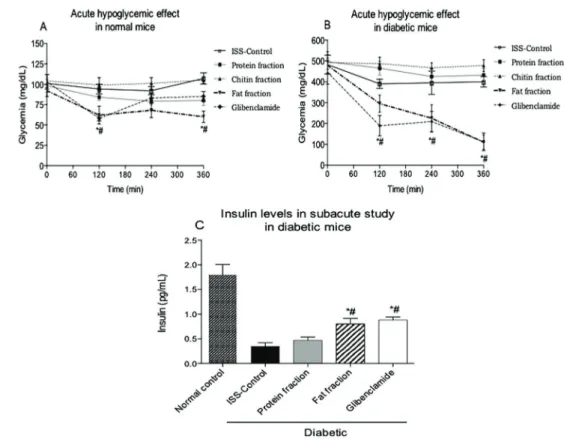
The hypoglycemic effects of a single administration of protein, fat or chitin to normal mice are shown in Figure 1A. Only the fat and glibenclamide treatments caused a significant reduction in glycemia at 120 and 360 min compared to the initial glycemia values. Evaluation of the hypoglycemic effect of the components of U. dermes-toidesin alloxan-induced diabetic mice revealed that the fat and glibenclamide treatments caused a significant reduction in glycemia when compared to both the basal glycemia level and to the diabetic control group treated with ISS (Figure 1B). The protein and chitin treatments did not show any hypoglycemic effects in normal or diabetic mice.
Hypoglycemic effect from daily administration of the fractions
triglyceride levels increased by 50%. Consumption of the protein fraction also caused a significant reduction in glycemia, but there were no significant differences in the group that received the protein fraction compared with the normal control group (Table 1). Finally, chitin
treatment (16 mgkg–1
day–1) was found to cause 100% lethality after 15 days of treatment. The body weight did not change significantly in any of the treated groups compared with the initial body weight (data not shown).
Figure 1.Acute hypoglycemic effect in normal mice (A). Acute hypoglycemic effect in alloxan-induced diabetic mice (B). Serum insulin levels in the subacute study in normal and alloxan-induced diabetic mice over 30 days (C). Mice were treated with either saline (control), protein, chitin or fat fractions (lipid) isolated fromUlomoides dermestoides, or glibenclamide. Data are reported as means±SE (n=6). *Po0.05 compared to basal glycemia (or insulin normal control).#Po0.05 compared to isotonic saline solution (ISS)-treated control (ANOVA and Tukey-Kramerpost hoctest).
Table 1.Effect of the daily administration for 30 days of the chitin, lipid, and protein fractions fromU. dermestoideson water intake, food intake, and biochemical parameters.
Parameter Glycemia (mg/dL)
Water intake (mL/mice)
Food intake (g/mice)
Cholesterol (mg/dL)
Triglycerides (mg/dL)
ALT (U/L)
AST (U/L)
Normal 135±5.5 8.2±0.6 7.2±1.4 100±4.2 70±3.8 8.4±3.8 5.9±1.6 Diabetic (ISS) 593±2.1* 53.5±4.3* 16.7±3.3* 105±3.8 71±9.7 7.2±3.8 198.3±3.3*
Chitin 598±7.8* 59.4±2.9* 17.6±4.6* – – – –
Fat 120±6.1# 18.5±4.6*# 6.9±2.4# 108±6.8 142±2.9*# 6.8±2.1 15.0±3.3* Proteins 386±16.4*# 31.3±2.7* 9.6±0.7 110±7.4 82±6.4 59.1±3.8*# 22.5±4.5*#
Glibenclamide 552±8.1* 50.2±8.7* 14.2±1.9* 110±5.2 78±6.0 5.9±9.8 23.8±4.8
Histological changes in the liver and pancreas
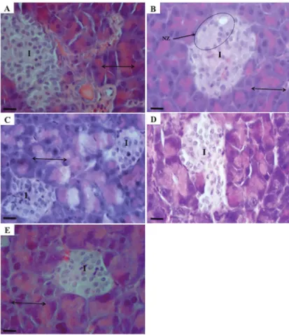
Micrographs of longitudinal pancreas sections from mice in the different treatment groups are shown in Figure 2. The general structure of the pancreas can be clearly observed in normal mice (Figure 2A), with an exocrine zone consisting of epithelial pancreatic acinus and an endocrine zone of pancreatic islets. Histological analysis showed qualitative and quantitative changes in the typical pancreas architecture after alloxan treatment, particularly in the islets of Langerhans (Figure 2B), with this group displaying necrotic changes in the pancreatic islets. In addition, nuclear changes, karyolysis, and a reduction in the number of islet cells were observed (reduced number of cells per zone).
In Figure 2C, pyknotic nuclei are evident in animals treated with the protein fraction of U. dermestoides. It is remarkable that with the fat fraction, recovery of the alloxan-induced alterations in pancreatic architecture was observed (Figure 2D). A small number of pyknotic nuclei, improved karyolysis, and a reduction in necrosis sugges-tive of progression to normal pancreatic morphology were evident. Pancreas samples from mice in the glibenclamide group also showed pyknotic cells, in addition to similar damage to the islets caused by alloxan and regeneration
of the central islet region (Figure 2E). Remarkably, the fat fraction increased the number of islets compared to the ISS-treated diabetic control group (41.8% vs 11.5%), as well as the number of cells in the islets and the density of the islets (Table 2). In Figure 3, it is evident that all diabetic groups with or without treatment (Figure 3B–E) exhibited
hyperemia.
In the liver histological analysis (Figure 4), hepatocytes of normal mice showed a well-defined nucleus and cytoplasm with cell margins, and the lobule central vein, hepatocytes plates, and the sinusoidal spaces can be clearly observed (Figure 4A). The liver slices from control diabetic mice showed sinusoidal dilatation, a loss of cytoplasmic integrity with indistinct cell margins, and a loss of nuclear staining intensity, nuclear function, and cell death (Figure 4B). The group treated with the protein fraction showed impaired nucleus architecture and mild liver steatosis (Figure 4C). Treatment with the fat fraction showed recovery of the close hepatic cords in the central lobule vein, a well-defined nucleus and cytoplasm, and apparent recovery of cellular function (Figure 4D). Gli-benclamide treatment caused changes in the hepatic lobule, including loss of hepatic cords andfibrotic zones (Figure 4E).
GC-MS analysis of the fat fromU. dermestoides
The GC-MS analysis of the fat fraction of U. dermestoides (Table 3) revealed the presence of 31 different fatty acids (FA), representing 91% of the total components in the fat. The major FA fraction identified was a mixture of linoleic and oleic acids (40.9%) followed by hexadecanoic (palmitic) (31.9%) and octadecanoic (9.3%) acids.
Effect of the fat fraction fromU. dermestoideson
PPARcandGLUT4expression
In vitrostudies in 3T3-L1 adipocytes performed in our laboratory showed that cell viability was unaffected by treatment withU. dermestoidesfat at different concentra-tions (data not shown). Therefore, concentraconcentra-tions of 10 and 100mg/mL were selected for this experiment. As shown in Figure 5A and B, 100 mg/mL of fat increased the mRNA Table 2.Histomorphometric analysis of pancreas of alloxan-induced diabetic mice administered isotonic saline solution (ISS) (diabetic control), proteins and fat fromU. dermestoides, and glibenclamide for 30 days.
Treatment Islets density (islets/387212.92 mm2) Percent of islets Islet area (mm2) Number of cells/islet
Normal control 10±2.0 100±1.2 10891.3±164.5 55±8
ISS Diabetic 2±0.7* 11.5±4.7* 10137.3±250.0 15±3*
Proteins 2±0.8* 11.5±4.7* 5977.2±424.5*# 12±2*
Fat 6±1.5* 41.8±3.2*# 12320.2±2820.5 31±3*#
Glibenclamide 1±0.6* 9.8±3.3* 8500.9±1363.5* 14±2*
Data are reported as means±SE (n=6). ISS: isotonic saline solution. *Po0.05vsnormal control;#Po0.05vsdiabetic control (ANOVA and Tukey-Kramerpost hoctest).
expression ofPPARg(75%) andGLUT4(66.6%) compared
with the control, and these effects were larger than those observed with pioglitazone, a molecule with PPARg-agonist activity.
Discussion
Protein, fat, and chitin were found to be the most abundant chemical components in adultU. dermestoides beetles. The protein and lipid fractions had a hypoglycemic effect in the acute experiment; however, in the subacute experiment, only the fat fraction demonstrated a hypogly-cemic effect, with an increase in serum insulin as well as a significant reduction in the consumption of food and water. The cholesterol and liver transaminase (ALT and AST) levels did not change, but an increase in triglycerides was
observed when mice were supplemented with the fat fraction.
both of which have been associated with anti-diabetic, anti-atherosclerotic, and anti-obesity effects as well as with immune-modulator properties (24). In addition, these fatty acids can act as ligands in transcription factors, functioning as metabolic regulators (25). Nevertheless, it is necessary to make trimethylsylyl derivatives of the fat fraction, as there are likely a number of less volatile derivates contained in the fat fraction that would be observed whether the trimethylsilyl or tert-butyldimethyl-silyl derivatives are analyzed (26). This should be further explored in additional studies.
Fatty acids represent an important source of metabolic energy, and can function as physiological signaling mole-cules. Some fatty acids act as ligands for the peroxisome proliferator-activated receptors (PPAR), especially PUFAs, which directly influence the transcriptional activity of genes encoding these receptors (25).
In the alloxan-induced diabetic mice treated with ISS (diabetic control), histological analysis showed disruption of the liver cords and sinusoidal dilation, both of which are associated with obstruction of venous outflow. These results are in agreement with those reported by Samadder et al. (27) and Saadoun et al. (28). Furthermore, sinusoidal dilatation is also considered an important marker of infl am-mation and cellular damage in certain diseases such as hypertension, tumors, and diabetes (29,30). Our results showed that animals treated with the protein fraction ofU. dermestoidesor glibenclamide exhibited liverfibrotic regeneration, which should be confirmed in further studies. Excessive protein intake can cause an overload for the kidneys and liver, both of which are responsible for removing waste substances (ammonia, urea, and uric acid) generated by excessive protein intake (31). In addition, excess protein disrupts liver tissue as the production of plasma proteins is reduced while transaminase levels are elevated. This may result in hepatotoxicity, inflammation Table 3.Composition of the fat fromU. dermestoides.
Retention time % Identified compound
3.702 0.012 Undecane
3.984 0.004 Octanoic acid
5.973 0.008 Decanoic acid
7.678 0.051 Dodecanoic acid
9.221 1.556 Tetradecanoic acid 9.889 0.221 Pentadecanoic acid 10.326 0.151 9-12-hexadecadienoic acid 10.420 1.255 9-hexadecanoic acid 10.806 31.910 Hexadecanoic acid 11.235 0.395 Heptadecanoic acid 11.972 40.957 Linoleic and Oleic acid 12.057 9.280 Octadecanoic acid 12.289 0.062 9-nonadecanoic acid
12.443 0.112 Nonadecanoic acid
12.657 0.073 7,10,13-eicosatrienoic acid 12.794 0.323 11.14-eicosenoic acid 12.837 0.637 11-eicosenoicacid
12.991 0.718 Eocisanoic acid
13.343 0.064 Tetracosane
13.891 1.002 Pentacosane
14.037 0.443 Docosanoic acid
14.868 0.718 Heptacosane
14.997 0.078 Tetracosanoic acid
15.314 0.051 Octacosane
15.597 0.099 Cholesta-3,5-diene
15.777 0.457 Nonacosane
15.914 0.021 Hexacosanoic acid
16.642 0.248 Hentriacontane
16.779 0.054 Octacosanoic acid
17.448 0.192 Tritriacontane
18.416 0.114 Pentatriacontane
8.735 Unidentified
and chronic liver damage, and could eventually lead to hepatic steatosis, fibrosis, cirrhosis or non-alcoholic fatty liver disease (32).
The fat fraction fromU. dermestoidesameliorated the changes in liver histology induced by alloxan, with reduced sinusoidal dilatation and improved tissue organization observed in this group. This fat may play an important role in cell permeability, improving communication between cells and transport of proteins or receptors embedded in the membrane of liver cells (33). Like the lipid fraction, the protein fraction also exhibited a hypoglycemic effect, although mice that consumed the protein fraction also showed increased liver transaminases as a sign of liver damage. Thisfinding is consistent with the liver damage observed in the micrographs. In contrast, the fat of U. dermestoidesimproved the hepatic architecture and promoted expression of PPARg and GLUT-4, which
might partly explain the positive effects of ingestion of U. dermestoideson glycemia in diabetic subjects.
Interestingly, in the pancreas, the fat fraction from U. dermestoides also augmented the number of islets, the cell density and regenerated the altered pancreatic architecture induced by alloxan. In the present investiga-tion, the vasodilatation observed in the pancreas of mice in the group treated with this fat could be due to a pancreatic response to increase bloodflow to the cells in order to obtain more nutrients. The higher cell density could lead to the activation of factors involved in vein dilation to satisfy the nutritional needs of the cells (34). In conjunction, these results may be indicative of islet regeneration. However, it is necessary to measure differ-entiation markers in future studies to clarify the nature of the new cells generated in the islets and to determine their origin.
Recent in vivo studies have shown that at least two transcription factors, Pax4 and Arx, can mediate transdif-ferentiation from a-cells to b-cells. It has been demon-strated that a-cells can continuously regenerate and convert into b-cells through overexpression of the tran-scription factor Pax4, which represses the master regula-tory transcription factor Arx of a-cells, thereby stimulating the conversion ofa-cells intob-cells (35). In addition, some compounds including g-aminobutyric acid (GABA) and artemisinin antimalarial drugs can act as inducers of this cellular conversion (36). However, whether this process of
cellular trans-differentiation in islets of Langerhans can be affected by the fat from U. dermestoides should be determined in further studies.
In relation to chitin, the main component of the exoskeleton of crustaceans and insects, its use is generally associated with beneficial effects (37). Our study showed that a dose of 16 mgkg–1
day–1 of chitin extracted from U. dermestoides induced 100% lethality after 15 days of treatment. Its ingestion has been associated with inflammatory and allergic processes, which may cause death in diabetic subjects (38). Nevertheless, further toxicological studies with chitin isolated from U. dermestoides are mandatory. Other compounds of U. dermestoides, such as the benzoquinone volatile compounds isolated from the defensive secretions of U. dermestoides, have also been reported to cause cytotoxicity and genotoxicity in cultured cells (7,9), whereas phenolic compounds exhibited a free radical scavenging activity and antiproliferative effect in human lymphocytes (39,40).
In conclusion, the fat fraction from U. dermestoides produced a hypoglycemic effect in normal and diabetic mice, with evidence of regeneration of pancreatic islets, a liver-protective effect, and insulin-sensitizing properties. In addition, while other studies have associated the lipids fromU. dermestoideswith anti-irritant and central nervous system depressing effects (14,21), this is thefirst study to support the beneficial effects of U. dermestoides fat in diabetes treatment.
Acknowledgments
The authors thank the Laboratorio Divisional de Biología Molecular of the DCBS at Universidad Autónoma Metropolitana for technical support, Dr. Jesus Romero Napoles (Instituto de Fitosanidad, Colegio de Postgra-duados Montecillo, Estado de México, México) for the taxonomic identification ofU. dermestoides, and Dr. Marco M. Gonzalez Chavez and Dr. Maria Salud Perez. The present study was supported by a grant from CONACyT awarded to E.I.J. Villagomez (290671) as part of his Ph.D. degree (Doctorado en Ciencias Biológicas y de la Salud from DCBS at Universidad Autónoma Metropolitana), and partially supported by a PROMEP-SEP grant (project UAM-PTC-218).
References
1. Baynes HW. Classification, pathophysiology, diagnosis, and management of diabetes mellitus.J Diabetes Metab2015; 6: 541–549, doi: 10.4172/2155-6156.1000541.
2. Chawla A, Chawla R, Jaggi S. Microvascular and macro-vascular complications in diabetes mellitus: distinct or continuum?Indian J Endocrinol Metab2017; 20: 546–551, doi: 10.4103/2230-8210.183480.
3. NCD Risk Factor Collaboration. Worldwide trends in diabetes since 1980: a pooled analysis of 751 population-based studies with 4.4 million participants. Lancet 2016; 387: 1513–1530, doi: 10.1016/S0140-6736(16)00618-8. 4. Azhar S. Peroxisome proliferator-activated receptors,
5. Mendoza-Meza DL, España-Puccini P. Cytotoxic and genotoxic activity of phenolic fractions from Ulomoides dermestoidesFairmaire, 1893 (Coleoptera, Tenebrionidae), in HaCat Cells.Rev Esp Cienc Quim Biol2016; 19: 83–91, doi: 10.1016/j.recqb.2016.06.001.
6. Deloya-Brito GG, Deloya C. Substances produced by the beetleUlomoides dermestoides(Chevrolat, 1878) (Insecta: Coleoptera: Tenebrionidae): inflammatory and cytotoxic effect.Acta Zoo Mex2014; 30: 655–661, doi: 10.21829/azm. 2014.30384.
7. Villaverde ML, Girotti SJ, Mijailovsky SJ, Pedrini N, Juarez P. Volatile secretions and epicuticular hydrocarbons of the beetle Ulomoides dermestoides. Comp Biochem Physiol Part B Biochem Mol Biol2009; 154: 381–386, doi: 10.1016/ j.cbpb.2009.08.001.
8. Santos RC, Lunardelli A, Caberlon E, Bastos CM, Nunes FB, Pires MG, et al. Anti-inflammatory and immunomodula-tory effects ofUlomoides dermestoideson induced pleurisy in rats and lymphoproliferation in vitro.Inflammation2010; 33: 173–179, doi: 10.1007/s10753-009-9171-x.
9. Crespo R, Villaverde ML, Girotti JR, Güerci A, Juarez MP, de Bravo MG. Cytotoxic and genotoxic effects of defense secretion ofU. dermestoideson A549 cells.J Ethnophar-macol2011; 136: 204–209, doi: 10.1016/j.jep.2011.04.056. 10. Association of Official Analytical Chemists (AOAC). Official
methods of analysis. 14th edn. Washington: AOAC; 1988. 11. Hernandez-Nuñez CM, Varo-Arguello WE, Leyva-Reyes N,
Ramirez-Barragan CA, Delgado-Fornue E, Andrade Ortega JA. Utilización de residuos de cáscara de camarón para la obtención de quitina blanqueada: propuesta de un trata-miento de alcalino-ácido y ozono. Avances en la Investiga-ción Científica. Centro Universitario de Ciencias Biológicas y Agropecuarias (CUCBA-México). 2008. p. 659–666. 12. Tzompa-Sosa DA, Yi L, van Valenberg HJF, van Boekel
MAJS, Lakemond CMM. Insect lipid profile: aqueous versus organic solvent-based extraction methods,Food Res Inter 2014; 62: 1087–1094, doi: 10.1016/j.foodres.2014.05.052. 13. Watson SA. Structure and composition. In: Watson AS,
Ramstad PE (editors), Corn: Chemistry and Technology. St. Paul: American Association of Cereal Chemists; 1987. p. 53–60.
14. Tobón FA, Gutiérrez ZGP, Mejia GML. Evaluation of neuropharmacological profile of oil fromUlumoides dermes-toides(Coleoptera: Tenebrionidae). Rev Colomb Entomol 2011; 37: 251–255.
15. Etuk EU. Animals models for studying diabetes mellitus. Agric Biol J N Am 2010; 1: 130–134, doi: 10.1.1.212. 9006&rep=rep1&type=pdf.
16. Prophet EB, Millis B, Arrington JB, Sobin L. AFIP laboratory methods in histotechnology. 1st edn. Washington American Registry of Pathology; 1992.
17. Presnell JK, Schreibman MP (Editors). Humason’s animal tissue techniques. 5th edn. Baltimore: Johns Hopkins Uni-versity Press; 1997.
18. Garcia-Macedo R, Sanchez-Munoz F, Almanza-Perez JC, Duran-Reyes G, Alarcon-Aguilar F, Cruz M. Glycine increases mRNA adiponectin and diminishes pro-inflammatory adipo-kines expression in 3T3-L1 cells.Eur J Pharmacol2008; 587: 317–321, doi: 10.1016/j.ejphar.2008.03.051.
19. Mosmann T. Rapid colorimetric assay for cellular growth and survival: application to proliferation and cytotoxicity assays.
J Immunol Methods1983; 65: 55–63, doi: 10.1016/0022-1759(83)90303-4.
20. Hidalgo-Figueroa S, Ramirez-Espinosa JJ, Estrada-Soto S, Almanza-Perez JC, Roman-18.Ramos R, Alarcon-Aguilar FJ, et al. Discovery of thiazolidine-2,4-dione/biphenylcarbo-nitrile hybrid as dual PPARa/gmodulator with antidiabetic effect: in vitro, in silico and in vivo approaches.Chem Biol Drug2013; 81: 474–483, doi: 10.1111/cbdd.12102. 21. Mendoza MDL, Saavedra AS. Chemical composition and
anti-irritant capacity of whole body extracts of Ulomoides dermestoides(Coleoptera, Tenebrionidae).Rev Fac Quim Farm2013; 20: 41–48.
22. Sears B, Perry M. The role of fatty acids in insulin resistance. Lipids Health Dis2015; 14: 121–129, doi: 10.1186/s12944-015-0123-1.
23. Siri-Tarino PW, Chiu S, Bergeron N, Krauss R. Saturated fats versus polyunsaturated fats versus carbohydrates for cardiovascular disease prevention and treatment.Annu Rev Nutr2015; 35: 517–543, doi: 10.1146/annurev-nutr-071714-034449.
24. Lehnen TE, Ramos da Silva M, Camacho A, Marcadenti A, Machado LA. A review on effects of conjugated linoleic fatty acid (CLA) upon body composition and energetic metabo-lism.J Int Soc Sports Nut 2015; 12: 36–46, doi: 10.1186/ s12970-015-0097-4.
25. Varga T, Czimmerer Z, Nagy L. PPARs are a unique set of fatty acid regulated transcription factors controlling both lipid metabolism and inflammation.Biochim Biophys Acta2011; 1812: 1007–1022, doi: 10.1016/j.bbadis.2011. 02.014.
26. Halket JM, Waterman D, Przyborowska A, Patel RK, Fraser PD, Bramley PM. Chemical derivatization and mass spectral libraries in metabolic profiling by GC/MS and LC/MS/MS.J Exp Bot2005; 56: 219–243, doi: 10.1093/jxb/ eri069.
27. Samadder A, Chakraborty D, Arnab D, Sundar BS, Bhadra K, Khuda BA. Possible signaling cascades involved in attenuation of alloxan-induced oxidative stress and hyper-glycemia in mice by ethanolic extract ofSyzygium jambo-lanum: drug-DNA interaction with calf thymus DNA as target. Eur J Pharm Sci 2011; 44: 207–217, doi: 10.1016/j.ejps. 2011.07.012.
28. Saadoun D, Cazals HD, Denninger MH, Boudaoud L, Pham BN, Mallet V, et al. Association of idiopathic hepatic sinusoidal dilatation with the immunological features of the antiphospholipid syndrome. Gut 2004; 53: 1516–1519, doi: 10.1136/gut.2003.037135.
29. Watanabe N, Takashimizu S, Nishizaki Y. Kojima S, Kagawa T, Matsuzaki S. An endothelin a receptor antagonist induces dilatation of sinusoidal endothelial fenestrae implications for endothelin-1 in hepatic microcirculation.J Gastroenterol2007; 42: 775–782, doi: 10.1007/s00535-007-2093-1.
30. Sathya A, Siddhuraju P. Protective effect of bark and empty pod extracts fromAcacia auriculiformisagainst paracetamol intoxicated liver injury and alloxan induced type II diabetes. Food Chem Toxicol 2013; 56: 162–170, doi: 10.1016/j.fct. 2013.02.031.
32. Lowery LM, Devia L. Dietary protein safety and resistance exercise: what do we really know?J Int Soc Sports Nutr 2009; 6: 1–7, doi: 10.1186/1550-2783-6-3.
33. van der Vusse GJ, van Bilsen M, Glatz JF, Hasselbaink DM, Luiken JJ. Critical steps in cellular fatty acid uptake and utilization.Mol Cell Biochem2002; 239: 9–15, doi: 10.1023/ A:1020538119691.
34. Dai C, Brissova M, Reinert RB, Nyman L, Liu EH, Thompson C, et al. Pancreatic islet vasculature adapts to insulin resistance through dilation and not angiogen-esis.Diabetes2013; 62: 4144–4153, doi: 10.2337/db12-1657.
35. Ben-Othman N, Vieira A, Courtney M, Record F, Gjernes E, Avolio F, et al. Long-term GABA administration induces alpha cell-mediated beta-like cell neogenesis. Cell 2017; 168: 73–85, doi: 10.1016/j.cell.2016.11.002.
36. Jin Li, Casteels T, Frogne T, Ingvorsen C, Honore, Courtney M, et al. Artemisinins target GABAAreceptor signaling and
impairacell identity.Cell2017; 168: 86–100, doi: 10.1016/j. cell.2016.11.010.
37. Koide SS. Chitin-chitosan: properties, benefits and risks. Nutr Res1998; 18: 1091–1101, doi: 10.1016/S0271-5317 (98)00091-8.
38. Reese TA, Liang HE, Tager AM, Luster AD, Van Rooijen N, Voehringer D, et al. Chitin induces accumulation in tissue of innate immune cells associated with allergy.Nature 2007; 447: 92–97, doi: 10.1038/nature05746.
39. Mendoza MDL, Maury-FC. Evaluation of total phenol content and free radical scavenging activity of hydrometha-nolic extracts from Ulomoides dermestoides (Coleoptera: Tenebrionidae).Rev Asoc Col Cienc2013; 25: 135–141. 40. Dávila VJP, Duarte MHE, López ACA, Pérez AE, Zagal