Indira Moraes Gomes Cavalcanti Wander José da Silva
Silvia Carneiro de Lucena Camila Cordeiro Pousa Altair Antoninha Del Bel Cury
Department of Prosthodontics and Periodontology, Piracicaba Dental School, Univ of Campinas - UNICAMP, Piracicaba, SP, Brazil.
Corresponding Author: Altair Antoninha Del Bel Cury E-mail: [email protected]
Influence of substratum position and
acquired pellicle on
Candida albicans
biofilm
Abstract: The purpose of this study was to evaluate the inluence of the
substratum position and the saliva acquired pellicle (AP) on Candida
al-bicans bioilm development. Poly (methylmethacrylate) (PMMA) disks were fabricated and randomly allocated to experimental groups: HNP (disks placed in a horizontal position and uncoated by pellicle), VNP (disks placed in a vertical position and uncoated by pellicle), HCP (disks placed in a horizontal position and coated by pellicle), and VCP (disks placed in a vertical position and coated by pellicle). Disks were placed in a 24-well plate and a suspension of 107 cells/mL of Candida albicans was
added to each well for bioilm development. The plates were aerobically incubated at 35°C. The bioilms were evaluated at 1.5 (adhesion time point), 24, 48, 72, and 96 hours. The number of viable cells was quanti-ied in terms of the colony-forming units per milliliter (CFU/mL). Meta-bolic activity was measured by the XTT assay. The bioilm structure was analyzed by scanning electron microscopy. The data were analyzed by
three-way ANOVA followed by Tukey’s test, with signiicance set at 5%.
The vertical groups showed less bioilm formation and lower metabolic activity than the horizontal groups (p < 0.05). Signiicant differences in cell viability and metabolic activity were observed between the adhesion and other time points (p < 0.05), but these variables were not affected by the presence of the pellicle (p > 0.05). It can be concluded that the sub-stratum position inluenced bioilm development.
Descriptors: Bioilms; Saliva; Candida albicans.
Introduction
Candida albicans bioilm is an organized community enclosed in an extracellular matrix attached to biotic tissues and abiotic surfaces.1
When exposed to the oral environment, prosthetic materials can be a viable substratum for Candida colonization and in an unbalanced oral situation, host factors, favorable substratum and the presence of bioilm contribute to the development of denture-related stomatitis.1,2
Candida has been shown to adhere to poly (methylmethacrylate) (PMMA), the material used to fabricate dentures.1,2 Several factors, such as
the surface roughness (Ra), material composition, and exposed area, have
been implicated in its colonization.2-4 Among the cited factors, the exposed
area has not been well explored and it is important to point that the area exposed to bioilm development is connected to the substratum position
Declaration of Interests: The authors certify that they have no commercial or associative interest that represents a conflict of interest in connection with the manuscript.
Submitted: Nov 13, 2012
such as either horizontally or vertically. The amount of bioilm formed in a substratum horizontally posi-tioned can be inluenced by gravity, which facilitates the deposition of cells onto the surface. In contrast in the vertical position, gravity may impair the cell adhe-sion.2,4 Although the substratum position is known to
be related to bioilm formation, how it inluences bio-ilm development has not been thoroughly reported.
Another factor that inluences bioilm develop-ment is the presence of an acquired pellicle (AP) on the denture material surface. The AP is a condition-ing ilm that forms immediately after the substratum is exposed to the oral environment4,5 by the selective
adsorption of peptides and proteins from the saliva. PMMA surfaces may be covered by this acellular ilm, permitting Candida to adhere directly to the PMMA or AP.5,6 Numerous in vitro studies have
dis-cussed how the substratum and AP inluence bioilm formation,4,7 but contradictory results have been
re-ported.3,6,8,9 Some studies have shown that the
pres-ence of an AP reduces the adherpres-ence of C. albicans
on the acrylic resin surface,3,9 whereas other studies
did not observe such differences.3,7,10 The presence
of a saliva pellicle can alter the substratum proper-ties, such as the surface free energy, because the ilm composition changes the surface reactivity, which provides different receptor sites for the adherence of microorganisms.5 Moreover, R
a can be modiied by
an AP once it has masked the substratum roughness,4
which may also inluence the adhesion of C. albicans. Given the scarcity of information about the substratum position and the contradictory results about the saliva AP, the purpose of this study was to evaluate the inluence of substratum position and the presence of the AP on C. albicans bioilm development.
Methodology
Experimental design
This in vitro study had a randomized and blinded
design. Disks of PMMA resin with a standardized Ra
were fabricated according to the manufacturer’s in-structions and randomly divided into 4 experimental groups:
• HNP (disks placed in a horizontal position and uncoated by the saliva pellicle),
• VNP (disks placed in a vertical position and
un-coated by the saliva pellicle),
• HCP (disks placed in a horizontal position and coated by the saliva pellicle), and
• VCP (disks placed in a vertical position and coat-ed by the saliva pellicle).
The C. albicans reference strain was reactivated and allowed to develop bioilms on disks, which were analyzed at 1.5, 24, 48, 72, and 96 hours. The number of viable cells was expressed in CFU/mL, the bioactivity was determined by XTT (sodium
3′-[1-(phenylaminocarbonyl)-3,4-tetrazolium]-bis
(4-methoxy-6-nitro) benzene sulfonic acid hydrate) assay and the bioilm structure was analyzed by scanning electron microscopy (SEM).
Preparation of PMMA disks
Disks were fabricated with PMMA resin (QC-20 PMMA; Dentsply, Weybridge, UK) that was polymer-ized in a hot water bath with a metal matrix (10 mm diameter, 2 mm thickness), according to the manufac-turer’s instructions. The disks were immersed in dis-tilled water at 37°C for 12 hours for residual mono-mer release.11 They were ground with progressively
smooth aluminum oxide papers (320, 400, and 600 grit) in a horizontal polisher (model APL-4; Arotec, São Paulo, Brazil), thoroughly rinsed, and ultrasoni-cally cleaned (Thornton T740; Thornton-Inpec Ele-trônica Ltda., Vinhedo, Brazil) for 10 minutes twice to remove any contaminants from the surface. The disks were disinfected with a 0.5% NaClO solution for 5 minutes and dried under aseptic conditions. For groups VNP and VCP, the disks were positioned ver-tically in wells with holders that allowed both sur-faces to be colonized by cells. For groups HNP and HCP, the disks were positioned horizontally at the bottom of each well, such that only the surface ex-posed to the cells was able to be colonized.
Ra measurements
The Ra of the resin disks was measured with a
107 cells/mL. A 2 mL volume of the suspension was
added to each well of a 24-well plate containing an experimental disk. The plate was incubated under agitation at 35°C for 1.5 hours17 (adhesion). Then,
the disks were gently washed twice with 2 mL of PBS and 2 mL of new YNB medium with 100 mM glucose. The disks were added to a new 24-well plate and incubated for 24, 48, 72, and 96 hours, with a gentle wash every 24 hours before changing the medium.
Biofilm analysis
Biofilm viability. After each time point, each disk
was inserted into a polypropylene tube with 3 ml of PBS and sonicated at 7 W for 30 seconds for bioilm collection. A 20-µL aliquot of the homogenized sus-pension was serially diluted, plated on Sabouraud dextrose agar, and incubated aerobically for 24 to 30 hours at 35°C.18 The colony-forming units (CFU)
were counted with an optical microscope (Leitz Or-tholux; LeitzWetzlar, Germany) at a magniication
of 10× and expressed in CFU/mL.
Bioactivity analysis. Bioilm bioactivity was
analyzed by the XTT reduction assay, as previ-ously described.16,17 Briely, XTT solution was
pre-pared by dissolving XTT salt (Sigma-Aldrich) to a inal concentration of 1 mg/mL in PBS containing 200 mM glucose. The solution was ilter-sterilized and stored frozen at −20°C until use. A 0.4 mM solution of menadione (Sigma-Aldrich) in acetone was prepared before each assay. The XTT solution was mixed with the menadione solution at a ratio of 20:1 (v/v). The resin disks with bioilm were placed in a 24-well plate, and 2 mL of the XTT solution were added to each well. The plates were covered with aluminum foil and incubated at 35°C in the dark under agitation for 3 hours. Afterwards, the solution was centrifuged, and 1.5 mL were trans-ferred to a cuvette for reading on a spectrophotome-ter (Beckman Coulspectrophotome-ter, Indianapolis, USA) recorded at 490 nm.
Structural analysis. The disks were rinsed twice
in phosphate buffer and placed in a 24-well plate for subsequent dehydration and ixation. The disks were ixed in Karnovsky (PBS; pH 7.2) solution overnight, dehydrated in a series of ethanol washes (60%, 70%, readings were made, and the mean value was
calcu-lated.12 The average (mean ± SD) R
a for all disks was
0.31 ± 0.02 mm.
Acquired pellicle
Five healthy volunteers participated in this study, which was approved by the Research and Ethics Committee of FOP/UNICAMP (170/2009). The volunteers provided their written informed consent for participation. Whole stimulated saliva was col-lected from all volunteers. None of the volunteers were using antibiotics, mouth rinses, or medications that are known to affect the salivary composition or low.
Saliva was collected during masticatory stimula-tion with a lexible ilm (Parailm M; American Can Co., Neenah, USA). A 50 mL volume of saliva was clariied in a polypropylene tube by centrifugation at 10,000 g for 5 minutes at 4°C.13,14 For each
ex-periment, the same volume of saliva was collected at the same time of day to standardize the circa-dian rhythm. The supernatant was iltered through a 0.22 µm membrane ilter (Corning Inc., Corning, USA) and used immediately. Under aseptic condi-tions, each disk of the VCP and HCP experimental groups was placed inside a 24-well plate with 2 mL of saliva. The plate was incubated for 60 minutes at 35°C in an orbital shaker.15
Biofilm assay
Strain reactivation. Bioilm assays were
per-formed with a C. albicans reference strain (ATCC 90028). Prior to each experiment, the strain was grown aerobically on Sabouraud dextrose agar (Dif-co Laboratories, Detroit, USA) at 35°C for 48 hours, inoculated in yeast nitrogen base (YNB) broth me-dium (Difco Laboratories) that was supplemented with 50 mM glucose, and incubated aerobically at 35°C overnight in an orbital shaker (Model NT 151, Kline Shaker; Nova Tecnica Laboratory, Piracicaba, Brazil). The cells were harvested and washed twice with phosphate-buffered solution (PBS, Sigma-Al-drich, St. Louis, USA).16
Biofilm development. The cells were
80%, 90% for 5 minutes and 100% for 10 minutes), dried under aseptic conditions and gold-sputtered for analysis by SEM (Leo 435 VP, Carl Zeiss SMT, Oberkochen, Germany) at 15 kV. Images were ob-tained at 1,000× magniication.
Statistical analysis
Statistical analysis was performed with SAS 9.0 software (SAS Institute, Cary, USA). Dependent vari-ables were substratum position, time and presence of AP. Response variables were CFUs and bioactiv-ity. The normality of the error distribution and the degree of nonconstant variance were checked for each response variable. The cell counts were trans-formed by logarithm [log10(χ)], as suggested by the
software. The data were analyzed by three-way analysis of variance (ANOVA), followed by Tukey’s HDS test for comparisons. The signiicance level was set at 5%.
Results
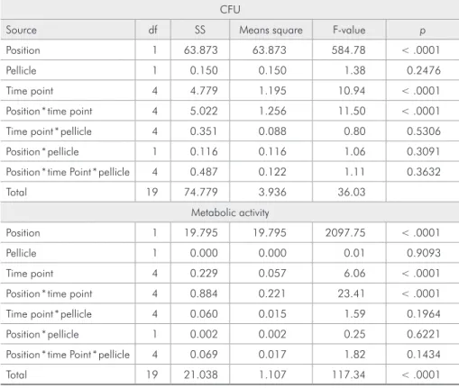
Three-way ANOVA showed statistically signii-cant differences for position, time, and interactions
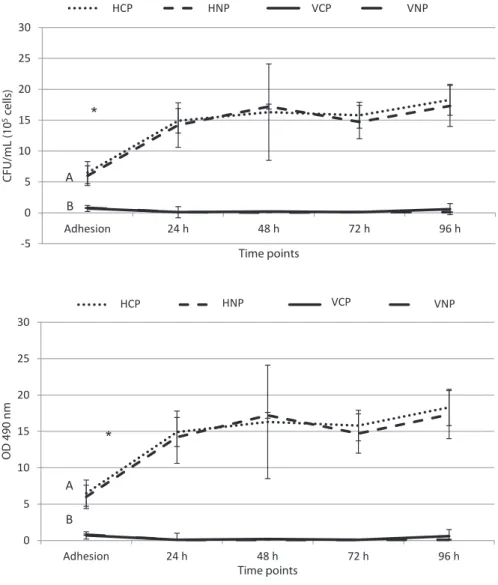
between position and time. No signiicant differ-ences were observed for the pellicle or interactions between pellicle and position, pellicle and time, or their combination (Table 1). The amount of bioilm formed was affected by the substratum position for all time points (p < .0001). Vertical disks showed less bioilm formation than horizontal disks (p < .0001). The adhesion time point differed from the other time points for both groups (p < .0001). However, presence of the AP did not affect bio-ilm formation in the same position (p = .2476) (Figure 1).
The metabolic activity of horizontal groups was higher compared to that of the vertical groups (p < .001). The metabolic activity at the adhesion time point was higher compared to that at all other time points (p < .001) (Figure 2). This variable was not affected by the presence of AP (p = .9093). In accordance with the amount of bioilm formed and the metabolic activity, the SEM images showed more bioilm for the horizontal group. A difference in the amount of bioilm was also observed between the ad-hesion time point and other time points (Figure 3).
CFU
Source df SS Means square F-value p
Position 1 63.873 63.873 584.78 < .0001
Pellicle 1 0.150 0.150 1.38 0.2476
Time point 4 4.779 1.195 10.94 < .0001
Position * time point 4 5.022 1.256 11.50 < .0001
Time point * pellicle 4 0.351 0.088 0.80 0.5306
Position * pellicle 1 0.116 0.116 1.06 0.3091
Position * time Point * pellicle 4 0.487 0.122 1.11 0.3632
Total 19 74.779 3.936 36.03
Metabolic activity
Position 1 19.795 19.795 2097.75 < .0001
Pellicle 1 0.000 0.000 0.01 0.9093
Time point 4 0.229 0.057 6.06 < .0001
Position * time point 4 0.884 0.221 23.41 < .0001
Time point * pellicle 4 0.060 0.015 1.59 0.1964
Position * pellicle 1 0.002 0.002 0.25 0.6221
Position * time Point * pellicle 4 0.069 0.017 1.82 0.1434
Total 19 21.038 1.107 117.34 < .0001
Discussion
Although the substratum position is known to inluence microbial colonization, this variable has not been well characterized. Therefore, the purpose of this in vitro study was to evaluate whether the substratum position and the presence of AP would inluence C. albicans bioilm formation. The re-sults showed that the substratum position inlu-enced the amount of bioilm formation. Although the exposed area of the vertically positioned disks was twice the exposed area of the horizontally po-sitioned disks, the number of viable cells on the vertical disks was less than on the horizontals. This result was supported by the reduced metabolic ac-tivity of the bioilm formed in the vertical position compared to the horizontal position and by the
SEM images.
These results can be explained by physical fac-tors, such as gravity, which may impair cell tion in the vertical position but facilitate the deposi-tion horizontally.19 Previous studies have simulated
microgravity conditions to show that gravity inlu-ences bioilm development, suggesting that grav-ity should be considered as an important factor in bioilm development studies.20,21 Future studies are
needed to determine whether the adhesive mecha-nisms of C. albicans are inluenced by the substra-tum position and whether factors other than gravity are involved in the reduced bioilm formation of the vertical position.
The presence of the AP did not inluence the amount of bioilm formed in either position or the
-5 0 5 10 15 20 25 30
Adhesion 24 h 48 h 72 h 96 h
CFU/m
L (10
5 c
e
ll
s)
Time points
PH NPH VP NPV
*
A
B
HCP HNP VCP VNP
0 5 10 15 20 25 30
Adhesion 24 h 48 h 72 h 96 h
OD
490 n
m
Time points
PH NPH PV NPV
A
B
*
HCP HNP VCP VNP
Figure 1 - CFU (105 cells/mL) for
each time point in the presence or absence of acquired pellicle in the horizontal and vertical positions
(mean ± SD; n = 9). HCP, HNP:
Disks placed horizontally and coated or uncoated by saliva pellicle. VCP, VNP: Disks placed vertically and coated or uncoated by saliva pellicle. Uppercase letters indicate significant differences between horizontal (A) and vertical (B) groups. *Significant differences between adhesion and other time
points. Tukey’s test (p< 0.05).
Figure 2 - Metabolic activity optical density (O.D; 490 nm;
mean ± SD; n = 9). HCP, HNP:
Disks placed horizontally and coated or uncoated by saliva pellicle. VCP, VNP: Disks placed vertically and coated or uncoated by saliva pellicle. Uppercase letters indicate significant differences between horizontal (A) and vertical (B) groups. *Significant differences between adhesion and other time
metabolic activity. It might be hypothesized that the substratum position is a more important factor for bioilm formation than the AP. Previous stud-ies of salivary AP without the inluence of the sub-stratum position have noted signiicant differences, suggesting that saliva is an important modifying factor for surface properties and, consequently, for bioilm development.7,9,15,22 However, divergent
indings regarding the role of the AP on bioilm formation have been reported,3,5,6,10 which may be
due to the use of different methodological proto-cols and saliva collection methods (stimulated or unstimulated), conditions that can change the com-position and viscosity of saliva6,10,13 and inluence
bioilm development.6,13
A previous study8 using stimulated saliva
veri-ied that the presence of an AP increased C. albi-cans adherence to acrylic resin, which is not con-sistent with our results. In studies using whole and unstimulated saliva, no effect was observed on bioilm formation in the presence of an AP.3,7,10
Regardless of whether stimulated or unstimulated saliva is used, the presence of an AP is related to the modiication of substratum properties, such as the surface free energy and surface reactivity, which provides different receptor sites for micro-organisms.5,22 However, in the present study, the
vertical positioning may have overcome the effects
Figure 3 - SEM visualization of biofilm developed on the resin disk surfaces. (A) Disk placed in a horizontal or (B) vertical
posi-tion, at the adhesion time point (1.5 hours). (C) Disk placed in a horizontal position or (D) vertical position, after 72 hours of
of the AP. Further studies are needed to investigate whether other dental materials with different sur-faces properties can produce the same results for C. albicans bioilm formation.
Conclusion
Considering the limitations of this study, it can be concluded that the substratum position
inlu-enced the development of C. albicans bioilm.
References
1. Sereviratne CJ, Jin L, Samaranayake LP. Biofilm lifestyle of Candida: a mini review. Oral Dis. 2008 Oct;14(7):582-90. 2. Luo G, Samaranayake LP. Candida glabrata, an emerging
fungal pathogen, exhibits superior relative cell surface hydro-phobicity and adhesion to denture acrylic surfaces compared with Candida albicans. APMIS. 2002 Sep;110(9):601-10. 3. Zaperini CA, Machado AL, Vergani CE, Pavarina AC,
Giam-paolo ET, Cruz NC. Adherence in vitro of Candida albicans to plasma treated acrylic resin. Effect of plasma parameters, surface roughness and salivary pellicle. Arch Oral Biol. 2010 Oct;55(10):763-70. Epub 2010 Jul 27.
4. Radford DR, Sweet SP, Challacombe SJ, Walter JD. Adherence of Candida albicans to denture-base materials with different surface finishes. J Dent. 1998 Sep;26(7):577-83.
5. Pereira-Cenci T, Del Bel Cury AA, Crielaard W, Ten Cate JM. Development of candida-associated denture stomatitis: new insights. J Appl Oral Sci. 2008 Mar-Apr;16(2):86-94. 6. Pereira-Cenci T, Cury AA, Cenci MS, Rodrigues-Garcia
RC. In vitro Candida colonization on acrylic resins and denture liners: influence of surface free energy, roughness, saliva, and adhering bacteria. Int J Prosthodont. 2007 May-Jun;20(3):308-10.
7. San Millan R, Elguezabal N, Regulez P, Moragues MD, Quin-dos G, Ponton J. Effect of salivary secretory IgA on the adhe-sion of Candida albicans to polystyrene. Microbiology. 2000 Sep;146(Pt 9):2105-12.
8. Vasilas A, Molina L, Hoffman M, Haidaris CG. The influence of morphological variation on Candida albicans adhesion to denture acrylic in vitro. Arch Oral Biol. 1992 Aug;37(8):613-22. 9. Moura JS, Silva WJ, Pereira T, Del Bel Cury AA, Rodrigues
Garcia RCM. Influence of acrylic resin polymerization meth-ods and saliva on the adherence of four Candida species. J Prosthet Dent. 2006 Sep;96(3):205-11.
10. Jin Y, Samaranayake LP, Samaranayake Y, Yip HK. Biofilm formation of Candida albicans is variably affected by saliva and dietary sugars. Arch Oral Biol. 2004 Oct;49(10):789-98. 11. Oliveira VM, Leon BL, Del Bel Cury AA, Consani S. Influ-ence of number and position of flasks in the monomer release,
Knoop hardness and porosity of a microwave-cured acrylic resin. J Oral Rehabil. 2003 Nov;30(11):1104-8.
12. Verran J, Maryan CJ. Retention of Candida albicans on acrylic resin and silicone of different surface topography. J Prosthet Dent. 1997 May;77(5):535-9.
13. Aps JK, Martens LC. Review: the physiology of saliva and transfer of drugs into saliva. Forensic Sci Int. 2005 Jun 10;150(2-3):119-31.
14. Thein ZM, Samaranayake YH, Samaranayake LP. Characteris-tics of dual species Candida biofilms on denture acrylic surfaces. Arch Oral Biol. 2007 Dec;52(12):1200-8. Epub 2007 Aug 6. 15. Nikawa H, Nishimura H, Makihira S, Hamada T, Sadamori
S, Samaranayake LP. Effects of serum concentration on Can-dida biofilm formation on acrylic surfaces. Mycoses. 2000 May;43(3-4):139-43.
16. da Silva WJ, Seneviratne J, Parahitiyawa N, Rosa EA, Sama-ranayake LP, Del Bel Cury AA. Improvement of XTT assay performance for studies involving Candida albicans biofilms. Braz Dent J. 2008;19(4):364-9.
17. Gomes PN, Silva WJ, Pousa CC, Del Bel Cury AA. Bioactiv-ity and cellular structure of Candida albicans and Candida glabrata biofilms grown in the presence of fluconazole. Arch Oral Biol. 2011 Nov;56(11):1274-81. Epub 2011 May 6. 18. Ramage G, Tomsett K, Wickes BL, Lopez-Ribot JL, Redding
SW. Denture stomatitis: a role for Candida biofilms. Oral Surg Oral Med Oral Pathol Oral Radiol Endod. 2004 Jul;98(1):53-9. 19. Soll DR. Candida commensalism and virulence: the evolution
of phenotypic plasticity. Acta Trop. 2002 Feb;81(2):101-10. 20. McLean RJ, Cassanto JM, Barnes MB, Koo JH. Bacterial
biofilm formation under microgravity conditions. FEMS Mi-crobiol Lett. 2001 Feb 20;195(2):115-9.
21. Lynch SV, Mukundakrishnan K, Benoit MR, Ayyaswamy PS, Matin A. Escherichia coli biofilms formed under low-shear modeled microgravity in a ground-based system. Appl Envi-ron Microbiol. 2006 Dec;72(12):7701-10. Epub 2006 Oct 6. 22. Sipahi C, Anil N, Bayramli E. The effect of acquired salivary