Agda Maria de Moura Marcia André
Margareth Torrecillas Lopez Reinaldo Brito e Dias
Departmentof Maxillofacial Surgery, Prosthesis and Traumatology, Faculdade de Odontologia, Univ de São Paulo - USP, São Paulo, SP, Brazil.
Corresponding Author: Agda Maria de Moura E-mail: [email protected]
Prevalence of caries in Brazilian
children with cleft lip and/or palate,
aged 6 to 36 months
Abstract: The aim of this study was to assess the prevalence of caries in children with cleft lip and/or palate aged 6–36 months of life. This study was conducted at the University of São Paulo School of Dentistry, Brazil. A total of 143 children were selected (73 male, 70 female), all of whom had at least two erupted teeth. The children were distributed as follows: 88 had cleft lip and palate, 35 had cleft palate, 20 had cleft lip. Den-tal caries disease was diagnosed according to criteria set by the World Health Organization. It was observed that 18.9% of the study children had caries. No correlation between prevalence of caries and gender or type of cleft was observed. Mean dmf-t was 0.47. The prevalence of car-ies was higher in the upper arch, and the most affected tooth was the central incisor (p = 0.001). Children with cleft lip and/or palate did not have high caries indices.
Descriptors: Dental Caries; Prevalence; Cleft Lip; Cleft Palate; Infant.
Introduction
Cleft lip and/or palate are among the most prevalent congenital mal-formations in humans, and are inluenced by complex genetic and envi-ronmental factors, with relative recurrence.1 The international literature reports epidemiological indices of cleft lip and/or palate ranging from 0.87 to 1.03 per 1000 births.2 These indices are similar to those reported in studies carried out in Brazil.3
Although not a major cause of mortality in developed countries, cleft lip and/or palate (CL/P) does cause considerable morbidity among af-fected children and imposes a substantial inancial risk for families with a concomitant societal burden.4
CL/P is correlated with esthetic, functional and psychological prob-lems, and calls for early preventive interventions involving a multidisci-plinary team.5
The parents of cleft patients have higher expectations regarding the effectiveness of surgical treatment. They fail to realize the close connec-tion between oral health or speech rehabilitaconnec-tion and correcconnec-tion of the malocclusion. Therefore, it is dificult to make them understand that early caries and dental loss can affect the success of surgical treatment, orthodontic treatment and speech therapy.5
Studies have reported that children with cleft lip and/or palate are at a high risk of dental caries. Zhu et al.6, Al-Wahadni et al.7, and Stec-Slonic Declaration of Interests: The authors
certify that they have no commercial or associative interest that represents a conflict of interest in connection with the manuscript.
Submitted: Aug 28, 2012
et al.8 observed that patients with a cleft condition, not only are at a high risk of caries, but also have an even higher prevalence of caries than individuals without cleft.
The classic study of Lauterstein and Men-delsohn,9 as well as the studies conducted by Lucas
et al.10, King et al.11 and Tannure et al.12, found no signiicant differences in caries between children with and without cleft.
Regarding the type of cleft, Ankola et al.13 ob-served that children with cleft lip were more sus-ceptible to tooth decay than those with cleft palate. When comparing the cleft lip only or cleft palate only with the cleft lip and palate, the authors no-ticed that the latter showed a higher prevalence of caries. Zhu et al.6 found a higher prevalence of car-ies in cleft lip with or without palate. Byan et al.5 observed a higher percentage of caries in bilateral, as compared to unilateral, cleft lip and palate, and a higher prevalence of rampant caries in cleft palate. The prevalence of caries seems to be distributed evenly between the genders of children with cleft. This inding was observed in other studies (Byan et al.5; Neves et al.14).
There is relatively little information on the ex-perience of dental caries in preschool children with cleft, and the literature15 indicates that children usu-ally show a lower index of caries at a younger age rather than at an older age.
The purpose of the present study was to assess the prevalence of dental caries in children 6 to 36 months old, with cleft lip and/or palate, and to cor-relate the prevalence of caries with the cleft type, gender and age of the patients.
Methodology
This study was carried out by performing a trans-versal study at the University of São Paulo School of Dentistry. A project was evaluated and approved by the Ethics Committee at this university prior to con-ducting the study. The appropriate written consent was obtained from the children’s parents/guardians. The sample size was 143 children with cleft lip and/ or palate, who had been seen at the University of São Paulo School of Dentistry outpatient clinic since their irst weeks of life.
Inclusion criteria: • cleft children,
• without multiple abnormalities, • aged 6–36 months,
• with at least two erupted teeth, and
• with access to an optimally luoridated public water supply.
The children with cleft lip and/or palate usually go to the clinic for cleft and not caries treatment. In addition to receiving guidance in regard to per-sonal diet, children (or the parents/guardians of very young children) are also given speciic oral hygiene education to improve their hygiene as of their irst days of life. A reliable control group with the same proile and the same environmental factors could not be established.
The children were divided into ive age groups: • 6–12 months,
• 13–18 months, • 19–24 months, • 25–30 months and • 31–36 months.
According to the Spina Classiication,16 the cleft was divided into three types:
• cleft lip (CL), including cleft lip with or without cleft alveolus;
• cleft palate (CP), including cleft soft palate with or without cleft hard palate; and,
• cleft lip and palate (CLP).
Intraoral examination was performed by the same examiner (Kappa 1.0), following a systematic pattern: the quadrants of the mouth were examined clockwise, beginning at the most posterior upper right tooth.
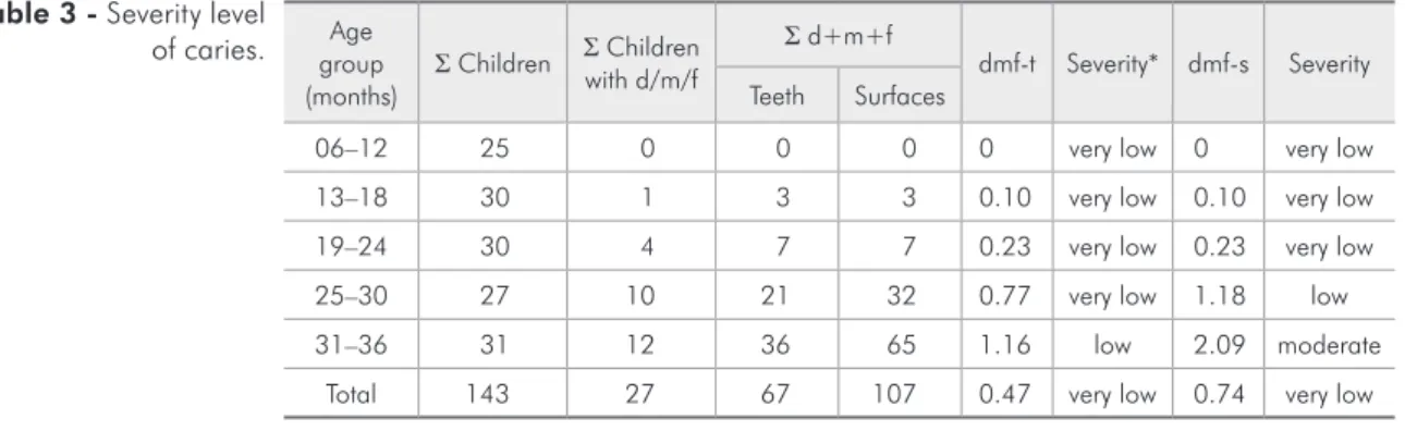
between these two variables (p < 0.001) (Table 2). The mean dmf-t was 0.47, with very low severity according to FDI18 (Table 3). Of the teeth examined, 62 were decayed and 5 were illed; no teeth had been extracted. Caries disease was found to be more prevalent in the upper as opposed to the lower arch. In the upper arch, caries disease was signiicantly more prevalent in the central incisors (p = 0.044), whereas, in the lower arch, caries disease was more prevalent in the irst molars (p < 0.001).
Discussion
The low index of caries found in this study is a signiicant result in terms of the dental health of children with cleft. This evidence demonstrates that when children with cleft lip and/or palate par-ticipate in prevention programs and receive luoride supplementation, they can achieve lower indices of The data obtained were analyzed statistically
us-ing the chi-squared test, adjusted by Fisher’s statis-tical method, in order to establish the relationship among caries prevalence and gender, age and cleft type. The difference was considered statistically sig-niicant when the p value was less than 0.05.
Results
Table 1 shows the distribution of the study popu-lation, comprised of 143 cleft patients. The patients were classiied according to gender, age and cleft type.
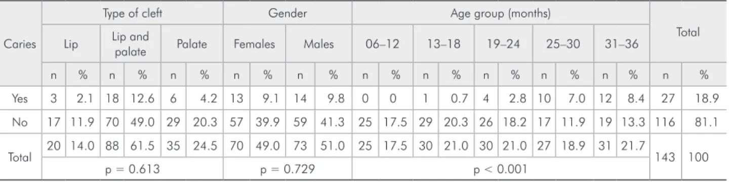
The study observed that 18.9% of the children had caries. The prevalence of caries was similar in both males and females (Table 1) and did not corre-late with the type of cleft (p = 0.613) (Table 2).
The number of children with caries increased with age, and signiicant correlation was observed
Table 1 - Sample distribution according to cleft type, gender and age, expressed in absolute numbers and percentages.
Age group (months)
Cleft type
Total
Cleft lip Cleft lip and palate Cleft palate
Females Males Females Males Females Males
n % n % n % n % n % n % n %
06–12 3 2.1 2 1.4 9 6.3 6 4.2 2 1.4 3 2.1 25 17.5
13–18 1 0.7 2 1.4 8 5.6 10 7.0 5 3.5 4 2.8 30 21.0
19–24 3 2.1 2 1.4 9 6.3 9 6.3 1 0.7 6 4.2 30 21.0
25–30 1 0.7 1 0.7 8 5.6 11 7.7 6 4.2 0 0.0 27 18.9
31–36 2 1.4 3 2.1 7 4.9 11 7.7 5 3.5 3 2.1 31 21.7
Total 10 7.0 10 7.0 41 28.7 47 32.9 19 13.3 16 11.2 143 100
20 (14.0%) 88 (61.6%) 35 (24.5%)
Table 2 - Correlation between dental caries and cleft type, gender and age group expressed in absolute numbers and percent-ages.
Caries
Type of cleft Gender Age group (months)
Total Lip Lip and palate Palate Females Males 06–12 13–18 19–24 25–30 31–36
n % n % n % n % n % n % n % n % n % n % n %
Yes 3 2.1 18 12.6 6 4.2 13 9.1 14 9.8 0 0 1 0.7 4 2.8 10 7.0 12 8.4 27 18.9
No 17 11.9 70 49.0 29 20.3 57 39.9 59 41.3 25 17.5 29 20.3 26 18.2 17 11.9 19 13.3 116 81.1
Total 20 14.0 88 61.5 35 24.5 70 49.0 73 51.0 25 17.5 30 21.0 30 21.0 27 18.9 31 21.7 143 100
caries than the general population, despite the high risk of caries promoted by a cleft disorder.
Cleft children require adjustments to be made in diet and breastfeeding. Parents/guardians are counseled to give cleft children milk, juice and foods in general more frequently. This may lead to the accumulation of food debris. However, this fre-quency is essential to provide the nourishment and weight gain that are central to the development of the child.5 After reconstructive surgery, the remain-ing tissue, scar retraction and persistent istulas, as well as dental anomalies of number, position, shape and structure, are risk factors for caries.19 They may pose an obstacle to ensuring effective oral hy-giene,19,20 usually overlooked because of parental anxiety.
Dental caries disease is currently deined as a biosocial disease, the prevention of which is not limited to speciic measures. The parents of cleft lip and/or palate children seen at the clinic of the De-partment of Maxillofacial Surgery, Prosthesis and Traumatology of the University of São Paulo School of Dentistry as of the irst weeks of life are instruct-ed on the importance of oral health and how to maintain it. Special focus is given to the risk factors for caries in children with cleft lip and/or palate. In addition, the parents are instructed on how to avoid other risk factors, such as inadequate diet and poor oral hygiene.
Wong and King15 reported that there are few studies investigating oral care in cleft children, and also found various inconsistencies in these stud-ies. They concluded that the prevalence of caries
may be higher in cleft individuals than in the gen-eral population. The authors suggested that the care given to these children should follow a protocol in which priority is given to oral care prior to surgical procedures.
Based on the caries risk proposed by Fejerskov,21 it could be inferred that children with cleft lip and/ or palate are at a high risk for caries. However, only 18.9% of the children investigated in the present study had caries (Table 2), which was a surprisingly positive inding.
Clinically, it was observed that the cleft children of this research, seen at the clinic of the Depart-ment of Maxillofacial Surgery, Prosthesis and Trau-matology of the University of São Paulo School of Dentistry, had a low prevalence of caries, and lower indices than those found in the general population. In the present study, the mean dmf-t was 0.47. Our indings are not in accordance with another study carried out in Brazil,14 which reported higher mean
dmf-t indices. According to Bönecker et al.22, the mean caries indices for the general population have decreased in the last decade. Therefore, the indings of the present report may be explained by this gen-eral reduction in the caries indices.
A study investigating the initial acquisition of
S. mutans reported that it occurs at approximately 26 months of age,23 whereas another study report-ed that initial S. mutans colonization may occur as early as 14 months of age if there is greater sucrose consumption.24 Bokhout et al.25 reported a case of
S. mutans colonization in an 18-month-old child. In the present study, a 16-month-old child show-Age
group
(months) Σ Children
Σ Children with d/m/f
Σ d+m+f
dmf-t Severity* dmf-s Severity Teeth Surfaces
06–12 25 0 0 0 0 very low 0 very low
13–18 30 1 3 3 0.10 very low 0.10 very low
19–24 30 4 7 7 0.23 very low 0.23 very low
25–30 27 10 21 32 0.77 very low 1.18 low
31–36 31 12 36 65 1.16 low 2.09 moderate
Total 143 27 67 107 0.47 very low 0.74 very low
*FDI.
ing early S. mutans colonization had three decayed teeth. However, even if cleft children are colonized at the same age as noncleft children, they may have a lower prevalence of caries because tooth eruption in cleft individuals is delayed, especially at the af-fected site.19,26 It is likely that teeth with delayed eruption are less exposed to risk factors, and that cleft children have a lower chance of developing caries at this age. It is noteworthy to mention that several one-year-old children were not included in our study because they did not have any erupted teeth.
Some authors have reported that S. mutans is vertically transmitted from mother to child.25,27 Therefore, it has been suggested that prevention programs should include the education of family members, focusing especially on the habits of the mother.28
The analysis of the results for caries develop-ment showed that the prevalence of caries increased with age. The literature is unanimous in reporting the correlation between the prevalence of caries and age in both cleft5,8,13 and noncleft children.22 Our study population was divided into ive age groups, namely 06–12 months, 13–18 months, 19–24 months, 25–30 months and 31–36 months.
The number and percentage of children in each of the aforementioned groups who developed caries were 0 (0.0%), 1 (0.7%), 4 (2.8%), 10 (7.0%) and 12 (8.4%), respectively.
Caries was observed in 27 children (Table 2). In the present study, the prevalence of caries was similar between males and females (Table 2), which is in accordance with the indings of other stud-ies.5,14 Studies carried out in Brazil involving non-cleft individuals have also reported similar preva-lence of caries among males and females.22
Assessing the prevalence of caries according to cleft type, a greater number of children with cleft lip and palate developed caries. This could be due to a higher prevalence of this type of cleft. However, statistical analysis showed no signiicant correlation between caries prevalence and cleft type (Table 2), which corroborates the results of other studies.13
With regard to the distribution of caries, a high-er prevalence was obshigh-erved in the upphigh-er central in-cisors, followed by the upper lateral incisors and the lower irst molars. This is in accordance with the indings of Neves et al.14, who reported a higher prevalence of caries in the left upper central inci-sors. It is known that left unilateral cleft is the most prevalent type of cleft, and that the teeth adjacent to the cleft are the most susceptible to caries, since they are more affected by dental anomalies of po-sition and shape. Studies investigating children in the general population have also reported a higher prevalence of caries in the upper central incisors and lower irst molars.22
The low caries indices found in the present study could be attributed to the fact that the children we assessed have been undergoing treatment (for the cleft, not for caries) at the clinic of the Department of Maxillofacial Surgery, Prosthesis and Traumatol-ogy of the University of São Paulo School of Den-tistry, as of their irst weeks of life.
The optimal level of luoride in the water of São Paulo (0.7 mg/L), as well as parental education re-garding breastfeeding, diet, oral hygiene and para-functional habits, may have contributed to the low indices of caries that we found. Parental education on cleft-related issues has been given at the clinic of the Department of Maxillofacial Surgery, Pros-thesis and Traumatology of the University of São Paulo School of Dentistry in recent years. Monthly appointments are scheduled until the child is two years old. The results of this educational effort, in-volving the families of children with cleft lip and/or palate, provide further evidence that prevention is central to the oral health and appropriate develop-ment of cleft children.
Conclusion
References
1. Dixon MJ, Marazita ML, Beaty TH, Murray JC. Cleft lip and palate: understanding genetic and environmental influences. Nat Rev Genet. 2011 Mar;12(3):167-78.
2. Derijcke A, Eerans A, Carels C. The incidence of oral clefts: a review. Br J Oral Maxillofac Surg. 1996 Dec;34(6):488-94. 3. França CMC, Locks A. Incidência das fissuras lábio-palatinas
de crianças nascidas na cidade de Joinville (SC) no período de 1994 a 2000. J Bras Ortodon Ortop Facial. 2003 Sep-Oct;8(47):429-36.
4. Wehby G, Cassell CH. The impact of orofacial clefts on quality of life and healthcare use and costs. Oral Dis. 2010 Jan;16(1):3-10.
5. Byan Z, Du M, Bedi R, Holt R, Jin H, Fan M. Caries preva-lence and oral health behavior in Chinese children with cleft lip and/or palate. Pediatr Dent. 2001 Sep-Oct;23(5):431-4. 6. Zhu WC, Xiao J, Liu Y, Wu J, Li JY. Caries experience in
individuals with cleft lip and/or palate in China. Cleft Palate Craniofac J. 2010 Jan;47(1):43-7.
7. Al-Wahadni A, Alhaia EA, Al-Omari MA. Oral disease status of a sample of Jordanain people ages 10 to 28 with cleft lip and/or palate. Cleft Palate Craniofac J. 2005 May;42(2):304-8. 8. Stec-Slonic M, Szczepanska J, Hirschfelder U. Comparison of
caries prevalence in two populations of cleft patients. Cleft Palate Craniofac J. 2007 Sep;44(5):532-7.
9. Lauterstein AM, Mendelsohn M. An analysis of the car-ies experience of 285 cleft palate children. Cleft Palate J. 1964 Jul;1(29):314-9.
10. Lucas VS, Gupta R, Ololade O, Gelbier M, Roberts GJ. Dental health indices and caries associated microflora in children with unilateral cleft lip and palate. Cleft Palate Craniofac J. 2000 Sep;37(5):447-52.
11. King NM, Wong WL, Wong HM. Caries experience of chi-nese children with cleft lip and palate. 2012 Mar 1. Epub ahead of print.
12. Tannure PN, Costa MDE, Küchler EC, Romanos IC, Gran-jeiro JM, Vieira AR. Caries experience in individual with cleft lip and palate. Pediatr Dent. 2012 Mar-Apr;34(2):127-31. 13. Ankola AV, Nagesh L, Hegde P, Karibasappa GN. Primary
den-tition status and treatment needs of children with cleft lip and/ or palate. J Indian Soc Pedod Prev Dent. 2005 Jun;23(2):80-2. 14. Neves LT, Gomide MR, Costa B, Ciamponi AL. Compor-tamento da doença cárie de portadores de fissuras lábio pa-latinas entre 7 e 66 meses. Pesqui Odontol Bras. 2002 Ago-Sep;16(supl):185-7.
15. Wong FWL, King NM. The oral health of children with clefts – a review. Cleft Palate Craniofac J. 1998 May;35(3):248-54.
16. Spina V, Psillakis JM, Lapa FS, Ferreira MC. Classificação das fissuras lábio-palatinas. Sugestão de modificação. Rev Hosp Clin Fac Med São Paulo. 1972;27(1):5-6.
17. World Health Organization. Oral health surveys. Basic meth-ods. 4th ed. Geneva: World Health Organization; 1997. 18. Global goals for oral health by the year 2000. Fédération
Dentaire Internationale. Int Dent J. 1982 Mar;32(74):74-7. 19. Cheng LL, Moor SL, Ho CTC. Predisposing factors to
den-tal caries in children with cleft lip and palate: a review and strategies for early prevention. Cleft Palate Craniofac J. 2007 Jan;44(1):67-78.
20. Lai MC, King NM, Wong HM. Abnormalities of maxillary anterior teeth in Chinese children with cleft lip and palate. Cleft Palate Craniofac J. 2009 Jan;46(1):58-64.
21. Fejerskov O. Changing paradigms in concepts on dental caries: consequences for oral health care. Caries Res. 2004 May-Jun;38(3):182-91.
22. Bönecker M, Ardenghi TM, Oliveira LB, Sheiham A, Mar-cenes W. Trends dental caries in 1- to 4-yeard-old children in a Brazilian city between 1997 and 2008. Int J Paediatr Dent. 2010 Mar;20(2):125-31.
23. Caufield PW, Cutter GR, Dasanayake AP. Initial acquisition of mutans streptococci by infants: evidence for a discrete window of infectivity. J Dent Res. 1993 Jan;72(1):37-45.
24. Mohan A, Morse DE, O’Sullivan DN, Tinanoff N. The re-lationship between bottle usage/content, age, and number of teeth with mutans streptococci colonization in 6-24-month-old children. Community Dent Oral Epidemiol. 1998 Feb;26(1):12-20.
25. Bokhout B, Van Loveren C, Hofman FX, Buijs F, Van Lim-beek J. Prevalence of streptococcus mutans and lactobacilli in 18-month-old children with cleft lip and/or palate. Cleft Palate Craniofac J. 1996 Sep;33(5):424-8.
26. Duque C, Dalben GS, Aranha AMF, Carrara CFC, Gomide M, Costa B. Chronology of deciduous teeth eruption in chil-dren with cleft lip and palate. Cleft Palate Craniofac J. 2004 May;41(3):285-9.
27. Bönecker MJS, Ardenghi TM, Trindade CP, Cury P. Trans-missão vertical de streptococcus mutans e suas implicações. JBP Rev Ibero Am Odontopediatr Odontol Bebê. 2004 May-Jun;7(37):297-303.