Fabricio Batistin Zanatta(a) Bruno Rodrigues Giacomelli(a) Patricia Pasquali Dotto(a) Vânia Regina Camargo Fontanella(b)
Cassiano Kuchenbecker Rösing(c)
(a)Centro Universitário Franciscano (UNIFRA), Santa Maria, RS, Brazil.
(b)Universidade Luterana do Brasil (ULBRA), Canoas, RS, Brazil.
(c)Universidade Federal do Rio Grande do Sul (UFRGS), Porto Alegre, RS, Brazil.
Corresponding author: Fabricio Batistin Zanatta
R. Tiradentes, 76, apto. 801 - Centro Santa Maria - RS - Brazil
CEP: 97050-730
E-mail: [email protected]
Received for publication on Jun 22, 2010 Accepted for publication on Sep 20, 2010
Comparison of different methods
involved in the planning of clinical crown
lengthening surgery
Abstract: There is little material in the literature that compares biologi-cal width measurements in periapibiologi-cal and bite-wings radiographs with clinical measurements. The purpose of this study was to compare mea-surements of biological width taken by three different methods which are frequently used for planning periodontal surgery – periapical radio-graph, bite-wing radiograph and transperiodontal probing – with the trans-surgical measurements. Thirty-four sites from twenty-one subjects were analyzed. The intra-class correlation coeficients between measure-ments obtained trans-surgically (gold standard) and those obtained by transperiodontal probing, periapical radiography and bite-wing radiog-raphy were determined. Average measurements were compared using the Wilcoxon test at a signiicance level of 0.05. Also, the frequency distri-bution of differences between test measurements and the gold standard was calculated. The results showed that transperiodontal probing (mean 2.05 mm) was the most accurate measurement, as compared to the gold standard (mean 1.97 mm), with no statistically signiicant difference ob-served. On the other hand, periapical and bite-wing radiographic mean values (1.56 mm and 1.72 mm, respectively) were smaller than the gold standard, with statistically signiicant differences (p < 0.05). It was con-cluded that transperiodontal probing was the most accurate measure-ment, as compared to the gold standard, followed by that obtained with the bite-wing radiograph. The clinical relevance of these results could be that planning for crown lengthening surgery should, preferably, include transperiodontal probing.
Descriptors: Periodontics; Planning Techniques; Crown Lengthening; Surgery, Oral; Radiology.
Introduction
When planning clinical crown lengthening (CCL) surgeries, it is nec-essary to observe if the periodontal biological width has been infringed. This width comprises the distance between the bottom of the histological gingival sulcus and the alveolar bone crest, disregarding the histological epithelium, since it can be included in prosthetic and restorative proce-dures.1
A classic study by Gargiullo et al.2 measured these distances in
epithe-lium (0.97 mm), and connective tissue attachment (1.07 mm), which, in total, comprise 2.73 mm. Ad-ditional evidence, obtained with similar methodolo-gy, conirmed these indings with small variations.3-6
However, it has been understood that these values involved a high degree of variation among subjects.4
It has been argued that disturbance of biologi-cal width by restorations may result in chronic in-lammation, pocket installation and, eventually, in attachment loss and bone resorption.7-9 Therefore,
CCL has been indicated in order both to prevent the inlammatory reaction and to allow for correct re-storative/prosthetic technical procedures.
Taking into consideration the possible problems linked to the disturbance of biological width, it is important that an accurate diagnostic method be applied. According to Reed and Polson,11 bite-wing
radiographs are probably the best for establishing an accurate radiographic scenario of alveolar bone height. On the other hand, parallel periapical ra-diographs seem better for determining the extent of the level of all the periodontal bone. Some studies suggested that radiographs tend not to be accurate, and that different radiographic techniques should be studied for their accuracy.12-14 Additionally, a
clini-cal method has been proposed: transperiodontal probing, which could avoid radiation exposure and provide more precision.
The clinical applicability of this information is twofold:
1. it indicates when biological width is infringed, and
2. it allows more precise planning of the surgical procedure, i.e., if there is need or not to assess the alveolar bone (in order to perform osteotomy). Therefore, the aim of this study was to compare three different methods for diagnosing the invasion of periodontal biological width – periapical radiog-raphy, bite-wing radiography and transperiodon-tal probing – with the real surgical measurements. The hypothesis to be tested is that transperiodontal probing is at least as accurate as the radiographic measurements, compared to surgical measurement.
Materials and Methods
The sample for the present study comprised
twenty-one eligible participants, from a total of thirty-six initially screened subjects, ranging be-tween 21 and 60 years of age (mean age 41 ± 10.68) and referred for CCL surgery at the Dental School of the Franciscan University Center, in Brazil. The research protocol was approved by the Ethical Com-mittee of the Franciscan University Center (CONEP: Number 1246 / CEP/UNIFRA: Number: 078.2008-2).
Inclusion criteria: The participating subjects should be over 18 years, have need of CCL in an interproximal site with probable osteotomy, in order to allow adequate restorative rehabilitation.
Exclusion criteria: The subjects with general-ized periodontal inlammation, those who exhibited a systemic condition that contraindicated the surgi-cal procedure, and individuals who did not agree to sign the consent form were excluded from the study.
After inclusion, individuals underwent CCL sur-gery. Following the removal of all carious tissue (if necessary), in order to determine the real terminus of sound dental tissue, two radiographs were taken for each area involved: 1 (one) periapical radiograph was taken using the bisecting-angle technique, and 1 (one) bite-wing radiograph.
All radiographs were taken by the same exam-iner, using radiographic indicators (Indusbello, Londrina, Paraná, Brazil) and an X-ray machine (Dabi Atlante Spectro 70X, Ribeirão Preto, São Paulo, Brazil), with exposure time of 0.4 s in both upper and lower jaws. Periapical type ilms (Kodak Insigth, Manaus, Amazonas, Brazil) were used, and processing was accomplished manually, employing unused chemical substances and following the time-temperature table in order to standardize the radio-graphs for the measurements.
obtained.
A modiied Widman lap was elevated, soft tis-sue (gingiva and granulation) was removed, the root surfaces were planed and the wound was rinsed with saline. The trans-surgical measurement of the distance from the most apical portion of the proxi-mal cavity/defect to the bone crest was then per-formed. Next, the routine surgery was carried out with osteotomy according to the necessity, sutured, and concluded with post-operative instructions.
Radiographic measurements
Each periapical and bite-wing radiograph was analyzed in an X-ray illuminator covered by a black paper. This was used in order to concentrate all the light of the device onto the radiograph to be analyzed. Using a digital caliper (Jomarca, Stain-less Hardened, Nieuw, Vennep, Netherlands) the measurement from the most apical portion of the proximal defects/cavities to the bone crest was made using the periapical and bite-wing radiographs. All clinical and radiographic measurements were made by the same examiner.
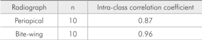
Pre-experimental training and calibration Prior to making the radiographic measurements, the examiner attended a training session given by an oral radiologist for the measurement of the peri-apical and bite-wing radiographs. Calibration was subsequently carried out using 10 periapical and 10 bite-wing radiographs. Finally, agreement between the measurements of the same radiographs was ob-tained at two different time-points (2-week interval) and compared using the intraclass correlation coef-icient (Table 1).
Regarding the trans-surgical and transperiodon-tal measurements, one trained examiner made the measurements; however, calibration igures were calculated.
Statistical Analysis
Mean values of radiographic measurements, taken from periapical and bite-wing radiographs, as well as the measurements obtained by transperi-odontal probing, were obtained and compared with those of the trans-surgical measurements (gold stan-dard) using the Wilcoxon signed test at a .05 level of signiicance. The intraclass correlation coeficient among the measurements obtained with the three test methods and the gold standard was also calcu-lated.
Results
The mean age of the 21 patients included in the study was 41 years-old (± 10.68). Table 2 shows the distribution of teeth and surfaces examined in the present study. It can be observed that the 34 teeth evaluated comprised 32 molars and 2 premolars. Regarding tooth surfaces, the sample comprised 19 distal and 15 mesial surfaces.
The results of the comparison among the mean values of the measurements obtained by transperi-odontal probing, and from periapical and bite-wing radiographs in relation to the trans-surgical mea-surements (gold standard) are shown in Table 3. Also, correlations of each method, compared to the gold standard, are demonstrated. It was observed that the mean trans-surgical measurement (gold standard) was 1.97 mm. Statistically signiicant dif-ferences were observed when all measurements were compared to the gold standard. The mean closest to this measurement was the transperiodontal assess-ment (2.05 mm). When the intraclass correlation coeficient was calculated, transperiodontal mea-surement was the most accurate as compared to the trans-surgical assessment, followed by the bite-wing and periapical measurements, respectively.
Table 1 - Intra-examiner calibration to the radiograph mea-sures.
Radiograph n Intra-class correlation coefficient
Periapical 10 0.87
Bite-wing 10 0.96
Table 2 - Absolute and percentage distribution of the teeth and sites evaluated.
Teeth
Total (%) Molar Pre-molar
Sites
Distal 19 0 19 (56%)
Mesial 13 2 15 (44%)
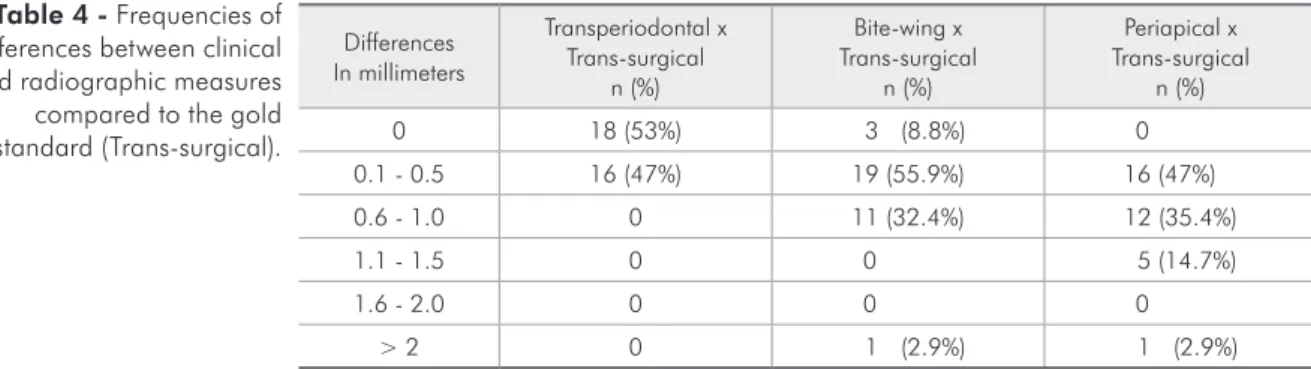
Table 4 shows the means and frequencies of the differences found between each method and the gold standard. It can be observed that there were no differences between the mean transperiodontal mea-surements and the gold standard in 53% of the sites, and that the difference ranged between 0.1 mm and 0.5 mm in 47% of the sites. On the other hand, there were no differences between the bite-wing and gold standard measurements in only 8.8% of the sites, while 55.9% differed by at most 0.5 mm, 32.4% by at most 1 mm, and 2.9% by more than 2 mm. Finally, none of the periapical measurements coincided with the gold standard, and differences of up to 0.5 mm, 1 mm, 1.5 mm, and of more than 2 mm were observed in 47%, 35.4%, 14.7% and 2% of sites, respectively.
Discussion
The present study clearly demonstrated that the diagnostic measurement closest to the real measure-ment (gold standard) was the transperiodontal prob-ing. This approach exhibited the highest frequency of measurements coincident with the gold standard.
Furthermore, when the measurements differed, these differences were not greater than .5 mm and the intra-class correlation coeficient was 0.92, very close to a perfect agreement. Transperiodontal prob-ing has not been a widely used method, especially taking into consideration that it requires local an-esthesia. However, once the necessity for surgery is determined, the dentist can take advantage of either the previous anesthesia, for removing the carious tissue (when necessary); or even, the anesthesia ad-ministered prior to the surgery. This would certainly allow for better planning of the surgical procedure. Festugatto et al.13 presented results similar to the
ones found in this study. Their results indicated greater accuracy for probing as compared to the ra-diographs. However, the periapical radiographs in the study by Festugatto et al.13 were periapical with
parallelism. It should be noted that this radiograph is not widely used in clinical practice, and the pres-ent study analyzed radiographic approaches which are more frequently used in clinical practice. Other studies, with different objectives, also compared clinical to radiographic situations and have reported
Differences In millimeters
Transperiodontal x Trans-surgical
n (%)
Bite-wing x Trans-surgical
n (%)
Periapical x Trans-surgical
n (%)
0 18 (53%) 3 (8.8%) 0
0.1 - 0.5 16 (47%) 19 (55.9%) 16 (47%)
0.6 - 1.0 0 11 (32.4%) 12 (35.4%)
1.1 - 1.5 0 0 5 (14.7%)
1.6 - 2.0 0 0 0
> 2 0 1 (2.9%) 1 (2.9%)
Table 4 - Frequencies of differences between clinical and radiographic measures compared to the gold standard (Trans-surgical).
Methods Mean ± SD
CI 95% of mean (lower – upper)
Median
(P25-P75) A x D B x D C x D
Transperiodontal (A) 2.05 ± 0.94X* (1.73 - 2.38)
2.00 (1.68 - 2.5)
0.92# 0.61 0.84 Periapical (B) 1.56 ± 0.90
T* (1.18 - 1.86)
1.62 (0.89 - 2.32)
Bite-wing (C) 1.72 ± 0.85W* (1.32 - 2.00)
1.85 (1.12 - 2.24)
Trans-surgical (D) 1.97 ± 0.96 Y* (1.63 - 2.30)
2.00 (1.37 - 2.50)
*Different letters show statistically significant differences by the Wilcoxon signed test (p < 0.05). #Intra-class correlation coefficient between clinical and radiographic measurements compared to gold standard.
observations that the radiograph normally underes-timates bone loss as compared to the clinical situa-tion.15-20
On the other hand, in cases where proximal cavi-ties in adjacent teeth are present, transperiodontal probing could be dificult to perform. In this way, radiographs may help with diagnosis and planning. However, radiographs themselves have the limita-tion of two-dimensional representalimita-tion of a three-dimensional structure, which can distort the real measurements to be analyzed. In this study, the means of both types of radiographic measurements showed statistically signiicant differences when compared to the gold standard by underestimating it. From a clinical point of view, this means that the biological width of the periodontium would be shown as being invaded by the restoration and CCL, with osteotomy frequently indicated; yet in fact, the surgical procedure either would not be needed, or at least could be limited to soft tissues. Nevertheless, the bite-wing radiographic measurement was far closer to the gold standard than the periapical ra-diographic measurement. This was detected by the comparison of means, frequency distribution of dif-ferences and in the intraclass correlation coeficient. These indings were corroborated by Pimentel et al.14 who compared bite-wing and periapical
radio-graphic measurements to trans-surgical measure-ments, and found the highest distortions for the periapical method. This fact could be explained due to the use of the bisecting-angle technique, where the bone crest is projected in the oclusal/incisal di-rection, resulting in an overestimation of the inva-sion of biological width. Other studies, that com-pared bite-wing and periapical radiographs, also reported similar results.12, 20, 22
Conclusions
Taking into consideration the design of the pres-ent study, it can be concluded that none of the meth-ods employed reproduce the trans-surgical measure exactly. However, transperiodontal probing exhib-ited the best results, followed by the bite-wing and periapical radiographs, respectively. Therefore, for diagnosis of the invasion of biological width and surgical planning, transperiodontal probing is rec-ommended.
Acknowledgements
The authors wish to thank the volunteers who participated in this research. Drs. Zanatta, Dotto, Giacomelli, Fontanella and Rösing report no con-licts of interest related to this study.
References
1. Bragger U, Lauchenauer D, Lang NP. Surgical lengthening of the clinical crown. J Clin Periodontol. 1992 Jan;19(1):58-63. 2. Gargiulo AW, Wentz FM, Orban B. Mitotic activity of human oral epithelium exposed to 30 per cent hydrogen peroxide. Oral Surg Oral Med Oral Pathol. 1961 Apr;14(4):474-92. 3. Ingber JS, Rose LF, Coslet JG. The “biologic width”--a concept
in periodontics and restorative dentistry. Alpha Omegan. 1977 Dec;70(3):62-5.
4. Tarnow DP, Magner AW, Fletcher P.The effect of the distance from the contact point to the crest of bone on the presence or absence of the interproximal dental papilla. J Periodontol. 1992 Dec;63(12):995-6.
5. Tristão GC. Espaço biológico: Estudo histométrico em perio-dontia clinicamente normal em humanos [tese]. São Paulo: Universidade de São Paulo, Faculdade de Odontologia; 1992. 57 p.
6. Vacek JS, Gher ME, Assad DA, Richardson AC, Giambarresi LI. The dimensions of the human dentogingival junction. Int J Periodontics Restorative Dent. 1994 Apr;14(2):154-65. 7. Valderhaug J, Birkeland JM. Periodontal conditions in patients
5 years following insertion of fixed prostheses. Pocket depth and loss of attachment. J Oral Rehabil. 1976 Jul;3(3):237-43. 8. Schätzle M, Land NP, Anerud A, Boysen H, Bürgin W, Löe
H.. The influence of margins of restorations on the periodontal tissues over 26 years. J Clin Periodontol. 2001 Jan;28(1):57-64.
9. Padbury JA, Elber R, Wang HL. Interactions between the gingiva and the margin of restorations. J Clin Periodontol. 2003 May;30(5):379-85.
11. Reed BE, Polson AM. Relationships between betwing and periapical radiographs in assessing crestal alveolar bone levels. J Periodontol 1984 Jan;55(1):22-7.
12. Miranda DAO, Gomes Filho IS, Trindade SC, Flores PSC, Ro-drigues CL, Miranda CB, et al. Avaliação da distância entre a junção cemento-esmalte e a crista óssea alveolar no estudo comparativo das técnicas radiográficas interproximal e periapi-cal do paralelismo. Rev Periodontia 1999 Jul-Set;11(2): 25-32. 13. Festugatto FE, Daudt FARL, Rösing CK. Aumento de coroa
clínica: comparação de técnicas de diagnóstico de invasão do espaço biológico do periodonto. Rev Periodontia. 2000 Jan-Jun;13(1):42-9.
14. Pimentel JO, Filho AMMF, Mota OML, Pereira SLS, Lima DLFL, Carlos MX. Estudo comparativo entre a avaliação radiográfica e transcirurgica no diagnóstico da invasão do espaço biológico periodontal. Rev Periodontia. 2006 Jan-Jun;16(1):11-15. 15. Sonick M. Esthetic crown lengthening for maxillary anterior
teeth. Compend Contin Educ Dent. 1997 Aug;18(8):807-12. 16. Ziada H, Irwin C, Mullally B, Byrne PJ, Allen E. Periodon-tics: 5. Surgical crown lengthening. Dent Update. 2007 Oct;34(8):462-4.
17. Lee EA. Aesthetic crown lengthening: classification, biologic rationale, and treatment planning considerations. Pract Proced Aesthet Dent. 2004 Nov-Dec;16(10):769-78; quiz 780. 18. Merigo CR. Análise radiográfica dos níveis ósseos
interproxi-mais em radiografias periapicais convencionais e periapicias milimetradas: Um estudo comparativo [dissertação]. Canoas: Universidade Luterana do Brasil, Curso de Odontologia, 1998. 63 p.
19. Eickholz P, Kim TS, Benn DK, Staehle HJ. Validity of radio-graphic measurement of interproximal bone loss. Oral Surg Oral Med Oral Pathol Oral Radiol Endod. 1998 Jan;85(1):99-106.
20. Kiliç AR, Efeoglu E, Yilmaz S, Orgum T. The relationship between probing bone loss and standardized radiographic analysis. Periontal Clin Investig. 1998 Jan;20(1):25-32. 21. Reed BE, Polson AM. Relationships between bitewing and
periapical radiographs in assessing crestal alveolar bone levels. J Periodontol. 1984 Jan;55(1):22-7