INSTITUTO SUPERIOR DE CIÊNCIAS DA SAÚDE
EGAS MONIZ
MESTRADO INTEGRADO EM CIÊNCIAS FARMACÊUTICAS
MICRORNAS IN PATHOLOGY AND AS THERAPEUTIC
TARGETS IN GENE THERAPY
Trabalho submetido por
Tiago Miguel Dourado Santos
para a obtenção do grau de Mestre em Ciências Farmacêuticas
Trabalho orientado por Doutora Alexandra Maia e Silva
The research included in this monograph could not have been performed without the support of many persons. I would like to extend my gratefulness first and foremost to my thesis advisor Doctor Alexandra Maia e Silva for mentoring me during the last months. She gave me the opportunity of study the amazing world of microRNAs, one of the most noteworthy discoveries in molecular biology over the last decade.
I would also like to extend my appreciation to Doctor Maria Luísa Cyrne for her critical comments.
I would additionally like to thank Doctor John Rossi and Doctor Phillip Zamore for the revision process that has lead to this document.
Resumo
Para uma grande variedade de doenças ainda não existe um tratamento eficaz. Uma compreensão aprofundada dos mecanismos moleculares de doença e terapias específicas eficazes são ainda necessárias para várias doenças potencialmente fatais.
Na última década os microRNAs foram descobertos como reguladores chave de milhares de genes a nível pós-transcricional tanto no estado fisiológico normal como em situação patológica. Estes pequenos RNAs não codificantes são altamente conservados entre os animais e a sua expressão inapropriada tem sido associada a várias doenças, tais como, cancros, doenças neurodegenerativas, autoimunes e cardiovasculares. Com base nestas observações, a terapia com miRNAs está a ser desenvolvida por várias companhias farmacêuticas com o objetivo de aumentar a resposta à doença e elevar as taxas de cura. As estratégias terapêuticas baseadas na modulação da expressão e função dos miRNAs, nomeadamente, o bloqueio e a restituição dos miRNAs foram estudadas nos últimos anos. Uma vez que os miRNAs atuam como moléculas-chave afetando diversos processos celulares através da regulação de diferentes genes, é expetável que as terapias com miRNAs sejam particularmente efetivas em doenças heterogéneas que não podem ser tratadas com um único agente terapêutico. No entanto, os efeitos fora do alvo são esperados como resultado da natureza pleiotrópica dos microRNAs.
Apesar do fato de estarem a decorrer vários programas de descoberta de fármacos, o mais avançado desses programas está ainda em ensaio clínico de fase 2 para o tratamento da infeção pelo vírus da hepatite C. Um esforço adicional necessita de ser realizado para trazer essas abordagens terapêuticas baseadas em microRNAs para a prática clínica.
For a diverse range of diseases there are no effective treatments. A refined understanding of the underlying molecular mechanisms of disease and effective targeted therapies are still required for several life-threatening disorders.
In the past decade, microRNAs have been discovered as master regulators of thousands of genes at the post-transcriptional level in both normal physiological conditions and in disease. These small non-coding RNAs are highly conserved among animals and their inappropriate expression has been linked to a variety of diseases, such as, cancer, neurodegenerative, autoimmune and cardiovascular diseases. Based on these remarks, miRNA-based therapies are being developed by several pharmaceutical companies with the principle to enhance disease response and elevate cure rates. Therapeutic strategies based on modulation of miRNA expression and function, namely, miRNA blocking and miRNA replacement therapies have been studied in recent years. Once, miRNAs act as key molecules affecting many cellular processes through the regulation of different genes, therapies based on miRNAs are expectable to be particularly effective in heterogeneous diseases tha t cannot be treated by a single therapeutic agent. However, off- target effects are expected as a result of pleiotropic nature of microRNAs.
Despite the fact of many drug discovery programs are ongoing, the most advanced of these programs are yet in phase 2 clinical trials for the treatment of hepatitis C virus infection. An additional effort need to be made to bring these microRNA-based approaches to the clinic.
Table of contents
Acknowledgements ...2
Resumo...3
Abstract...4
Table of contents ...5
List of figures...7
List of tables ...8
List of abbreviations and acronyms ...9
1. Historical introduction to microRNAs...15
2. The microRNA biogenesis pathway ...18
3. microRNAs and cancer ...22
3.1. microRNAs as oncogenes or tumor suppressors ...22
3.2. microRNAs targeting the hallmarks of cancer...24
3.2.1. microRNAs regulate immune responses in cancer...25
3.2.2. microRNAs control angiogenesis in cancer ...26
3.2.3. microRNAs regulate inflammation in cancer ...27
3.2.4. microRNAs target growth suppressors in cancer ...27
3.2.5. microRNAs regulate tissue invasion and metastasis ...28
3.2.6. microRNAs control cellular metabolism ...28
3.2.7. microRNAs regulate the limitless replicative potential of cancer cells ...30
3.2.8. microRNAs control genomic instability of cancer cells ...30
3.2.9. microRNAs regulate apoptosis in cancer ...31
3.2.10. microRNAs regulate uncontrolled proliferation of cancer cells ...31
4. microRNAs and neurodegenerative diseases ...33
4.1. Alzheimer´s Disease ...33
4.2. Parkinson´s disease ...34
4.3. Amyotrophic lateral sclerosis ...35
4.4. Huntington´s disease ...35
5.1. Psoriasis...37
5.2. Rheumatoid arthritis ...38
5.3. Systemic lupus erythematosus ...38
6. microRNAs and cardiovascular diseases ...40
7. Strategies for microRNA-based therapies ...41
7.1. microRNA replacement therapy...42
7.1.1. microRNA replacement therapy for liver cancer ...42
7.1.2. microRNA replacement therapy for lung cancer ...43
7.2. microRNA blocking therapy...43
7.2.1. Anti-microRNA oligonucleotides ...43
7.2.2. microRNA sponges ...47
7.2.3. microRNA-masking antisense oligonucleotide technology ...47
7.2.4. Small-molecule inhibitors of microRNAs...48
8. Routes to in vivo delivery of microRNAs ...49
8.1. Intratumoral ...49
8.2. Intramuscular ...51
8.3. Intraperitoneal ...52
8.4. Intravenous ...53
8.5. Intranasal ...54
8.6. Intracerebroventricular ...54
9. miRNAs and pharmacogenomics...56
10. Concluding remarks and future directions ...58
List of figures
Figure 1 – Number of non-coding RNA publications during the last thirteen years.. ... 15
Figure 2 – Schematic overview of miRNA biogenesis... 19
Figure 4 – The role of miRNAs in the hallmarks of human cancers ... 24
Figure 5 – miRNA targeting amyloid pathway in Alzheimer´s Disease. ... 34
Figure 6 – Multiple functions of miR-208a in the heart ... 40
Figure 7 - Strategies for miRNA-based therapies... 41
Figure 8 – Design of chemically modified antimiR oligonucleotides ... 44
Figure 9 – Silencing miRNAs with antagomiRs.. ... 46
Figure 10 – Antagomir-10b treatment to prevent lung metastases spread of breast cancer in mice. ... 46
Figure 11 – Routes to in vivo delivery of therapeutic miRNAs... 49
Figure 12 – Representative images of mice from the two groups (miR-control and synthetic miR-708) before treatment (day 30) and after treatment (day 58). Intratumoral delivery of miR-708 leads to regression of tumors in a renal cancer xenograft mouse model. Subcutaneous tumors are indicated by arrows. ... 50
Figure 13 – Antitumorigenic effect of miR-34b in vivo ... 51
Figure 14 - Bioluminescent imaging at 4 weeks post intrasplenic injection of 1x10⁴ melanoma cells into mice. Mice were randomized into two groups, receiving either anti-miR-182 or negative control anti- miR administered by intraperitoneal injection twice weekly. In vivo luciferase imaging showed that mice treated with anti- miR-182 had a lower burden of liver metastases compared with control ... 53
Table 1 – Some miRNAs involved in diverse human cancers with altered expression levels………23
Table 2 – miRNAs and the hallmarks of cancer……….32 Table 3 – Limitations and advantages of direct microRNA-based therapeutic
approaches………...58
List of abbreviations and acronyms
2´-O-Me 2´-O-methyl
AAV Adeno-associated virus
ACPA Anti-citrullinated protein antibody AD Alzheimer´s disease
AGO Argonaute protein
ALS Amyotrophic lateral sclerosis AMOs Anti- miRNA oligonucleotides
Apaf-1 Apoptotic protease activating factor 1 API5 Apoptosis inhibitory protein 5
APLP2 Amyloid precursor- like protein 2 APP Amyloid precursor protein ATM Ataxia Telangiectasia Mutated ATXN1 Ataxin 1
Aβ β-amyloid
BACE1 Beta-secretase 1
BCRP Human breast cancer resistance protein
BM Bone marrow
CCA Cholangiocarcinoma circRNAs Circular RNAs
CSF Cerebrospinal fluid
DGCR8 DiGeorge syndrome critical region gene 8 DMD Duchenne muscular dystrophy
DNMT DNA methyltransferase DSB Double-strand break EBV Epstein-Barr virus
EMT Epithelial- mesenchymal transition EOC Epithelial ovarian cancer
ESCC Esophageal squamous cell carcinoma EXP5 Exportin 5
FAF1 Fas-associated factor 1 fALS familial ALS
FGFR2 Fibroblast growth factor receptor 2 FOXO3 Forkhead box O3
GC guanine-cytosine
GLUT3 Glucose transporter member 3 HBV Hepatitis B virus
HCC Hepatocellular carcinoma HCV Hepatitis C virus
HD Huntington’s disease
HIF-1α Hypoxia- inducible factor 1 alpha
Hnf1b Hepatocyte nuclear factor 1 homeobox b HNSCC Head and neck squamous cell carcinoma HOXD10 Homeobox D10
HPV Human papillomavirus
Htt Huntingtin
i.c.v. Intracerebroventriculary
ICAM2 Intercellular adhesion molecule 2 IID Iatrogenic immunodeficiency
IL Interleukin
IRAK1 Interleukin-1 receptor-associated kinase 1 KSHV Kaposi´s sarcoma-associated herpesvirus Ldbr Lariat debranching enzyme
Leprdb/db Mice homozygous for the diabetes db mutation of the leptin receptor Limk1 LIM kinase-1
LNA Locked nucleic acid
LSCC Laryngeal squamous cell carcinoma
LT Large T-antigen
MCC Merkel cell carcinoma MCV Merkel cell polyomavirus MED13 Transcription subunit 13
miR- masks miRNA-masking antisense oligonucleotides
miRNA microRNA
miRNA* Passenger strand
MITF-M Microphthalmia-associated transcription factor-M
MM Multiple myeloma
MMP Matrix metalloproteases
MYCN v- myc avian myelocytomatosis viral oncogene neuroblastoma derived homolog
ncRNA non-coding RNA
NDs Neurodegenerative diseases NF-H Neurofilament heavy subunit NK Natural killer
NLE Neutral lipid emulsion NPC Nasopharyngeal carcinoma NSCLC Non-small cell lung carcinoma NSCLC Non-small cell lung carcinoma oncomiR Oncogenic miRNA
ORF Open reading frame
OSCC Oral squamous cell carcinoma
PD Parkinson’s disease
PLL Polylysine
Pol II RNA polymerase II Pol III RNA polymerase III PolyQs Polyglutamines pre-miRNA precursor- miRNA
pri- miRNA primary miRNA transcript PTEN Phosphatase and tensin homolog PTMA Prothymosin-alpha
RA Rheumatoid arthritis RCC Renal cell carcinoma Rcor 1 REST corepresor 1
RECK Reversion- inducing-cysteine-rich protein with kazal motifs
RF Rheumatoid factor
Rgs2 G-protein signaling 2
RhoC Ras homolog gene family member C RISC RNA- induced silencing complex RNA Ribonucleic acid
RNase Ribonuclease sALS sporadic ALS
STAT1 Signal transducer and activator of transcription-1 TDP43 TAR DNA-binding protein 43
TF Transcription factor Th2 cells Type 2 CD4+ lymphocytes TIMP3 Metalloproteinase inhibitor 3
TPF Trypaflavine
TS Tumor suppressor
tsmiR Tumor suppressive miRNA UTR Untranslated region
1. Histor ical intr oduction to micr oRNAs
1. Historical introduction to microRNAs
For several years geneticists have expected that human genome contained a greater number of protein-coding genes than simpler life forms, like Caenorhabditis elegans (Wright & Bruford, 2011). However, genomic sequencing has demonstrated that humans, mice and C. elegans share approximately an identical number of protein-coding genes. This finding suggests that diversity of cell types and tissues found in complex organisms depends of the non-coding RNAs (ncRNAs) considered for many years as “junk” DNA (Costa, 2010; Taft, Pang, Mercer, Dinger, & Mattick, 2010).
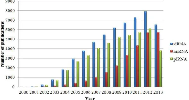
Currently there are mainly three types of small ncRNAs, to be exact, small-interference RNAs (siRNAs), piwi- interacting RNAs (piRNAs) and micro RNAs (miRNAs). During the last decade we have witnessed a near-exponential spread of scientific papers dedicated to regulatory RNAs as shown in Figure 1. All of these ncRNAs have the ability to target genes and silence their expression. Transcriptome analyses of small ncRNAs have brought to light new types of RNA molecules that do not fit into well-established classes (e.g. small nucleolar RNAs, snoRNAs) (Aalto & Pasquinelli, 2012).
Figure 1 – Number of non-coding RNA publications during the last thirteen years. Records were retrieved using PubMed database. The search terms used were: siRNA, miRNA and piRNA. Note that 2013 data we re only collected until 20 October.
50% of all protein-coding genes and therefore disruption in their expression is linked with several human diseases (Krol, Loedige, & Filipowicz, 2010).
In 1993, Victor Ambros a developmental biologist together with his co-workers Rosalind Lee and Rhonda Feinbaum described in Cell, the first miRNA in C. elegans, called lin-4. They found that lin-4 gene did not encode a protein but produced a pair of small transcripts that are complementary to 3´UTR region of lin-14 mRNA, which suggested an antisense regulatory mechanism (R. C. Lee, Feinbaum, & Ambros, 1993).
In C. elegans nematode, the heterochronic genes lin-4, lin-14, lin-28 and lin-29 control temporal postembryonic development, termed “larva-to-adult switch”. The switch is controlled by regulatory interactions between these genes, in the early stages
lin-14 and lin-28 negatively regulate lin-29 and consequently prevent early switching, whereas in the later stage lin-4 inhibits lin-14 and lin-28 and activates lin-29 which promotes switching (Ambros, 1989). Why have scientists used the C. elegans over other animal models to study the regulation and function of miRNAs? Well, it is inexpensive to cultivate, easy to manipulate physically, transparent at every stage of their life cycle, has two sexes (hermaphrodite and male), their development cycle is clear and their larval development is rapid (Corsi, 2006; J. Liu, Yang, & Ai, 2013).
Seven years after the discovery of the first miRNA, Reinhart et al. (2000) discovered the second miRNA in C. elegans. They demonstrated that let-7 is a heterochronic switch gene that contains a small RNA of ~21nt length, complementary to 3´UTR region of lin-14, lin-28, lin-41, lin-42 and daf-12 heterochronic genes. Let-7 loss-of-function causes a return to larval cell fates during adult stage, whereas let-7
gain-of- function causes precocious expression of adult fates during larval stage (Reinhart et al., 2000; Roush & Slack, 2008). Furthermore, lin-41 is negatively regulated by let-7 and negatively regulates lin-29 (Slack et al., 2000).
1. Histor ical intr oduction to micr oRNAs
2. The microRNA biogenesis pathway
The production of mammalian miRNAs begins with the transcription of one miRNA gene by RNA polymerase II (Po l II) or less frequently by RNA polymerase III (Pol III), to produce a capped and polyadenylated long primary miRNA transcript (pri-miRNA) with a stem- loop structure (hairpin) which contains the mature miRNA sequence in the stem (Graves & Zeng, 2012). A typical pri- miRNA hairpin comprises an imperfect stem of ~33bp, a loop and flanking RNA segments at its base that are critical for processing (Han et al., 2006).
There are two possible pathways of miRNA biogenesis, based on how the pri-miRNAs are processed [Figure 2]. In the canonical pathway, the miRNA gene is localized in intergenic region and in the mirtron pathway, the miRNA gene is in the intron of a protein-coding gene (Graves & Zeng, 2012).
In the canonical pathway, the pri- miRNA is cropped, in the nucleus, by the nuclear RNase III-type protein Drosha along with a cofactor DiGeorge syndrome critical region gene 8 (DGCR8) to form a truncated hairpin precursor- miRNA (pre-miRNA) of ∼65 nt, which has 2-nt 3´ overhangs (Xiaoxiao Zhang & Zeng, 2010). The microprocessor complex that comprises Drosha and its cofactor, DGCR8, is crucial for pri- miRNA processing, which in turn is a critical step in miRNA biogenesis because it will define the mature miRNAs sequence (Han et al., 2006). The 6-11nt that flank a pri-miRNA hairpin (5´or 3´ side) are unstructured by microprocessor complex at the distance ~11 bp from the ssRNA-dsRNA junction. An important consideration is that Drosha not cleave pri- miRNAs without its cofactor, DGCR8. Thus, RNA binding protein DGCR8 specifically recognizes the pri- miRNA and assists the cleavage by Drosha (Faller et al., 2010).
2. The micr oRNA biogen esis pathway
Figure 2 – Schematic overview of miRNA biogenesis. (A) Canonical pathway of miRNA biogenesis and
The guide strand (or miRNA) is loaded into RNA- induced silencing complex (RISC), whereas the passenger strand is degraded. The guide strand is extremely important because it is responsible for translational inhibition and target destabilization of target mRNAs and is involved in several diseases when its expression is misregulated. But then, is the passenger strand always degraded or can it be a potential regulatory molecule? The miRNA* strand destiny depends on phylogenetic conservation. Well-conserved miRNA* strands can eventually play a significant roles in regulation network (L. Guo & Lu, 2010). Despite the mechanism of guide strand selection not being fully understood, it was suggested that the guanine–cytosine (GC) frequencies and pairing information of miRNA:miRNA* duplex are essential to the selection of guide strand (D. Ma et al., 2011).
The core components of the miRNA- induced silencing complex (miRISC) is a miRNA-loaded into one of four Argonaute proteins (AGO1-4), which targets and silences the mRNAs in the 3´ untranslated regions (UTRs). There are other proteins, no less important, like PABP, CCR4-NOT and PAN2-PAN3 deadenylase complex linked to miRNA- mediated gene silencing (Fabian & Sonenberg, 2012).
The specificity of miRNAs to mRNAs targets is determined by the sequence complementary between nucleotides 2-8 on the 5´end of the miRNA (“seed sequence”) and the 3´ unstranslated region (UTR) of the mRNA. There are three well studied mechanisms by which miRNAs regulate gene expression, na mely, endonucleolytic cleavage, inhibition of translation initiation and mRNA degradation by deadenylation. When mRNA/miRNA match is perfect (or near perfect), the mechanism of gene silencing that occurs is through endonucleolytic cleavage of the scissile phosphate of the nucleotide paired to the 10th and 11th nucleotides of the guide RNA (Beezhold, Castranova, & Chen, 2010).
2. The micr oRNA biogen esis pathway
In the last years, the biochemical and functional properties of mirtrons have been studied in D. melanogaster, C. elegans and vertebrates. There are three classes of splicing-derived miRNAs in mammals, to be precise, conventional mirtrons, 5´-tailed mirtrons and 3´-tailed mirtrons. In conventional mirtrons biogenesis, splicing and debranching defines both ends of pre- miRNA hairpin, whereas "tailed" mirtrons contain unstructured extensions at their 5´ tails (5´-tailed mirtrons) and 3´ tail (3´-tailed mirtrons). 3´-tails exists in Drosophila and are trimmed by RNA exosome, whilst vertebrate 5´-tails are trimmed by an enzyme not yet discovered (Ladewig, Okamura, Flynt, Westholm, & Lai, 2012).
3. microRNAs and cancer
3.1.microRNAs as oncogenes or tumor suppressors
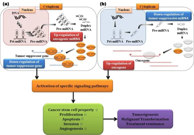
The abnormal expression of miRNAs is implicated in human tumorigenesis. The upregulation of oncogenic miRNAs (oncomiRs) leads to tumor develop ment by negatively targeting tumor suppressor proteins or proteins that control cell differentiation and apoptosis. On the other hand, the downregulation of tumor suppressive miRNAs (tsmiRs) leads to tumor development by upregulation of oncogenic proteins [Figure 3] (Ahmad et al., 2013).
A single miRNA has the ability to regulate multiple targets, so the function of miRNAs are also wide-ranging, including regulation of cancer stem cell properties, tumor proliferation, apoptosis, invasion and angiogenesis (Mizoguchi et al., 2013).
3. micr oRNAs and cancer
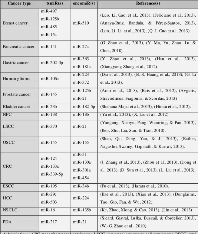
Growing number of tsmiRs and oncomiRs are being involved in different types of human cancers. Some of the most usually dysregulated miRNAs are summarized in Table 1.
Table 1 – So me miRNAs involved in d iverse human cancers with a ltered e xpression levels .
Cancer type tsmiR(s) onc omiR(s) Reference(s)
Breast cancer miR-497 miR-125b miR-485 miR-15a miR-510
(Luo, Li, Gao, et a l., 2013), (Fe lic iano et al., 2013), (Anaya-Ruiz, Bandala, & Pére z-Santos, 2013), (Luo, Li, Li, et al., 2013), (Q. J. Guo et a l., 2013).
Pancreatic cancer miR-141 miR-27a (G. Zhao et al., 2013), (Y. Ma, Yu , Zhao, Lu, & Chen, 2010).
Gastric cancer miR-202-3p miR-363 miR-181a
(Y. Zhao et al., 2013), (Hsu et al., 2013), (Xiangyang Zhang et al., 2012).
Hu man glio ma miR-106a
miR-223 miR-372
(Da i et al., 2013), (B.-S. Huang et al., 2013), (G. Li et al., 2013).
Prostate cancer miR-145 miR-125b miR-21
(Amir et al., 2013), (Re is et al., 2012), (Avgeris, Stravodimos, Fragoulis, & Scorilas, 2013)
Bladder cancer miR-23b miR-182-5p (Shahana Majid et a l., 2013), (Hirata et al., 2012). NPC miR-138 miR-18b (Yu et a l., 2013), (X. Liu et a l., 2012).
LSCC miR-370 miR-21 (Yungang, Xiaoyu, Pang, Wenming, & Pan, 2013) , (Ren, Zhu, Liu, Sun, & Tian, 2010).
OSCC miR-145 miR-155 (Shao, Qu, Dang, Yao, & Ji, 2013) , (Rather, Nagashri, Swa my, Gopinath, & Ku ma r, 2013) .
CRC miR-124 miR-133a miR-339-5p miR-31 miR-130a miR-301a miR-454
(J. Zhang et al., 2013), (Zhou et al., 2013), (Dong et al., 2013), (D. Sun et al., 2013), (L. Liu et al., 2013).
ESCC miR-195 miR-34b (Fu et al., 2013), (Ha rata et al., 2010).
HCC miR-29c
miR-503 miR-224
(Bae et al., 2013), (Xiao et al., 2013), (Donglaima , Tao, Gao, Fan, & Wu, 2012).
NSCLC miR-16 miR-135b (Ke, Zhao, Xiong, & Cao, 2013), (Lin et al., 2013).
PDA miR-217 miR-21 (Sicard, Gay ral, Lu lka, Buscail, & Cordelier, 2013) , (W.-G. Zhao et al., 2010).
3.2.microRNAs targeting the hallmarks of cance r
Human tumorigenesis is a multistep process that reflects changes in gene expression. In the year 2000, the cancer researchers Douglas Hanahan and Robert Weinberg published “The Hallmarks of Cancer”, that comprises six biological abilities acquired by cancer cells during the multistep development of human tumours (Hanahan & Weinberg, 2000). Two years ago, Weinberg and Hanahan proposed four new hallmarks as a result of scientific progress in the last decade. Actually, there are ten hallmarks that drive the transformation of normal cells to cancer cells, including, sustaining proliferative signaling, evading growth suppressors, activating invasion and metastasis, enabling replicative immortality, inducing angiogenesis, evading apoptosis, deregulating cellular energetics, genome instability and mutation, avoiding immune destruction and tumour-promoting inflammation (Hanahan & Weinberg, 2011). This work focused on the role of miRNAs in the hallmarks of human cancers as described by Hanahan and Weinberg [Figure 4].
Figure 4 – The role of miRNAs in the hallmarks of human cancers. (Ada pted from Ba la et al., 2012;
3. micr oRNAs and cancer
3.2.1. microRNAs regulate immune responses in cancer
The immune system is crucial to prevent tumor formation and progression, however, cancer cells have developed the faculty to escape the immune surveillance and proliferate. miRNAs are key mediators in immune system development and function in innate and adaptive immune responses. Dysregulation of these regulatory molecules can activate the occurrence of cancers in the immune system (Davidson-Moncada, Papavasiliou, & Tam, 2010).
The infectious agents, Epstein-Barr virus (EBV/human herpesvirus 4) and Kaposi’s sarcoma-associated herpesvirus (KSHV/human herpesvirus 8) are well known oncogenic viruses. They are mainly linked to lymphoproliferative diseases and lymphomas that occur in persons with HIV/AIDS or in those with iatrogenic immunodeficiency (IID) following solid organ transplantation (Carbone, Cesarman, Spina, Gloghini, & Schulz, 2009). A recent study shows that KSHV and EBV have functionally conserved miRNAs, respectively miR-K12-7 and miR- BART-5p, that downregulate MICB to avoid immune cell attack by natural killer (NK) cells (Nachmani, Stern-Ginossar, Sarid, & Mandelboim, 2009b). Human NK cells are innate immune lymphocytes that recognize abnormal cells, to be precise, tumor and virus-infected cells. NK cells are activated when stress-induced ligands (e.g. MICB) are expressed on the surface of abnormal cells and are recognized by activating NK cells receptors, like NKG2D. MiRNAs plays an important role in NK cell regulation and consequently in hematopoietic system (Leong, Sullivan, & Fehniger, 2012).
3.2.2. microRNAs control angiogenesis in cancer
Angiogenesis is a physiologic process that contributes to the formation of new blood vessels from pre-existing vasculature. In cancer cells, this process is accelerated because the overproduction or induction of pro-angiogenic factors in tumor microenvironment, such as, vascular endothelial growth factor (VEGF). Thus, in bone marrow (BM) deregulated angiogenesis is linked to disease progression and poor prognosis in multiple myeloma (MM) patients (Giuliani, Storti, Bolzoni, Palma, & Bonomini, 2011).
MM is a cancer of plasma B cells characterized by clonal proliferation of malignant plasma cells in the bone marrow microenvironment and extramedullary sites (e.g. cortical bone). The pathogenesis of MM results from a cascade of several genetic and microenvironmental events starting with monoclonal gammopathy of undetermined clinical significance (MGUS), followed by smoldering myeloma and ends with symptomatic myeloma. Current treatment for patients with MM has include autologous stem-cell transplantation and use of thalidomide, lenalidomide and bortezomib (Laubach, Richardson, & Anderson, 2011; Palumbo & Anderson, 2011).
Sun et al. (2013) recently discovered that miR-15a and miR-16 are down-regulated in ~70% of MM tumors, especially in advanced stage tumors, suggesting that downregulation of these miRNAs contributes to disease progression. This study found that both miRNAs are complementary to the VEGF-A 3´-UTR and that this interaction inhibits the overexpression of VEGF in MM cells. Thus, restoration of normal expression miR-15a and miR-16 can improve prognosis of MM patients by modulation of angiogenesis through targeting VEGF-A (C.-Y. Sun et al., 2013). This study is proof-of concept that miRNAs regulate the angiogenic process in cancer and that miRNAs could be used as an anti-angiogenic treatment therapeutic approach.
3. micr oRNAs and cancer
mRNA levels, showing that both miRNAs are inhibitors of tumor angiogenesis (He et al., 2013).
3.2.3. microRNAs regulate inflammation in cancer
Recent data reveal that some miRNAs are capable of regulating the inflammatory response in cancer, for example, in orodigestive periodontitis-related cancer. Periodontitis is an inflammatory disease that affects the oral periodontium, the set of tissues that support the teeth. This disease is the most common cause of tooth loss and several bacterial species are involved, namely, P orphyromonas gingivalis,
Tannerella forsythia and Treponema denticola (Darveau, 2010). Orodigestive cancer mortality is linked to periodontitis and P . gingivalis orodigestive colonization (Ahn, Segers, & Hayes, 2012).
Recent data suggest that miR-146a and miR-146b-5p are up-regulated in periodontal disease and consequently decrease pro- inflammatory cytokines (e.g. IL-1β, IL-6 and TNF-α) by inhibition of interleukin-1 receptor-associated kinase 1 (IRAK1) expression through direct binding to the 3´-UTR of IRAK1. Therefore, miR-146 is a negative regulator of immune response in periodontal inflammation (Y.-F. Xie et al., 2013b).
Other current study has exposed that miR-155 upregulation in monocytes of patients with hepatitis C virus (HCV) infection increases pro- inflammatory state and also suggests the possibility of use miR-155 as disease biomarker (Bala et al., 2012).
3.2.4. microRNAs target growth suppressors in cancer
Another study has demonstrated the role of miRNAs in growth/proliferation of paediatric brain cancer. Neuroblastoma is a pediatric tumor of the autonomic nervous system, affects ~10 children per million births and is the leading cause of cancer during the first year of life (Maris, 2010). miR-497 over-expression triggers the apoptosis in v-myc avian myelocytomatosis viral oncogene neuroblastoma derived homolog (MYCN) -amplified neuroblastoma cells by targeting the 3´-UTR of WEE1, a tyrosine kinase regulator of the cell cycle. This finding suggests that WEE1 can be a therapeutic target in neuroblastoma management (Creevey et al., 2013).
3.2.5. microRNAs regulate tissue invasion and metastasis
miRNAs have the ability to regulate metastatic potential of tumors and the outcomes among patients, particularly, in breast cancer and cholangiocarcinoma (CCA). In breast cancer, GATA 3 is a transcription factor (TF) that regulates epithelial cell differentiation in mammary gland and suppresses breast metastasis. GATA 3 gene is mutated in more than 10% of all breast cancers and the loss of GATA 3 expression is predictive of poor prognosis. This TF induces anti- metastatic miR-29b expression that inhibits metastasis by targeting a network of pro- metastatic regulators, such as, ANGPTL4, LOX and MMP9. This discovery, based on GATA3- miR-29b axis, brings a new therapeutic field to the treatment of breast cancer patients (Chou et al., 2013).
CCA is the most frequent biliary tract cancer characterized by being difficult to diagnose and classified into intrahepatic, perihilar and distal extrahepatic CCA (Blechacz, Komuta, Roskams, & Gores, 2011). It’s known that miR-21 is overexpressed in CCA cells. A recent study demonstrates that knockdown of miR-21 inhibit cellular invasion and metastasis and increases reversion- inducing-cysteine-rich protein with kazal motifs (RECK) protein levels. MiR-21 targets RECK, a metastasis suppressor gene, and promote cell invasion and metastasis (Q. Huang et al., 2013).
3.2.6. microRNAs control cellular metabolis m
3. micr oRNAs and cancer
processes, such as, glucose uptake, glycolysis, tricarboxylic acid cycle, insulin production, lipid metabolism and amino acid biogenesis (B. Chen et al., 2012). Here, I will explain the regulation of cellular metabolism by miRNAs in human bladder cancer and in HCV infection.
Bladder cancer is the most common malignancy of the urinary tract, however, the mortality rates have declined in recent years, probably due to improved diagnostic techniques and therapeutics (Cheung, Sahai, Billia, Dasgupta, & Khan, 2013). The main risk factors for bladder cancer include genetic and molecular abnormalities (e.g. oncogene activation and tumor suppressor gene inactivation), chemical and environmental exposures (e.g. cigarette smoking) and chronic irritation in patients w ith bladder catheters (e.g. Schistosoma haematobium infestation) (Kaufman, Shipley, & Feldman, 2009).
Glucose transport in bladder cancer is increased, due to upregulation of the high affinity glucose transporter member 3 (GLUT3). Fei et al. (2012) have for the first time discovered that miR-195-5p over-expression downregulates the GLUT3 protein level in bladder cancer. This finding suggests that miR-195-5p could function as a tumor suppressor in bladder cancer. Although, miR-195-5p expression is up-regulated in Chronic lymphocytic leukemia (CLL), meaning that this miRNA can play different roles in various cancers (Fei et al., 2012).
3.2.7. microRNAs regulate the limitless replicative potential of cancer cells Senescence and immortality are two physiological processes that control the replicative potential of cells. Senescence is a tumor suppression mechanism that occurs in response to cell stress (e.g. telomere dysfunction), whereas, immortality is the ability to escape senescence (Y. Kong, Cui, Ramkumar, & Zhang, 2011). Recently, it was discovered that some miRNAs have the ability to regulate the limitless replicative potential of melanoma and endothelial cells.
The transcription factor E2F1, a master regulator of the G1/S cell cycle transition
phase, when overexpressed, it triggers an oncogenic event that enhances the proliferation of melanoma cells. Dar et al. (2011) have demonstrated that overexpression of miR-205 in melanoma cells reduced E2F1 protein levels and, in turn, induced a senescent phenotype (Dar et al., 2011).
The atherosclerosis and coronary artery disease are age related diseases. The Silent information regulator 1 (SirT1) is an important regulator that promotes longevity and prevents disease by avoiding stress- induced senescence. miR-217 is gradually expressed in endothelial cells during aging and is a natural inhibitor of SirT1 during endothelial senescence. This fact opens a new opportunity to prevent endothelial dysfunction in metabolic diseases (Menghini et al., 2009).
3.2.8. microRNAs control genomic instability of cancer cells
Several studies have confirmed the role of miRNAs in the regulation of genomic instability in cancer cells (e.g. colon cancer). Ataxia Telangiectasia Mutated (ATM) protein is responsible for the repair of double-strand DNA breaks (DSB), an extremely cytotoxic DNA lesion. miR-18a is upregulated in human colorectal cancer (CRC) and negatively regulates ATM by targeting ATM 3´UTR. Thus, miR-18a has an oncogenic role in CRC through the attenuation of cellular repair (C.-W. Wu et al., 2013).
3. micr oRNAs and cancer
therapeutic agent in improving the efficacy of radiotherapy and chemotherapy in cancer patients (Y. Wang et al., 2011).
3.2.9. microRNAs regulate apoptosis in cance r
Apoptosis is programmed cell death that occurs in physiological and pathological situations and is characterized by a loss of equilibrium among cell division and the cell death (Wong, 2011). Various miRNAs participate in the apoptotic signaling pathway.
The hormone Prothymosin-alpha (PTMA) is an apoptotic inhibitor that binds to apoptotic protease activating factor 1 (Apaf-1). A recent study has suggested that miR-1 induces nasopharyngeal carcinoma apoptosis by targeting PTMA mRNA (C.-D. Wu et al., 2011).
The caspase inhibitor, X- linked inhibitor of apoptosis protein (XIAP), has a crucial role in stopping apoptotic cell death. Recently it was demonstrated that miR-24 downregulates XIAP expression by targeting 3´UTR of the XIAP mRNA. This discovery can possibly overcoming the apoptosis resistance problem in cancer cells (Y. Xie et al., 2013).
3.2.10. microRNAs regulate uncontrolled prolife ration in cancer cells
Current studies have proven that miRNAs have the ability to control the uncontrolled proliferation in different cancers, such as, gastric cancer and oral squamous cell carcinoma (OSCC).
Gastric cancer is the second cause of death worldwide, normally, it is diagnosed in advanced stages of disease and an early diagnostic is crucial for good prognos is (Takahashi, Saikawa, & Kitagawa, 2013). Phosphatase and tensin homolog (PTEN) acts as a tumor suppressor gene in human gastric cancer, however, its expression is decreased in gastric cancer cells. miR-214 is overexpressed in gastric cancer and negatively regulates PTEN. Knockdown of miR-214 inhibits proliferation of gastric cancer cells and improves the prognosis (Yang et al., 2013).
miR-125b is expressed. These results propose that miR-125b suppress cell proliferation and overcomes the radioresistance problem in OSCC (Shiiba et al., 2013).
The following Table 2 describes summarily several miRNAs and its predicted targets.
Table 2 – miRNA and the hall marks of cancer
Hallma rks of cancer Target (s) miRNA (s) Refe rence
Avoiding immune destruction
Host MICB miR-BA RT-5p
miR-K12-7 (Nach mani et al., 2009b) MCPy V large
T-antigen MCV-miR-M 1-5p (S. Lee et al., 2011)
Inducing a ngiogenesis
VEGF miR-15a
miR-16 (C.-Y. Sun et al., 2013) VEGF / HIF-1α miR-199a
miR-125b (He et a l., 2013)
Tumour-promoting infla mma tion
IRA K 1 miR-146a
miR-146b -5p (Y.-F. Xie et al., 2013b)
TNFα miR-155 (Ba la et a l., 2012)
Eva ding growth suppressors
IL-24 pro moter miR-205 (J. S. Kim et a l., 2013)
WEE1 miR-497 (Creevey et al., 2013)
Activa ting inva sion a nd meta sta sis
ANGPTL4
LOX / MMP9 miR-29b (Chou et al., 2013)
RECK miR-21 (Q. Huang et al., 2013)
Deregula ting cellula r meta bolism
GLUT3 miR-195-5p (Fei et al., 2012)
ApoB100 and others miR-27a (Shirasaki et a l., 2013)
Unlimited replica tive potential
E2F1 miR-205 (Dar et al., 2011)
SirT1 miR-217 (Menghini et al., 2009)
Genome insta bility a nd muta tion
ATM miR-18a (C.-W. Wu et a l., 2013)
H2A X miR-138 (Y. Wang et al., 2011)
Eva ding a poptosis
PTMA miR-1 (C.-D. Wu et a l., 2011)
XIAP miR-24 (Y. Xie et a l., 2013)
Susta ining prolifera tive signa ling
PTEN miR-214 (Yang et al., 2013)
4. micr oRNAs and neur odegener ative diseases
4. microRNAs and neurodegenerative diseases
Aging is one of the major factors that contribute to brain neurodegeneration. In a current Letter published in Nature, Liu et al. (2012) demonstrate that miR-34 is involved is age-associated diseases and long-term brain integrity in Drosophila. Thus, 34 upregulation suppresses neurodegeneration and extends lifespan, whereas, miR-34 downregulation triggers brain aging and late-onset degeneration (Nan Liu et al., 2012).
Recent studies showed that some miRNAs from patients with neurodegenerative diseases (NDs) are deregulated in the brain. For example, disruption of proteins required for miRNA biogenesis (e.g. loss of Dicer) induces neurodegeneration (Abe & Bonini, 2013). Several studies have proved that miRNAs are differentially expressed in subtypes of neurons and also regulate genes involved in NDs (Junn & Mouradian, 2010).
NDs are progressive disorders that include Alzheimer´s disease (AD), Parkinson´s disease (PD), Amyotrophic lateral sclerosis (ALS), Huntington´s disease (HD) and Spinocerebellar ataxias (SCAs) (Abe & Bonini, 2013). All of these diseases share a common feature, protein aggregates in different brain regions. The accumulation and aggregation of misfolded proteins in the brain leads to neuronal dysfunction and accelerates neurodegeneration (S.-J. Lee, Lim, Masliah, & Lee, 2011). Here will be discussed examples of scientific experiments displaying the role of miRNAs in neurodegenerative diseases.
4.1.Alzheimer´s Disease
AD is a neurologic illness characterized by continuous loss of neurons and consequently cognitive functions (e.g. memory) (Y. Huang & Mucke, 2012). Accumulation of β-amyloid (Aβ) peptide and abnormal filaments of Tau in brain establishes the disease. Despite progress with anti-amyloid strategies this disease remains incurable and leads to death ~9 years after AD diagnosis (Citron, 2010).
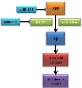
miR-195 in the central neural system and found that miR-miR-195 negatively regulates Beta-secretase 1 (BACE1) mRNA translation and consequently inhibits Aβ formation in vitro [Figure 5] (H.-C. Zhu et al., 2012). On the other hand, in vivo studies have strengthened the role of miRNAs in Aβ plaque formation. In a recent study, miR-153 downregulated amyloid precursor protein (APP) [Figure 5] and its ortholog amyloid precursor- like protein 2 (APLP2) in transgenic mouse model, suggesting miR-153 is a potential target in AD therapeutic management (Liang et al., 2012).
Figure 5 – miRNA targeting amyloid pathway in Alzheimer´s Disease. (Adapted from Holohan, Lahin,
Schneider, Fo roud, & Say kin, 2013).
4.2.Parkinson´s disease
Parkinson´s disease is a degenerative disorder of the central nervous system marked by death of dopaminergic neurons in substantia nigra, gliosis and the presence of eosinophilic intracytoplasmic inclusions, called Lewy bodies. These inclusions are abnormal aggregates of neuronal proteins, mainly alpha-synuclein (Margis, Margis, & Rieder, 2011).
4. micr oRNAs and neur odegener ative diseases
brains, however, miR-205 expression is decreased. miR-205 has the ability to target directly the 3´-UTR mRNA of LRRK2 gene and therefore suppresses its expression. Researchers propose that miR-205 has a therapeutic potential in PD and also as biomarker of the disease (Cho et al., 2012).
Alpha-synuclein oligomers are mediators of neurodegeneration in PD, as they confer toxicity to cells and initiate neuronal death (Kalia, Kalia, McLean, Lozano, & Lang, 2013). A recent study demonstrated that two miRNAs widely expressed in the brain, miR-7 and miR-153, bind directly to the 3´-UTR α-synuclein mRNA and repress α-synuclein expression. Lowering endogenous α-synuclein levels may represent an interesting approach for PD therapy (Doxakis, 2010).
4.3.Amyotrophic late ral sclerosis
Amyotrophic lateral sclerosis, also called motor neurone disease, is a neurodegenerative disorder of the motor neurons in brainstem, spinal cord and motor cortex. This illness can be grouped in familial ALS (fALS) or sporadic ALS (sALS). The main signs and symptoms are muscle atrophy, weakness, fasciculations and muscle spasticity that usually appear after 40 years of age. Proteins associated with ALS, such as, TAR DNA-binding protein 43 (TDP43) and RNA-binding protein FUS are involved in pre- mRNA splicing, RNA transport and RNA translation, suggesting that abnormal RNA metabolism may be crucial to the ALS pathogenesis (Robberecht & Philips, 2013).
Williams et al. (2009) showed that expressing miR-206, a skeletal muscle-specific miRNA, promotes nerve regeneration in ALS mouse model after acute nerve damage. This finding brings a potential target for ALS therapy (Williams et al., 2009). Another study demonstrated that miR-9 negatively regulates the neurofilament heavy subunit (NF-H) mRNA in spinal motor neurons. This neurofilament was previously implicated in ALS and thereby miRNA- mediated NF-H downregulation could slow or reverse neurodegenerative states (Haramati et al., 2010).
4.4. Huntington´s disease
in human body. Mutation in HD gene originates an expanded CAG-triplet repeat, which encodes polyglutamines (PolyQs) within Htt protein that results in neuropathologic changes in neostriatum and cerebral cortex. Tetrabenazine is the only approved drug for HD, so it is imperative to find new therapeutic approaches (Krobitsch & Kazantsev, 2011; Ross & Tabrizi, 2011).
A current study has demonstrated that miR-22 overexpression is potentially neuroprotective using in vitro models of HD. miR-22 has the ability to target several genes linked to HD, such as, histone deacetylase 4 (HDAC4), REST corepresor 1 (Rcor1) and regulator of G-protein signaling 2 (Rgs2). MiR-22 also reduces caspase activation by inhibiting pro-apoptotic protein expression (e.g. MAPK12/p38 and Tp53inp1) preventing neuronal apoptosis. These findings sustain the idea of enhanced miR-22 expression as therapeutic approach in HD (Jovicic, Zaldivar Jolissaint, Moser, Silva Santos, & Luthi-Carter, 2013).
4.5. Spinocerebellar ataxias
5. micr oRNAs and autoimmune diseases
5. microRNAs and autoimmune diseases
miRNAs play an important role in the regulation of innate and adaptive immune response, as even, in the immune cell development. Therefore, defective miRNA regulation in immune function has serious consequences, namely, immune cell cancers, loss of tolerance and development of autoimmunity, impaired adaptive immunity, inflammatory autoimmune disorders and dysregulation of antibody production (Pauley, Cha, & Chan, 2009). Various studies have revealed potential roles for miRNA regulation in autoimmune diseases, such as, psoriasis, rheumatoid arthritis (RA) and systemic lupus erythematosus (SLE).
5.1. Psoriasis
Psoriasis is an immune- mediated inflammatory disease that results from a complex interaction between genetic, immunological and environmental factors. This chronic disease affects the skin and joints and has a prevalence of 2-3% worldwide (Perera, Di Meglio, & Nestle, 2012). The epidermis of psoriasis patients is scaly and thick. The scales are a consequence of rapid maturation of keratinocytes and retention of nuclei in the stratum corneum (parakeratosis), whereas, the epidermal thickness occurs because the mitotic rate of the keratinocytes is augmented (acanthosis) (O. Nestle, H. Kaplan, & Barker, 2009).
The miRNA expression profile in psoriasis is different when compared with miRNA expression profile in healthy skin. The miR-125b is downregulated in psoriatic lesional skin, however, its target, Fibroblast growth factor receptor 2 (FGFR2) expressed in keratinocytes is upregulated in psoriatic epidermis. Xu et al. (2011) have demonstrated that miR-125b inhibits keratinocytes proliferation and foments terminal differentiation through downregulation of FGFR2. This discovery suggests miR-125b as a possible target for psoriasis therapeutic management (Xu et al., 2011).
5.2. Rheumatoid arthritis
Rheumatoid arthritis (RA) is an autoimmune disease of unknown cause characterized by synovitis and synovial hyperplasia (enlargement), autoantibody-positive for rheumatoid factor (RF) and anti-citrullinated protein antibody (ACPA), cartilage degradation and bone erosion (McInnes & Schett, 2011). Several studies have implicated specific miRNAs in the pathophysiology of RA, namely, miR-146a and miR-23b.
miR-146a is upregulated in CD4+ T cells of RA patients and has a positive correlation with TNF-α, a critical mediator of the inflammatory pathway in the rheumatoid joints. This miRNA negatively regulates Fas-associated factor 1 (FAF1) and suppresses T cell apoptosis. Therefore, increased miR-146a expression may be implicated in maintaining inflammation in RA. This discovery affords a promising novel therapeutic target in RA (J. Li et al., 2010).
Abnormal inflammatory responses, like elevated expression of proinflammatory cytokines in RA (e.g. TNF-α, IL-1β and IL-17), boosts chronic inflammation and tissue damage in autoimmune diseases. miR-23b is downregulated in RA patients, once, the cytokine IL-17 downregulates miR-23b expression in human fibroblast- like synoviocytes. On the other hand, miR-23b robustly restrains inflammation by decreasing activation of the NFκB pathway and inflammatory cytokine expression, such as, TNF-α, IL-1β and IL-17. These outcomes suggest miR-23b may be a new target for therapeutic intervention of inflammatory diseases (S. Zhu et al., 2012).
5.3. Systemic lupus erythe matosus
Systemic lupus erythematosus (SLE) is a multisystem autoimmune connective tissue disease characterized by a global loss of self- tolerance with production of pathogenic autoantibodies against nucleic acids and their binding proteins. Anomalous innate immune responses (e.g. release of inflammatory cytokines and autoreactive T and B cells) leads to production of autoantibodies and tissue injury (Choi, Kim, & Craft, 2012).
5. micr oRNAs and autoimmune diseases
indirectly downregulates DNA methyltransferase 1 (DNMT1) expression. DNMT1 downregulation leads to overexpression of autoimmune-related methylation-sensitive genes (e.g. CD11 and CD70) and by this means DNA hypomethylation in lupus CD4+ T cells. DNA hypomethylation triggers the onset and progression of SLE. The therapeutic use of miR-29b inhibitors to reverse the hypomethylation status could be a potential strategy for the treatment of SLE (Qin et al., 2013).
6. microRNAs and cardiovascular diseases
Expression levels of miRNAs is dysregulated in heart diseases, which suggests their involvement in cardiomyopathies, such as, arrhythmias, defects in ventricular septation, cardiac hypertrophy and myocyte hyperplasia (Callis & Wang, 2008).
Energy homeostasis results from equilibrium between energy storage (e.g. food intake) and energy expenditure (e.g. physical activity). The dysregulation of this biologic mechanism is associated with obesity, diabetes mellitus, hypertension, hyperlipidemia and cardiovascular diseases. The mediator of RNA polymerase II transcription subunit 13 (MED13) regulates energy homeostasis and is downregulated by miR-208a, a heart-specific miRNA encoded by an intron of the MYH6 (also known as MyHC-α) gene [Figure 6]. The overexpression of MED13 or the inhibition of miR-208a in mice leads to resistance to diet- induced obesity, improves systemic insulin sensitivity, resistance to metabolic syndrome and lower plasma lipid profile, whereas, deletion of MED13 contributes to diet- induced obesity and increases metabolic syndrome occurrence. This discover proposes that miR-208a inhibitors could represent an additional strategy in cardiovascular diseases therapy (Grueter et al., 2012; van Rooij & Olson, 2012).
7. Str ategies for micr oRNA-based ther apies
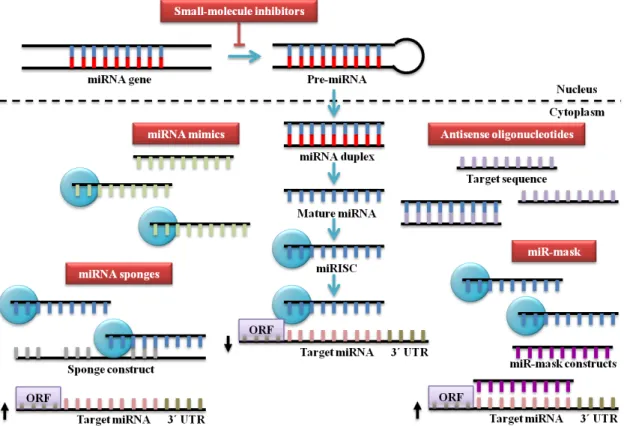
7. Strategies for microRNA-based the rapies
Therapeutic strategies based on modulation of miRNA expression and function can be divided in two main approaches: miRNA blocking (miRNA antagonists) and miRNA replacement (miRNA mimics) [Figure 7]. miRNA replacement consists of the delivery of miRNAs that are downregulated or deleted in tumours, whereas, miRNA blocking consists of the inhibition of miRNAs that are upregulated or overexpressed in tumours (Y. W. Kong, Ferland-McCollough, Jackson, & Bushell, 2012).
Two major problems have delayed the miRNA-based therapies in vivo. The first hurdle is the low stability of RNA in vivo due to ribonuclease (RNase)- mediated degradation. The second difficulty is to guarantee tissue-specific delivery and sustained target inhibition. Once one single miRNA can regulate multiple mRNA targets is difficult to avoid off-target effects (Y. W. Kong et al., 2012).
Figure 7 - Strategies for miRNA-based therapies. Bloc king oncomiRs can be achieved by the use of
7.1. microRNA replace ment the rapy
miRNA replacement is the reintroduction of a tumor-suppressor miRNA lost during carcinogenesis and the restoration of cellular programs commonly activated in normal cells that regulate oncogenic programs. For several years, the designation of a tumor suppressor was limited to protein-encoding genes but currently miRNA mimics also fit into the definition of tumor suppressor. miRNA mimics ha ve a few advantages compared to gene therapy regarding the delivery of DNA plasmid or viral vector with protein-encoding genes. miRN As mimics, contrary to the proteins, have low molecular weight, can be delivered systemically and are easily activated by crossing the cytoplasm layer of cancer cells (Bader, Brown, & Winkler, 2010).
miRNA mimic technology involves the design of a synthetic RNA molecule with the ability to enter into the complex RISC and regulate the same target genes as the endogenous miRNA. To improve half- lives, specificity of RNA molecule and activity, several sugar and phosphate modifications can be integrated in miRNA mimic, such as, 2´-O-methyl, 2´F, 2´NH2, 2´H, phosphorothioates and locked nucleic acids (LNAs)
(Bader, Brown, Stoudemire, & Lammers, 2011).
7.1.1. microRNA replace ment the rapy for liver cancer
Hepatocellular carcinoma (HCC) is a highly prevalent disease, affecting more than half a million people globally. The major risk factors include hepatitis B virus (HBV) or HCV previous infection, alcoholic liver disease and nonalcoholic fatty liver disease (El-Serag, 2011).
7. Str ategies for micr oRNA-based ther apies
AAV type 9 display favored tropism for cardiac tissue and AAV type 2 for skeletal muscle (N.-C. Lee et al., 2012; Nathwani et al., 2011; Qi et al., 2010).
7.1.2. microRNA replace ment the rapy for lung cance r
Lung cancer is the main cause of death cancer-related worldwide and 80% of lung cancers are classified as non-small cell lung carcinoma (NSCLC) (Farhat & Houhou, 2013). Both, miR-34a and let-7 are tumor suppressors that are downregulated in lung cancer. Trang and co-workers have verified that systemic delivery of miR-34a in
Kras-activated mouse model of NSCLC using a neutral lipid emulsion (NLE) reduced 60% of tumor area. This noteworthy discover supports the notion that miRNAs can regulate several targets because the proto-oncogene Kras is not directly repressed by miR-34a. Furthermore, let-7 and miR-34 have different tumor inhibition mechanisms: miR-34a reduces proliferation and increases apoptosis, whereas, let-7 only reduces proliferation. This suggests that combination therapy may enhance the therapeutic effect (Trang et al., 2011).
7.2.microRNA blocking therapy
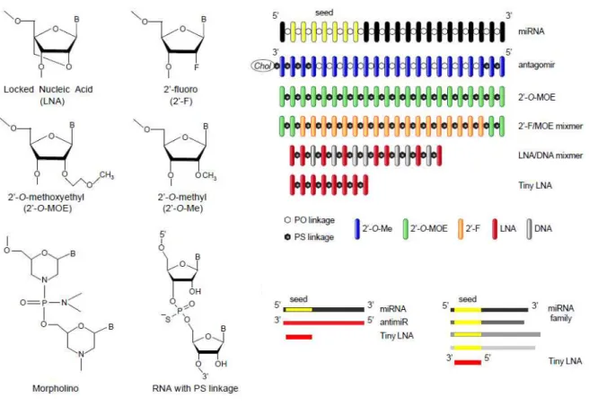
7.2.1. Anti-microRNA oligonucleotides
Figure 8 – Design of chemica lly mod ified antimiR o ligonucleotides . (Retrieved from Stenvang, Petri, Lindow, Obad, & Kauppinen, 2012).
The phosphorothioate backbone is the most extensively studied chemical modification that consists of replacement of one of the nonbridging oxygens by sulphur atom. Phosphorothioate oligonucleotides exhibit nuclease stability, easy synthesis, high solubility, significant antisense activity and are capable of activating RNase H activity (Dias & Stein, 2002). 2’-4’ LNA is a class of therapeutic agents in which the ribose sugar ring is locked by a oxymethylene bridge linking the 2´-O and 4´-C atoms (Veedu & Wengel, 2010). LNAs have notable characteristics, to be precise, high binding affinity regarding RNA or DNA, remarkable base pairing specificity, nuclease resistance, generally non-toxic and easily to manipulate (Veedu & Wengel, 2009).
7. Str ategies for micr oRNA-based ther apies
Therapeutic silencing of disease-associated miRNAs using LNA- modified antimiRs oligonucleotides has been studied in lymphomas. A recent study demonstrated the role of an LNA- modified antimiR in the silencing of miRNAs associated to human diseases, such as, lymphomas. Waldenstrom´s macroglobulinemia (WM) is an rare indolent lymphoma, frequent in elderly and characterized by IgM monoclonal protein infiltration in BM (Buske & Leblond, 2013). miR-155 plays a critical role in the pathogenesis of B-cell malignancies and is overexpressed in B cells of transgenic mice. Zhang and colleagues have recently established that systemic delivery of tiny LNA antimiR-155 oligonucleotide in a mouse xenograft model of WM reduces tumor growth. This result emphasizes the value of tiny LNA antimiR therapy in hematologic malignancies (Y. Zhang et al., 2012).
AntagomiRs are single-stranded RNA molecules containing cholesterol, conjugated via a 2´-O-methyl (2´-O-Me), complementary to the mature target miRNA and contain several phosphorothioate moieties. Phosphorothioate backbone linkages confers to the antagomiR, increased binding to plasma proteins and by that way decreases renal clearance, whereas, the cholesterol improves cellular uptake, in vivo
stability and stimulates hepatic uptake (van Rooij & Olson, 2012).
Figure 9 – Silencing miRNAs with antagomiRs . (Retrieved from Czech, 2006).
Therapeutic silencing of miRNAs is a promising approach to suppress tumorigenesis. In vivo and in vitro silencing of miR-10b with antagomiR-10b notably decreases miR-10b levels and increases homeobox D10 (HOXD10) protein levels, leading to a decreased expression of Ras homolog gene family, member C (RhoC), a pro-metastatic gene. Systemic treatment of tumor-bearing mice with antagomiR-10b suppresses lung metastasis but does not reduce primary breast cancer growth [Figure 10] (L. Ma et al., 2010). This remarkable study demonstrates that systemic administration of antagomiR-10b can efficiently target a tumor in vivo without major toxicity in mice. Therefore, prophylactic treatment of tumors that have not yet metastasized using miR-10b antagomiRs could be a novel therapy option in the near future (De Palma & Naldini, 2010).
7. Str ategies for micr oRNA-based ther apies
7.2.2. microRNA sponges
miRNA sponges or target mimics are competitive inhibitors that contain several binding sites for a family of endogenous miRNAs sharing a common seed. They can be expressed from chromosomal transgene insertions if the intent is a partial miRNA inhibition or lentiviral and retroviral sponge vectors if the aim is long term miRNA inhibition (Ebert & Sharp, 2010). In vitro experiments demonstrated that miRNA sponges derepressed miRNA targets as robustly as chemically modified AMOs (Ebert, Neilson, & Sharp, 2007).
Circular RNAs (circRNAs) firstly discovered in plants, results from a covalent coupling of the ends of a single RNA molecule. Nowadays, at least 2,000 human circRNAs have been identified (Memczak et al., 2013). A surprising recent study have demonstrated that a highly expressed circRNA in human and mouse brains operates as miR-7 sponge. It robustly suppresses miR-7, a central regulator of several cancers and Parkinson disease. Circular RNA sponge for miR-7 (ciRS-7) holds about 70 selectively conserved miR-7 target sites and is a promising candidate in neurological diseases and brain tumors therapies (Hansen et al., 2013).
7.2.3. microRNA-masking antisense oligonucleotide technology
miRNA-masking antisense oligonucleotides (miR- masks) are single-stranded
7.2.4. Small-molecule inhibitors of microRNAs
8. Routes to in vivo deliver y of micr oRNAs
8. Routes to in vivo delivery of microRNAs
Currently, there are several routes to delivery therapeutic miRN As in vivo, such as, intratumoral, intramuscular, intravenous, intraperitoneal, intranasal and intracerebroventriculary [Figure 11].
Figure 11 – Routes to in vivo delivery of therapeutic miRNAs. (Ada pted from Iorio & Croce, 2012;
Nana-Sinka m & Croce, 2011).
8.1. Intratumoral
Intratumoral injections have been exploited to delivery therapeutic miRs in many diseases, namely, renal cell carcinoma (RCC) and prostate cancer.
A recent study demonstrated that miR-708 is a tumor suppressor in RCC and its expression is attenuated about 50-60% in RCC patients. Using an in vitro assay verified that replacement of miR-708 expression in RCC cell lines suppresses tumorigenicity through a notable increase in apoptosis. In addition to their in vitro data, they evaluated the therapeutic potential of a miR-708 mimic in vivo. They inoculated subcutaneously a RCC cell line into a mouse model and thirty days later a palpable tumor with 100-150mm3 grow. Intratumoral delivery of miR-708 mimic every 3 days reduced significantly the tumor size [Figure 12]. This discovery reveals that miR-708 is a pro-apoptotic miRNA in renal cancer and therefore is a striking target for prognosis and therapy of RCC (Saini et al., 2011).
Figure 12 – Representative images of mice fro m the two groups (miR-control and synthetic miR-708)
before treatment (day 30) and after treat ment (day 58). Intratumora l delivery of miR-708 leads to regression of tumors in a renal cancer xenograft mouse model. Subcutaneous tumors are indicated by arrows. (Retrieved from Sain i et a l., 2011).
8. Routes to in vivo deliver y of micr oRNAs
antitumorigenic effect of miR-34b was checked in a prostate cancer xenograft mouse model. Intratumoral injection of a miR-34b mimic reduced the tumor volume (~12mm3) in established tumors, whereas, intratumoral injection of miR control (Cont- miR) increased the tumor volume (~136mm3) in established tumors [Figure 13]. Replacement of miR-34b in prostate cancer cells downregulates DNA methyltransferase (DNMT) and histone deacetylase (HDAC) inducing demethylation and active chromatin modifications. miR-34b also directly targets Akt proliferative pathway genes and epithelial- mesenchymal transition (EMT) markers generating antiproliferative effects and antimigratory/- invasive effects. These discoveries open a new horizon to targeting miR-34b and its epigenetic regulators for the treatment of prostate cancer (S. Majid et al., 2013).
Figure 13 – Antitumorigenic e ffect of miR-34b in vivo. (Ada pted from S. Majid et al., 2013).
8.2. Intramuscular
Intramuscular injections of miRNAs have been studied in cardiac and skeletal muscle of mouse models to discover new therapeutic approaches for human diseases.
(Ning Liu et al., 2012). Another study demonstrated that in vivo delivery of miRNAs facilitated recovery of skeletal muscle after injury. The muscle-specific miR-1, miR-133 and miR-206 play an essential roles in regulation of muscle development. Nakasa et al.
(2010) discovered that one single local injection of these miRNAs in rat tibialis anterior muscle accelerates muscle regeneration. This finding could represent a new approach in musculoskeletal disorders, namely in sports and traumatology medicine (Nakasa et al., 2010).
Myocardial infarction (MI), a major cause of death worldwide, is characterized by myocardial cell death owing prolonged ischemia (Thygesen et al., 2012). Hu et al.
(2010) have discovered that miR-210 can rescue cardiac function in a murine model of MI. The researchers have showed that intramyocardial injections of miR-210 precursor through a nonviral minicircle vector improved left ventricular function after MI. Furthermore, histological analysis suggests that miR-210 promotes neovascularisation and inhibition of cellular apoptosis in the heart. This finding proposes that miR-210 is potentially useful in convalesce of human myocardial infarction (Hu et al., 2010).
8.3. Intrape ritoneal
miRN As injected intraperitoneally have been studied in management of complicated diseases, such as, melanoma.
8. Routes to in vivo deliver y of micr oRNAs
Figure 14 - Bio lu minescent imaging at 4 wee ks post intrasplenic injection of 1x10⁴ melano ma ce lls into mice. M ice were randomized into two groups, receiving either anti-miR-182 or negative control anti-miR administered by intraperitoneal in jection twice wee kly. In vivo luciferase imag ing showed that mice treated with anti-miR-182 had a lower burden of liver metastases compared with control. (Retrieved from Huynh et al., 2011).
8.4. Intravenous
Intravenous injections of therapeutic miRNAs have been studied to target head and neck squamous cell carcinoma (HNSCC), obesity and type 2 diabetes.
Some miRNAs that are deregulated in metabolic tissues from obese mice may eventually promote the development of obesity and type 2 diabetes. miR-802 is overexpressed in high fat diet (HFD)-fed mice, obese humans and mice homozygous for the diabetes db mutation of the leptin receptor (Leprdb/db). Hepatocyte nuclear factor 1 homeobox b (Hnf1b) in a direct target of miR-802 and decreasing of Hnf1b in liver contributes to glucose intolerance, attenuate s insulin sensitivity and stimulates hepatic gluconeogenesis. Kornfeld et al. (2013) have found that intravenous injection of a miR-802 LNA oligonucleotide in HFD- fed mice results in lower serum insulin concentrations, glucose intolerance and insulin tolerance comparatively with control LNA oligonucleotide. These findings suggest that miR-802 inhibition could be used as therapeutic target in obesity and type 2 diabetes (Kornfeld et al., 2013).
8.5. Intranasal
Intranasal delivery of miRNAs to the lungs of murine model of asthma has been studied. Asthma is a chronic inflammatory disorder of the airways affecting 300 million people worldwide. Type 2 CD4+ lymphocytes (Th2 cells) and their cytokines (e.g. IL-13) play a key role in the pathogenesis of allergic asthma (Hansbro, Kaiko, & Foster, 2011). In a recent study, Kumar et al. (2011) found that intranasal delivery of let-7
mimic to the lungs of a murine model of asthma reduces IL-13 levels, an important cytokine responsible for inflammation and tissue remodelling in allergic asthma. Administration of let-7 mimic have also been conducted to attenuate asthma features, namely, airway hyperresponsiveness, airway inflammation and mucus metaplasia. These discoveries suggest that let-7 mimic therapy could be an attractive strategy to target inflammatory diseases like asthma (Kumar et al., 2011).
8.6. Intracerebroventricular
8. Routes to in vivo deliver y of micr oRNAs