CASE REPORT
Giant pilomatricoma in a patient with tuberous
sclerosis, both diagnosed in the adult life
Cristina Isabel Pinho Resende, Joana Gomes, Maria da Luz Duarte, Celeste Brito
Department of Dermatology and Venereology, Hospital de Braga, Braga, Portugal
Correspondence to Dr Cristina Isabel Pinho Resende,
cristinapresende@gmail.com
To cite:Resende CIP, Gomes J, Duarte M da L,
et al.BMJ Case Rep
Published online: [please includeDay Month Year] doi:10.1136/bcr-2013-010382
SUMMARY
Pilomatricoma is a relatively common benign skin neoplasm originating from the hair follicle matrix cells.
β-Catenin is a subunit of the cadherin protein complex.
It acts as an intracellular signal transducer that influences cell differentiation and proliferation. This protein was recently involved in the formation of hair follicle-related tumours, including pilomatricomas. Tuberous sclerosis (TS) is an inherited neurocutaneous disease, which is characterised by pleomorphic features involving many organs, hamartomas in multiple organ systems and by the fact that it is usually diagnosed early in life. We reported a case of a Caucasian patient with TS and a giant pilomatricoma, both diagnosed in the adult life.
BACKGROUND
Pilomatricoma is a benign neoplasm of follicular structure.1 2It commonly affects children and
ado-lescents, but it may develop at any age.2
The size of the tumour rarely exceeds 3 cm, although very large tumours were reported.2Most of these tumours occur in the head and the neck region, followed by upper extremities.2 The lower extremities are rarely affected.1
Tuberous sclerosis (TS) is a multisystem neurocu-taneous disease, which is characterised by the for-mation of benign tumours or hamartomas in multiple organ systems, including the brain, eyes, heart, skin, lung, liver, kidney and it is usually diag-nosed early in life.3 4
We reported a case of a Caucasian patient with TS and a giant pilomatricoma, both diagnosed in the adult life.
CASE PRESENTATION
A previously healthy 34-year-old caucasian man presented an asymptomatic slowly growing nodule on his right thigh. The nodule was associated with yellowish coloured papules and plaques on his left lumbar area and on occiput. These lesions appeared progressively over 9 years and there was no history of local trauma.
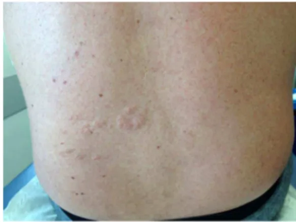
A clinical examination revealed a solitary, firm, non-tender nodule on the lateral surface of his right thigh, measuring about 6.5×2.8 cm (figure 1). The nodule was mobile and it was notfixed to the deep dermal layers. The examination also revealed mul-tiple yellowish-coloured, firm, non-tender papules and plaques, with diametres from 5 mm to 3 cm, on his left lumbar area and on occiput (figures 2 and 3). These lesions were isolated and confluent and they had an uneven surface, with a rough texture, resembling an orange peel. The remainder of the examination revealed the presence of eight hypopig-mented, well-demarcated, round and polygonal macules, with diametres from 0.5 to 2.0 cm. They were located on the abdomen and on the lower limbs. We also observed pink papules, with a smooth surface. They were symmetrically distribu-ted over centrofacial areas, suggesting facial angiofi -bromas. He also presented with numerous soft, pedunculated, skin coloured papules, located on neck, axillae and groin, suggestive of molluscum pendulum.
There are no nail, hair, teeth or mucosal changes to report.
In his personal history, it is important to report infantile spasms, developmental delay and mental retardation. This patient is not taking any chronic medication.
Figure 1 Nodule on the lateral surface of his right thigh.
Figure 2 Papules and plaques on his left lombar area.
Resende CIP,et al.BMJ Case Rep2013. doi:10.1136/bcr-2013-010382 1
His mother and his brother have a history of similar lesions, namely, numerous hypopigmented macules, facial angiofibromas and numerous molluscum pendulum. Moreover, they have peri-unguealfibromas on the toenails.
Clinical features suggested the diagnosis of TS. In light of this diagnosis, we continued our study.
Histopathological examination of excisional biopsy of a lesion taken from the right thigh displayed the typical features of a pilomatricoma (figure 4). In particular, it displayed small round basophilic cells adjacent to pale, eosinophilic, enucleated shadow cells. Focal areas of calcification were scattered through-out the tumour. A histological section of a lesion taken from the left lumbar area demonstrated that within the reticular dermis there were areas with randomly arranged dense, coarse collagen fibres of various sizes. Based on thesefindings, the diagnosis of collagenoma was made (figure 5).
Routine laboratory blood tests demonstrated no abnormal-ities. The echocardiogram was also within normal limits. The ultrasonographic examination revealed two small echogenic cysts in the left renal cortex.
Altogether, the clinical and pathological data suggested the diagnosis of a giant pilomatricoma in a patient with TS.
OUTCOME AND FOLLOW-UP
Our patient was referred to genetic counselling and he is also under vigilance in the dermatology, neurology, cardiology and oftalmology consultations.
DISCUSSION
TS is an autosomal dominant disorder with an incidence of 1–10 000 births.4The disease is caused by the mutation of one of two tumour-suppressor genes: TSC1 and TSC2, which encode hamartin and tuberin, respectively.3 5 Two-thirds of all
the cases are caused by sporadic mutations.2
The diagnosis of TS is based on clinical criteria.4 Genetic
testing is not required to make a diagnosis in patients who fulfil the criteria for definite TS, but it is helpful for family studies, defining reproductive risks for relatives.4Our patient was diag-nosed with TS because he had three major features for the clin-ical diagnosis of TS, namely facial angiofibromas, hypomelanotic macules and Shagreen patch. Histopathological examination of the lesion taken from the left lumbar area demonstrated the typical features of a collagenoma. The latter was compatible with a Shagreen patch, which is a connective tissue nevous composed of various amounts of vascular structures, adipose tissue, colla-gen, elastic fibre, smooth muscles and cutaneous appendages without increased vascularity.4The Shagreen patch mostly occurs
in the lumbosacral area. In addition, it typically begins to develop at the age of 2 years (approximately), affecting nearly half of the patients with TS. It is also worth noting that our patient also has a history of infantile spasms, which can occur in approximately 70% of infants with TS.4
Nearly every organ can be affected by TS and a multidiscip-linary approach is essential for an early, accurate diagnosis and proper management of the affected patients.4 5It is also
import-ant to provide genetic counselling to the affected individuals and families.4
We would also like to emphasise that our patient had a giant pilomatricoma in the right thigh. This is interesting because of the large size and the rare location of the pilomatricoma.
Pilomatricoma is a relatively common benign skin appendage neoplasm originating from the hair follicle matrix cells.1 2 6It has been observed in association with various disorders. However, to the best of our knowledge, in the literature, there is only one case, suggesting the association of pilomatricoma and TS.1According to the reported case, mutations of TS1 or Figure 3 Papules and plaques on occiput.
Figure 4 Small round basophilic cells adjacent to pale, eosinophilic, enucleated shadow cells.
Figure 5 Within the reticular dermis there were areas with randomly arranged dense, coarse collagenfibres of various sizes.
2 Resende CIP,et al.BMJ Case Rep2013. doi:10.1136/bcr-2013-010382
TS2 could contribute to β-catenin overactivity, resulting in the formation of a pilomatricoma.1
β-Catenin is a subunit of the cadherin protein complex that acts as an intracellular signal transducer. It is involved in signal-ling pathways that influence cell differentiation and prolifer-ation. It has recently been shown that β-catenin plays an important role in the formation of hair follicle-related tumours, including pilomatricomas.1 7–9 However, this is probably not the only explanation for the concomitance of TS and pilomatri-comas. If this was the only mechanism involved in this associ-ation, there would be much more cases described in the literature.
The treatment choice in cases of pilomatricoma is surgical excision.2 6Recurrences are rare after complete ressection.4 5
We presented this case because not only the diagnosis of TS and pilomatricoma in adult life is a rare phenomenon, but also because this case emphasises the rare concomitance of the two disease conditions, which could theoretically, be linked to each other.
Learning points
▸ TS is a multisystem neurocutaneous disease, which is
characterised by the formation of benign tumours or hamartomas in multiple organ systems. It is usually diagnosed early in life, although it can also be diagnosed in the adult life.
▸ Pilomatricoma is a relatively common benign skin
appendage neoplasm. It rarely exceeds 3 cm, although some very large tumours have been reported in the literature.
▸ Pilomatricoma and TS can occur simultaneously and there
could be a theoretical link between the two.
Acknowledgements We would like to thank Dr Ana Paula Vieira for helpful comments and suggestions.
Contributors CR contributed to the literature research, data collection, article writing and revision. JG contributed to the acquisition of data, interpretation of data and revision. MLD contributed to the interpretation of data and revision. CB has contributed to the acquisition of data and revision.
Competing interests None. Patient consent Obtained.
Provenance and peer reviewNot commissioned; externally peer reviewed.
REFERENCES
1 Krishna SM, Sacoolidge JC, Chiu MW. Anetodermic pilomatricoma in a patient with tuberous sclerosis.Clin Exp Dermatol2009;34:307–8.
2 Kovacic M, Rudic M, Nekic I,et al. Giant pilomatrixoma (benign calcifying epithelioma of Malherbe) of the neck and face.Dermatol Surg2007;33:340–3. 3 Jozwiack J, Wlodarski P. Hamartin and tuberin modulate gene transcription via
β-catenin.J Neurooncol2006;79:229–34.
4 Schwartz RA, Fernández G, Kotulska K,et al. Tuberous sclerosis complex: advances in diagnosis, genetics and management.J Am Acad Dermatol2007;57:189–202. 5 Jozwiack J, Jozwiack S, Wlodarski P. Possible mechanisms of disease development in
tuberous sclerosis.Lancet Oncol2008;9:73–9.
6 Perčin A, Kočer U, Ünlü R,et al. Giant pilomatrixoma in adult.Eur J Plast Surg
1996;19:215–17.
7 Kim YS, Shin DH, Choi JS,et al. The immunohistochemical patterns of theβ-catenin expression in pilomatricoma.Ann Dermatol2010;22:284–9.
8 Park SW, Suh KS, Wang HY,et al.β-catenin expression in the transitional cell zone of pilomatricoma.Br J Dermatol2001;145:624–9.
9 Mak BC, Kenerson HL, Aicher LD,et al. Aberrantβ-catenin signaling in tuberous sclerosis.Am J Pathol2005;167:107–16.
Copyright 2013 BMJ Publishing Group. All rights reserved. For permission to reuse any of this content visit http://group.bmj.com/group/rights-licensing/permissions.
BMJ Case Report Fellows may re-use this article for personal use and teaching without any further permission.
Become a Fellow of BMJ Case Reports today and you can: ▸ Submit as many cases as you like
▸ Enjoy fast sympathetic peer review and rapid publication of accepted articles ▸ Access all the published articles
▸ Re-use any of the published material for personal use and teaching without further permission
For information on Institutional Fellowships contact consortiasales@bmjgroup.com
Visit casereports.bmj.com for more articles like this and to become a Fellow
Resende CIP,et al.BMJ Case Rep2013. doi:10.1136/bcr-2013-010382 3