J Vasc Bras. 2012;11(4):324-328. Introduction
Angiomyolipomas (AML) are rare benign tumors
that account for 2% to 3% of all renal tumors1-7. hey are
twice more frequent among women1,5,6,8. Most are sporadic,
but 10% are associated with tuberous sclerosis complex (TSC)1-7,9-11.
AML are hamartomas that contain fat, blood vessels and muscle ibers1-13.
Selective arterial embolization (SAE), an efective and
safe treatment of renal angiomyolipomas (RAML),1,3,5,6,8,10,13
is used in the prophylaxis of complications of high risk tumors, to contain acute hemorrhages,1,2,4,6,9,11,14,15 to delay
invasion of the renal parenchyma1,2,4-6,8,10,11 and to reduce
bleeding during surgery as a preoperative adjuvant measure9,14,16.
he authors describe the case of a patient with bilateral RAML treated with endovascular superselective arterial embolization using microspheres.
Case report
A 55-year-old man presented with a major complaint of moderate and sporadic let lumbar pain. He denied any previous hematuria. His only systemic comorbidity was controlled hypertension.
He had been followed up by a urologist for one year due to a diagnosis of multiple RAML. Because of the
Abstract
We report a case of a patient with a major complaint of left lumbar pain, diagnosed with bilateral renal angiomyolipomas (AMLRs), with the most voluminous lesion of 6.2 cm in its largest diameter, underwent endovascular superselective arterial embolization with microspheres. he AMLRs are rare benign tumors. Most are sporadic, while a minority is associated with Tuberous Sclerosis Complex (ETC). he AMLRs larger than 4 cm must be treated due to higher risk of complications, especially hemorrhagic. A selective arterial embolization (EAS) is an efective and safe treatment for AMLRs.
Keywords: angiomyolipoma; kidney; embolization, therapeutic; microspheres.
Resumo
Relata-se um caso de um paciente com queixa principal de dor lombar à esquerda, portador de angiomiolipomas renais (AMLRs) bilaterais, com a lesão mais volumosa de 6,2 cm em seu maior diâmetro, submetido a tratamento endovascular por embolização arterial superseletiva com microesferas. Os AMLRs são tumores benignos raros. A maioria é esporádica, enquanto uma minoria está associada à Esclerose Tuberosa Complexa (ETC). Os AMLRs maiores do que 4 cm devem ser tratados devido ao maior risco de complicações, principalmente hemorrágicas. A embolização arterial seletiva (EAS) é um tratamento efetivo e seguro para os AMLRs.
Palavras-chave: angiomiolipoma; rim; embolização terapêutica; microesferas.
Endovascular treatment of renal angiomiolipoma by selective
arterial embolization
Tratamento endovascular de angiomiolipoma renal por embolização arterial seletiva
Renato Menezes Palácios1, Amanda Silva de Oliveira Góes1, Paloma Cals Albuquerque1,
Maurício Figueiredo Massulo Aguiar2, Flávio Roberto Cavalleiro de Macêdo Ribeiro3,
Adenauer Marinho de Oliveira Góes Junior4
Study carried out at the Hospital Ophir Loyola (HOL)- Belém-PA, Brazil. 1 Internos do curso de medicina, (UFPA), Belém-PA, Brazil.
2 Médico urologista, Belém-PA, Brazil.
3 Médico cirurgião vascular, angiorradiologista e cirurgião endovascular. Belém-PA, Brazil.
4 Médico cirurgião vascular, angiorradiologista e cirurgião endovascular. Professor da disciplina de habilidades médicas, curso de medicina da UFPA. Belém-PA, Brazil. Financial support: none
he devascularization of the “target tumor” and the perfusion of the rest of the let kidney were conirmed by an intraoperative control angiogram (Figure 4). Ater the procedure was complete, manual compression was applied to the puncture site for 20 minutes.
Immediately ater operation, the patient had moderate let lumbar pain, successfully controlled with analgesic drugs. No vomiting, nausea or fever was observed. he patient had no other complaints or intercurrent events, and was discharged about 24 hours ater the intervention.
He was referred to an urologist and returned to the
endovascular surgeon’s oice for evaluation on the 14th
postoperative day, when he reported no complaints.
A control MRI study was performed in the third postoperative month. Results revealed a reduction of the embolized RAML to 5.8 cm. he patient is currently under regular follow-up by his urologist and has reported no other episodes of let lumbar pain.
Discussion
In 1951, the term “angiomyolipoma” was coined
by Morgan2. Most RAML are diagnosed incidentally
because 60%12 are asymptomatic2,7,9,12,13. Although at an
unpredictable rate,14,15 tumors tend to grow6,12,14-16. greater frequency of pain and the size of the largest tumor,
he was referred to an endovascular surgeon to evaluate the possibility of embolization.
A previous MRI study showed nodular lesions with an important fat component; the lesions were found in the cortex of both kidneys, ive in the right and three in the let. Based on those imaging indings, a diagnosis of RAML was made. he largest lesion measured 6.2 cm in diameter and was found in the lower pole of the let kidney. In addition to RAML, MRI showed a simple renal cyst in the right kidney (Figure 1).
Preoperative laboratory tests were normal.
he patient underwent elective SAE for the largest RAML.
he procedure was conducted under local anesthesia and sedation. Vascular access was gained using the right common femoral artery approach, and a 5F introducer set was placed. An aortogram obtained with a 5F pigtail catheter showed that each kidney had only one artery with no signiicant atherosclerosis. It also showed hypervascular parenchymatous renal lesions, compatible with bilateral RAML.
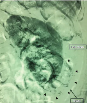
A 5F Cobra 2 catheter was used for selective catheterization of the let renal artery. An angiogram showed a patent renal artery without wall irregularities or signiicant tortuosity (Figure 2).
Superselective catheterization of the tumor feeding vessels was performed according to a road map arteriogram
and using an EmboCath® microcatheter and a Segway
guidewire (BioSphere Medical) (Figure 3). One vial of 300-500-micron Embosphere® (BioSphere Medical) was used for the embolization of feeding vessels.
Figure 1. MRI before embolization. A: Larger angiomyolipoma in lower left kidney pole. B: Simple renal cyst in right kidney. C: Renal angiomyo-lipoma in right kidney.
here is a correlation between RAML size and the
appearance of symptoms and complications1-9,14,16. he most
frequent sign, in 85% of the cases, is abdominal or lumbar pain1,6,7,10,13,16. A palpable abdominal mass6,7,12 is found in up to 53% of the cases,7 and anemia,6,7 in 21%7. Retroperitoneal
bleeding1-17 or macroscopic hematuria1,2,6,10,11 may also
occur. Large tumors may afect other organs and cause
anorexia1,10. Although unusual, renal parenchyma invasion
may occur and lead to renal failure1-3,8,10.
Ultrasound (US),2,6 CT2,6-9,11,15 or MRI2,7,8,14,15 studies are usually enough to establish a diagnosis and demonstrate the
presence of fat inside the renal mass1,2,6,7,11. Calciications,
typical of more aggressive tumors, are rare in RAML2,13.
In these cases, MRI should be used for the diferential diagnosis. For renal cell carcinomas, low signal intensity is found on T1, and high signal intensity, on T2, whereas the opposite is seen in the case of fat tissues2.
When bleeding, AML should be included in the diferential diagnosis of renal lesions, even if there is no
evidence of intralesional fat,14 as the presence of fat may
be masked by tumor hemorrhage6,14,15. Its radiological
aspect is typical, and a biopsy is rarely indicated2,13,14. As it is a hypervascular lesion, it may cause hemorrhage, which,
however, rarely afects treatment decisions2.
Angiograms show anomalous vascularization and the
formation of new vessels and microaneurysms2,4,7,9,11,15.
Vessels are more susceptible to aneurysms and rupture
because their walls have few normal elastic ibers,1,2,4,7,13
and their muscle layer is replaced with dense ibers,2 which
explains why the tumor may hemorrhage easily1,2,4,6,7,11,15,17.
Of all RAML, 94% are asymptomatic,1,2,8,16 and 60%
bleed spontaneously8.
Tuberous sclerosis complex (TSC), described by
Von Recklinghausen in 1862, is an autosomal dominant6
congenital disorder whose diagnosis is made according to
major and minor criteria2. Diicult-to-control seizures,
mental retardation and adenoma sebaceum2,6 are its three
classical signs, irst demonstrated by Campbell in 19052.
About 50% to 80% of the patients with TSC have RAML2,6.
Sporadic RAML is usually a single tumor. When
bilateral, as in our case, TSC2 should irst be ruled out,
which had already been done by the urologist in our case. he criteria for intervention are: diameter greater than 4 cm (3.5 cm for some authors)1-3,5,6,8,9,10,16 and pain,1,4,6,7,11,15-17 as in the case described here. Other indications are active
hemorrhage,1-17 changes in tumor characteristics,2-4 multiple
RAML, bilateral or unilateral if in a single kidney,2 and
patients with TSC4,6.
Embolization of these lesions was described over
20 years ago4 by Lalli et al.17 Currently, RAML are
Figure 4. Left kidney arteriogram after embolization. Devascularization of embolized tumor and perfusion of remaining renal parenchyma.
obstructed. he lack of particle homogeneity may also lead to inadequate penetration of the agent into the most distal points of the tumor vessels.
he calibrated microspheres are easy to handle. heir dilution in iodinated contrast medium and the use of zoom resources make it possible to control the low of the embolization agent during injection, and, because of their regular surface and size, they rarely obstruct the microcatheter. his agent was chosen for our case because of these characteristics.
here is no consensus in the literature about the superiority of any speciic embolization agent in the
treatment of RAML4,8. he choice should take into
consideration the surgeon’s familiarity with the embolization agent and its availability in the service where the treatment will be made.
Up to 32% of all tumors treated with SAE may continue
growing4,9. here is a positive association between the degree
of tumor volume reduction ater SAE and the percentage of
fat in the RAML8.
Tumor reduction should not be used as an isolated parameter when evaluating the eicacy of embolization. Assessments should take into consideration the
disappearance of symptoms,1,4,5,9,15 tumor growth
arrest1,4,5,9,13,15 and the absence of hemorrhage4,5,9,15.
Some postoperative complications of RAML
embolization are the postembolization syndrome1,2,4,9,13,15
(85%),2,13 renal abscess1,3,9 (5%),3 pleural efusion (3%)3 and
puncture site hematoma3,4,15. Lipiduria is a rare complication
assigned to the liquefactive necrosis of tumor fat tissue8.
Conclusions
In most cases, RAML are sporadic benign tumors. Tuberous sclerosis complex should be ruled out when the patient has multiple and bilateral tumors.
RAML larger than 4 cm should be treated because of the higher risk of complications, particularly hemorrhage.
Studies in the literature indicate that selective arterial embolization is safe and eicacious.
References
1. Sooriakumaran P, Gibbs P, Coughlin G, et al. Angiomyolipomata: challenges, solutions, and future prospects based on over 100 cases treated. BJU Int. 2009:105:101-106. http://dx.doi.org/10.1111/ j.1464-410X.2009.08649.x
2. Cerqueira M, Xambre L, Silva V, Prisco R, Santos R. Angiomiolipoma múltiplo bilateral esporádico - caso clinico. Acta Urol. 2003;20(3):63-68.
treated using embolization for diferent reasons: prevent spontaneous hemorrhage, stop active bleeding, delay
progressive tumor invasion of renal parenchyma,8,14 and
act as a preoperative adjuvant therapy to decrease bleeding during surgery16.
he main advantage of SAE over resection is the
preservation of the functional renal parenchyma2,4,5,13.
In cases of active hemorrhage, its rate of success reaches
86%2 and leads to gradual tumor reduction. Elective SAE
prevents hemorrhage2,4,6,9,13-15 in up to 94% of the cases14.
Hospital stay is usually shorter than 24 hours13.
In our case, embolization was chosen because it is a minimally invasive technique whose superselective nature preserves renal function, an important consideration in the treatment of patients that may have to undergo future interventions to treat multiple tumors.
Some of the embolization agents previously described are: gelfoam,4,9,14,17 polyvinyl alcohol (PVA) particles,4,8-10,13,14
alcohol,9,10,14,8,13,4,5,17 calibrated microspheres,4,8-10,14
coils,4,8,10,11,13,14,17 lipiodol5,8-10 and onyx,4 as well as the combined use of materials to improve the efect of embolization4,5,8,10,14,17.
Each agent has particular characteristics that deine advantages and disadvantages. Alcohol, for example, penetrates to the capillary level and promotes irreversible ischemia; its low cost makes it afordable for treatment in public health care settings in developing countries,
such as Brazil9. However, its embolization may be
unpredictable because of its radiolucency and luidity.
Although not necessary,8,10 some authors recommend the
use of occlusion balloon catheters for its infusion9 because
they make it possible to control its dispersion in the organ that receives embolization.
Another strategy is to mix alcohol and lipiodol, because lipiodol is radiopaque and, therefore, facilitates the accurate control of the low of material and increases the mixture’s
power of vascular occlusion5.
Coils should be used carefully because, once released, they block access to the most distal segments of the vessels that may have to be accessed again in early or late repeat interventions. Rupture of aneurysms in RMAL ater embolization of distal segments have been reported and may be explained by the fact that when the vessel is occluded distally to the aneurysm, the pressure on its walls increases, which predisposes to rupture. he implantation of coils inside the aneurysm or close to it may prevent its rupture4.
One of the disadvantages of PVA is the size and shape
of its irregular particles,9 and special attention should be
12. Lucky MA, Shingler SN, Stephenson RN. A case report of spontaneous rupture of a renal angiomyolipoma in a post-partum 21-year-old patient. Arch Gynecol Obstet. 2009;280:643-645. http://dx.doi. org/10.1007/s00404-009-0964-9
13. Faddegon S, So A. Treatment of angiomyolipoma at a tertiary care centre. he decision between surgery and angioembolization. Can Urol Assoc J. 2011;5(6):E138-E141. http://dx.doi.org/10.5489/ cuaj.10028
14. Incedayi M, Turba UC, Arslan B, et al. Endovascular therapy for patients with renal angiomyolipoma presenting with retroperitoneal haemorrhage. Eur Jvasc Endovasc Surg. 2010;39:739e744.
15. Dabbeche C, Chaker M, Chemali R, et al. Role of Embolization in renal angiomiolipomas. J Radiol. 2006;87:1859-67. http://dx.doi. org/10.1016/S0221-0363(06)74166-X
16. Shen H, Pan JH, Yan JN, et al. Resection of a giant renal angiomyolipoma in a solitary kidney with preoperative arterial embolization. Chin Med J. 2011;124(9):1435-1437.
17. Espinoza G, Miranda LC, Matias VAC, Fonseca JLT, Chagas VLA, Rocha FLD. Embolização pré operatória de tumores renais com microparticulas esféricas de tecnologia nacional (Spherus®-irst line brasil). Rev Col Bras Cir. 2008;35(1). http://dx.doi.org/10.1590/ S0100-69912008000100012
Correspondence
Adenauer Marinho de Oliveira Góes Junior Clínica Góes Rua Domingos Marreiros, 307, apto. 802 – Umarizal CEP 66055-210 – Belém (PA), Brazil Fone: (91) 8127-9656 E-mail: adenauer-junior@ibest.com.br
Authors’ contributions
Conception and design: AMOG, RMP, MFMA, FRCMR Analysis and interpretation: ASOG, PCA Data collection: RMP, AMOG Writing the article: RMP, AMOG Critical revision of the article: AMOG Final approval of the article*: AMOG Statistical analysis: not applicable Overall responsibility: AMOG *All authors have read and approved the inal version submitted to J Vasc Bras. 3. Ozkara H, Özkan B, Solok V. Management of renal abscess
formation after embolization due to renal angiomyolipomas in two cases. Int Urol Nephrol. 2006;38:427-429. http://dx.doi. org/10.1007/s11255-005-0253-x
4. Katsan K, Sabharwal T, Ahmad F, Dourado R, Adam A. Onyx embolization of sporadic angiomyolipoma. Cardiovasc Intervent Radiol. 2009;32:1291-1295. http://dx.doi.org/10.1007/s00270-008-9481-7
5. Chick C, Tan B-S, Cheng C, et al. Long-term follow-up of the treatment of renal angiomyolipomas after selective arterial embolization with alcohol. BJU Int. 2009;105:390-394. http://dx.doi. org/10.1111/j.1464-410X.2009.08813.x
6. Schneider-Monteiro ED, Lucon AM, Figueiredo AA, Rodrigues Junior AJ, Arap S. Bilateral giant renal angiomyolipoma associated with hepatic lipoma in a patient with tuberous sclerosis. Rev Hosp Clín Fac Med S Paulo. 2003;58(2):103-108. http://dx.doi. org/10.1590/S0041-87812003000200008
7. Peres L, Bader SL, Bueno AG, Espiga MC. Ruptura de Angiomiolipoma Renal Gigante. Relato de Caso. J Bras Nefrol. 2008;30(3):226-9.
8. Ishibashi N, Mochizuki T, Tanaka H, Okada Y, Kobayashi M, Takahashi M. A case of lipiduria after arterial embolization for renal angiomyolipomas. Cardiovasc Intervent Radiol. 2010;33:615-618. http://dx.doi.org/10.1007/s00270-010-9796-z
9. Lee S-Y, Hsu H-H, Chen Y-C,et al. Embolization of renal angiomyolipomas: shortterm and long-term outcomes, complications, and tumor shrinkage. Cardiovasc Intervent Radiol. 2009;32:1171-1178. http://dx.doi.org/10.1007/s00270-009-9637-0
10. Bora A, Soni A, Sainani N, Patkar D. Emergency embolization of a bleeding renal angiomyolipoma using polyvinyl alcohol particles. Diagn Interv Radiol. 2007;13:213-216.

