“Hot Cross Bun” Sign in Variant Creutzfeldt-Jakob
Disease
The “hot cross bun” sign refers to pontine cruciform hyperintensity on long TR sequences, which can be observed in multiple-system atrophy, spinocerebellar atrophy types 2 and 3,1and in parkinsonism secondary to vasculitis.2It has not been previously demonstrated in variant Creutzfeldt-Jakob disease (vCJD), to our knowledge.
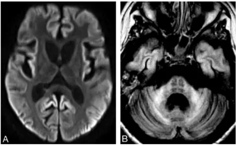
A 16-year-old girl, diagnosed with vCJD at age 14 years and de-scribed in detail elsewhere,3had progressive clinical deterioration, leading to a persistent vegetative state. MR imaging at 18 months after onset of the disease showed global brain atrophy and widespread re-stricted cortical diffusion (Fig 1A). Pontine cruciform hyperintensity was seen on fluid-attenuated inversion recovery images (Fig 1B).
It is believed that the “hot cross bun” sign results from pontine nuclei neuronal loss and pontocerebellar tract degeneration, with preserved corticospinal tracts. In vCJD, cerebellar involvement is prominent, characterized by marked neuronal loss, astrocytosis, and florid plaques.4In addition, spongiform changes have been detected in pontine nuclei.4Secondary degeneration of pontocerebellar tracts is therefore likely to occur, thus supporting our observation. The
presented case expands the differential diagnosis of neurodegenera-tive conditions in which the “hot cross bun” sign can be found.
References
1. Bu¨rk K, Skalej M, Dichgans J.Pontine MRI hyperintensities (“the cross sign”) are not pathognomonic for multiple system atrophy (MSA).Mov Disord
2001;16:535
2. Muqit MM, Mort D, Miskiel KA, et al.“Hot cross bun” sign in a patient with parkinsonism secondary to presumed vasculitis.J Neurol Neurosurg Psychiatry
2001;71:565– 66
3. Machado A, Soares H, Antunes H, et al.Variant Creutzfeldt-Jakob [corrected] disease: the second case in Portugal and in the same geographical region [ pub-lished erratum appears inJ Neurol Neurosurg Psychiatry2008;79:614].J Neurol Neurosurg Psychiatry2008;79:180 – 82
4. Ironside JW, Head MW, McCardle L, et al. Neuropathology of variant Creutzfeldt-Jakob disease.Acta Neurobiol Exp (Wars).2002;62:175– 82
J.P. Soares-Fernandes M. Ribeiro Department of Neuroradiology A´. Machado Department of Neurology Hospital de S. Marcos Braga, Portugal DOI 10.3174/ajnr.A1335
Fig 1.A, Axial diffusion-weighted image shows symmetric striatal and thalamic atrophy and widespread cortical high signal intensity. Apparent diffusion coefficient maps (not shown) revealed corresponding decreased cortical signal intensity, confirming restricted water diffusion.B, Axial fluid-attenuated inversion recovery image demonstrates pontine “hot cross bun” sign, atrophic and hyperintense middle cerebellar peduncles, global cerebellar atrophy, and cortical temporal lobe high signal intensity.
LETTERS