Letters to the Editor
Radiol Bras. 2016 Jul/Ago;49(4):267–276
267
0100-3984 © Colégio Brasileiro de Radiologia e Diagnóstico por Imagem
Letters to the Editor
Hemichorea-hemiballism: the role of imaging in diagnosing an unusual disorder in patients with nonketotic hyperglycemia
Dear Editor,
An 81-year-old man presented to the emergency room with a 4-day history of progressive confusion followed by frontal head-ache and left-sided choreiform movements. His medical history was remarkable for smoking, dyslipidemia, and poorly-controlled hypertension, with no previous diagnosis of diabetes mellitus (DM). On laboratory investigation, his serum glucose was 460 mg/dL and his glycated hemoglobin was 17.4% (consistent with a pro-longed period of undiagnosed DM). A computed tomography scan of the brain revealed hyperdensity of the right putamen without an associated mass effect (Figure 1), which suggested a diagno-sis of hyperglycemic hemichorea-hemiballism (HCHB). The patient was started on insulin, and few hours following glucose correction there was great improvement in his mental status and
a decrease in involuntary movements. On an unenhanced T1-weighted spin-echo magnetic resonance imaging (MRI) sequence obtained two weeks after initial presentation, there were hyperin-tense lesions, consistent with hyperglycemic HCHB, located in right putamen. Diffusion-weighted imaging showed no corre-sponding signal alterations. T2*-weighted imaging demonstrated bilateral punctiform hypointensities in the globus pallidus, which were presumably physiological in nature and did not match the unilateral T1 abnormality (Figure 2). The patient completely re-covered his previous cognitive and motor functions after glyce-mic control, being discharged without sequelae.
Ballistic and choreic movements are characterized by hyper-kinetic, random, involuntary movements in the proximal and dis-tal extremities, respectively(1,2). Because they usually occur
con-comitantly, the term HCHB was created to unify these signs into a clinical syndrome when presented unilaterally. Although HCHB syndrome is secondary to lesions in the basal ganglia, the source of the neuronal damage is controversial, the putative mechanisms including disruption of the blood-brain barrier, decreased thalamic gamma-aminobutyric acid input secondary to anaerobic metabo-lism, small hemorrhages in the striatal region, hyperviscosity re-lated to hyperglycemia, and Wallerian degeneration of putaminal white matter with protein desiccation(3,4).
Vascular cerebral lesions constitute the most common cause of HCHB(2). Hyperglycemia is considered an important, albeit
rare, risk factor for the development of HCHB, which is most com-monly seen in elderly female patients with uncontrolled DM. The predominance of Asian patients in the published data suggests an ethnic predisposition. The clinical course tends to vary depend-ing on the patient’s glycemic status—the hemiballism and hemi-chorea usually start together with the hyperglycemia, resolving after its correction(2,5).
Computed tomography findings of hyperglycemic HCHB include unilateral hyperdensity in the basal ganglia contralateral to the affected site. On T1-weighted MRI scans, the most common finding is signal hyperintensity in the caudate nucleus and puta-men, usually sparing the internal capsule(1,6). The apparent
dif-fusion coefficient and difdif-fusion-weighted MRI generally indicate
Figure 2. MRI findings two weeks after the initial presentation. A: Unenhanced T1-weighted spin-echo sequence showing a hyperintense lesion in the right putamen (arrowhead). B: Diffusion-weighted imaging sequence showing no restriction. C: T2*-weighted imaging showing bilateral hypointensities, presumably due to physiologic calcifications (arrows), in the globus pallidus.
A
B
C
Letters to the Editor
Radiol Bras. 2016 Jul/Ago;49(4):267–276
268
http://dx.doi.org/10.1590/0100-3984.2015.0037
Felipe Welter Langer1, Gustavo Suertegaray1, Daiane dos Santos1, Giordano Rafael Tronco Alves1, Carlos Jesus Pereira Haygert1
1. Hospital Universitário de Santa Maria (HUSM) – Universidade Federal de Santa Maria (UFSM), Santa Maria, RS, Brazil. Mailing address: Dr. Felipe Welter Langer. Departamento de Radiologia e Diagnóstico por Imagem, Hospital Universitário de Santa Maria – Universidade Federal de Santa Maria. Santa Maria, RS, Brazil, 97105-900. E-mail: [email protected].
restricted diffusion. There is typically no gadolinium enhancement. After glycemic correction, similarly to the clinical findings, such regions tend to return to normal signal intensity.
It is important to highlight the role of susceptibility-weighted imaging (SWI) in differentiating between changes seen in HCHB and areas of calcification or hemorrhage, which represent the most common differential diagnoses. Calcium and blood deposits both generally manifest as hyperintensities on T1-weighted images with corresponding hypointensities on T2*-weighted images and SWI; conversely, HCHB changes tend to present as unilateral hyperintensities on T1-weighted images with no matching changes on T2*-weighted images or SWI(7,8).
REFERENCES
1. Shan DE, Ho DMT, Chang C, et al. Hemichorea-hemiballism: an ex-planation for MR signal changes. AJNR Am J Neuroradiol. 1998;19:863– 70.
2. Postuma RB, Lang AE. Hemiballism: revisiting a classic disorder. Lan-cet Neurol. 2003;2;661–8.
3. Narayanan S. Hyperglycemia-induced hemiballismus hemichorea: a case report and brief review of the literature. J Emerg Med. 2012;43:442–4. 4. Wintermark M, Fischbein NJ, Mukherjee P, et al. Unilateral putaminal
CT, MR, and diffusion abnormalities secondary to nonketotic hypergly-cemia in the setting of acute neurologic symptoms mimicking stroke. AJNR Am J Neuroradiol. 2004;25:975–6.
5. Hawley JS, Weiner WJ. Hemiballismus: current concepts and review. Parkinsonism Relat Disord. 2012;18:125–9.
6. Zaitout Z. CT and MRI findings in the basal ganglia in non-ketotic hyperglycaemia associated hemichorea and hemi-ballismus (HC-HB). Neuroradiology. 2012;54:1119–20.
7. Chavhan GB, Babyn PS, Thomas B, et al. Principles, techniques, and applications of T2*-based MR imaging and its special applications. Radiographics. 2009;29:1433–49.
8. Hansford BG, Albert D, Yang E. Classic neuroimaging findings of nonketotic hyperglycemia on computed tomography and magnetic reso-nance imaging with absence of typical movement disorder symptoms (hemichorea-hemiballism). J Radiol Case Rep. 2013;7:1–9.
Anterior cerebral artery aneurysm rupture presenting as hemorrhage in the splenium of the corpus callosum
Dear Editor,
A 43-year-old, right-handed male presented with a three-day history of severe, holocranial headache. Three weeks prior, he had experienced another series of severe, pulsatile headaches accom-panied by fever, malaise, and paresthesia of the second and third digits of the left hand. The neurologic examination revealed apraxia of the left hand and constructional apraxia of the right hand, with-out sensorimotor or cerebellar deficits, consistent with callosal disconnection syndrome.
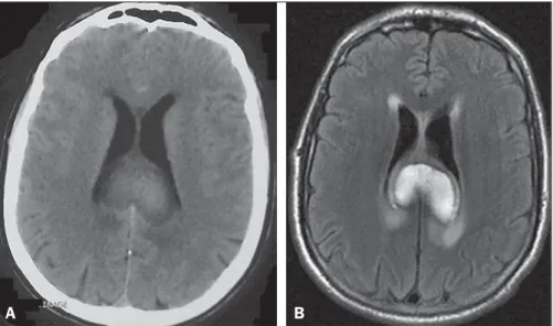
Non-contrast computed tomography and magnetic reso-nance imaging demonstrated a hematoma in the splenium of the corpus callosum (Figure 1). No subarachnoid blood was visual-ized. Cerebral angiography revealed evidence of recent aneurysm rupture at the junction of the A1 and A2 segments of the right anterior cerebral artery (ACA) and vasospasm of the distal right ACA (Figure 2A). The decision was made to embolize the aneu-rysm with detachable coils (Figure 2B). At the conclusion of the procedure, there was complete embolization of the aneurysm sac,
without disruption of the integrity of the intracranial arteries or defect in the brain parenchyma. The remainder of the hospital stay was uneventful, and the patient was discharged on post-ad-mission day 11 with a prescription for a 6-day tapered course of nimodipine. Angiography performed at 6 months of follow-up demonstrated that the coils remained in place within the aneu-rysm sac (i.e., the aneuaneu-rysm sac continued to be occluded).
Reports of remote intraparenchymal hemorrhage as a pre-senting finding of aneurysm rupture are rare(1). For example, in a
group of 460 patients with subarachnoid hemorrhage, Abbed et al.(2) reported 116 cases of intraparenchymal hematoma
forma-tion, none of which appeared to be proximal to the site of aneu-rysm rupture. In fact, our search of the literature revealed only isolated cases of remote focal hemorrhage. In 2002, Friedman et al.(3) described a ruptured anterior communicating artery
aneu-rysm associated with a perisylvian frontotemporal hematoma. Also in 2002, Lee et al.(4) described the case of a patient with ruptured
saccular ACA aneurysm that evolved to hemorrhage of the left putamen. In 2005, Paus et al.(5) reported an even more
perplex-ing case of anterior communicatperplex-ing artery aneurysm rupture, with adjacent subarachnoid hemorrhage and focal hematoma in the
Figure 1. Non-contrast computed tomography (A) and T2-weighted fluid attenuated inversion recovery mag-netic resonance imaging (B) demonstrating a large, heterogeneously enhancing mass in the splenium of the corpus callosum, consistent with a focal collec-tion of intraparenchymal blood. No evidence of sub-arachnoid hemorrhage is apparent.