2011 May-Jun;19(3):557-64
www.eerp.usp.br/rlae
Corresponding Author: Adriano Menis Ferreira
Universidade Federal de Mato Grosso do Sul. Departamento de Enfermagem Av. Ranulpho Marques Leal, 3220
Distrito Industrial
CEP: 79610-100 Três Lagoas, MS, Brasil E-mail: [email protected]
Condition of Cleanliness of Surfaces Close to Patients in an
Intensive Care Unit
Adriano Menis Ferreira
1Denise de Andrade
2Marcelo Alessandro Rigotti
3Maria Verônica Ferrareze Ferreira
4Surface cleaning is a well-known control procedure against the dissemination of
microorganisms in the hospital environment. This prospective study, carried out in an
intensive care unit over the course of 14 days, describes the cleaning/disinfection conditions
of four surfaces near patients. In total, 100 assessments of the surfaces were carried out after
they were cleaned. Three methods were used to evaluate cleanliness: a visual inspection,
an adenosine triphosphate (ATP) bioluminescence assay and testing for the presence of
Staphylococcus aureus and meticillin-resistant Staphylococcus aureus/MRSA. Respectively,
20%, 80% and 16% of the assessments by the visual method, ATP and the presence of
Staphylococcus aureus/MRSA failed. There were statistically significant differences (p<0.05)
between the rates of failure of the cleaning using the ATP method, compared to the visual
and microbiological methods. The visual inspection was not a reliable measure to evaluate
surface cleanliness. The results demonstrated that the adopted cleaning routine should be
reconsidered.
Descriptors: Staphylococcus aureus; Equipment Contamination; Cross Infection; Methicillin
Resistance; Housekeeping, Hospital.
1 RN, Post doctor in Nursing. Adjunct Professor, Universidade Federal de Mato Grosso do Sul, MS, Brazil. E-mail: [email protected].
2 RN, Free Lecture. Associate Professor, Escola de Enfermagem de Ribeirão Preto, Universidade de São Paulo, WHO Collaborating
Centre for Nursing Research Development, SP, Brazil. E-mail: [email protected].
3 RN, Masters’s Student in Nursing, Escola de Enfermagem de Ribeirão Preto, Universidade de São Paulo, WHO Collaborating Centre
for Nursing Research Development, SP, Brazil. E-mail: [email protected].
4 RN, Doctoral Student in Nursing, Escola de Enfermagem de Ribeirão Preto, Universidade de São Paulo, WHO Collaborating Centre
Condições de limpeza de superfícies próximas ao paciente, em uma unidade
de terapia intensiva
A limpeza das superfícies é reconhecidamente medida de controle da disseminação de microrganismos no ambiente hospitalar. Este estudo prospectivo, realizado em uma unidade de terapia intensiva, durante 14 dias, teve como objetivo descrever as condições de limpeza/desinfecção de quatro superfícies próximas do paciente. Cem avaliações das superfícies foram realizadas após o processo de limpeza. Utilizaram-se três métodos para avaliar a limpeza: inspeção visual, adenosina trifosfato (ATP) bioluminescência e presença de Staphylococcus aureus/MSRA. Respectivamente, 20, 80 e 16% das avaliações pelos métodos visual, ATP e presença de Staphylococcus aureus/MSRA foram consideradas
reprovadas. Houve diferenças estatisticamente signiicantes (p<0,05) entre as taxas de
reprovação da limpeza utilizando os métodos ATP, comparado ao visual e microbiológico.
A inspeção visual não se mostrou medida coniável para avaliar a limpeza das superfícies.
Os resultados demonstram que a rotina de limpeza adotada precisa ser revista.
Descritores: Staphylococcus aureus; Contaminação de Equipamentos; Infecção Hospitalar; Resistência a Meticilina; Serviço Hospitalar de Limpeza.
Condiciones de limpieza de supericies próximas al paciente en una
unidad de terapia intensiva
La limpieza de las supericies es reconocidamente una medida de control de la diseminación
de microorganismos en el ambiente hospitalario. Este estudio prospectivo, realizado en una unidad de terapia intensiva, durante 14 días, tuvo como objetivo describir las
condiciones de limpieza/desinfección de cuatro supericies próximas al paciente. Cien evaluaciones de las supericies fueron realizadas después del proceso de limpieza. Se
utilizaron tres métodos para evaluar la limpieza: inspección visual, adenosín trifosfato (ATP) bioluminiscencia y presencia de Staphylococcus aureus/MSRA. Respectivamente, 20%, 80% y 16% de las evaluaciones por los métodos: visual, ATP y presencia de Staphylococcus aureus/MSRA, fueron consideradas reprobadas. Hubo diferencias
estadísticamente signiicativas (p<0.05) entre las tasas de reprobación de la limpieza
utilizando los métodos ATP, comparado al visual y al microbiológico. La inspección visual
no se mostró una medida coniable para evaluar la limpieza de las supericies. Los resultados demostraron que la actual rutina de limpieza precisa ser modiicada.
Descriptores: Staphylococcus aureus; Contaminación de Equipos; Infección Hospitalaria; Resistencia a la Meticilina; Servicio de Limpieza en Hospital.
Introduction
Although the role of the healthcare environment in the spread of some infections is far from universally agreed upon, circumstantial evidence suggests that contaminated hospital environmental surfaces can be a risk factor for infection caused by some pathogens.
Cleaning consists of the removal of dirt or contaminants found on surfaces using mechanical (friction), physical (temperature) or chemical (sanitizing) means, during a given period of time. The cleaning of a patient’s hospitalization unit should be done on a daily basis, or whenever needed, being done before and not
at the same time as loor cleaning. The cleaning of
horizontal surfaces which have contact with patient’s and team’s hands deserve more attention, such as door handles, telephones, light switches, bed rails, nurse call buttons and others(8).
Cleaning has never been regarded as an evidence-based science and consequently receives little attention
from the scientiic community(9). Since there are
no scientiic standards to measure the effect of an
individual cleaner, or assess environmental cleanliness,
inding the evidence to beneit the control of infection
is further hampered(10). Cleaning is routinely monitored
by visual audits. While looking to see if a ward is ‘clean’ may aesthetically satisfy, it does not provide a reliable assessment of the infection risk for an individual patient in that ward(11). The organisms that cause infection are
invisible to the naked eye and their existence is not necessarily associated with any visible signs(10).
Sites that are frequently touched by hands are thought to present the greatest risk for patients, for instance, those surfaces situated right beside a patient(12-14). The responsibility for cleaning near-patient
sites commonly and frequently touched by hands does not always rest with the ward cleaners, however, since beds, drip stands, lockers and over-the-bed tables are more commonly cleaned by nurses(13-14). Nurses
are also responsible for the decontamination of more delicate clinical equipment. This overlapping of cleaning responsibilities has created some confusion; it has also meant that cleaning opportunities for some items are missed or abandoned(15).
Methods for monitoring the effectiveness of cleaning procedures include the visual assessment of
surfaces, application of luorescent dye to surfaces with
subsequent assessment of residual dye after cleaning, determination of aerobic colony counts, an indicator organism and detection of adenosine triphosphate (ATP) on surfaces. Few investigators have evaluated ATP bioluminescence methods for monitoring cleanliness in hospitals(9,11,16-17).
This study describes the conditions of cleanliness of surfaces using three different methods after routine daily cleaning.
Methods
A prospective study was carried out over a period of two weeks in January 2010 in a medical and general surgery Intensive Care Unit (ICU) in a Philanthropic Hospital. The study ICU has 10 beds for patients over 18 years of age. There was a 100% occupation level throughout the duration of the study.
The four environmental surfaces (bed rails, crank, bedside table, buttons of infusion pump) were selected for culturing after routine cleaning without notifying the cleaning staff (responsible for cleaning the ceilings,
walls and loors) or staff nurses (nursing technicians
and auxiliaries) in order to minimize changes in their behaviors. These objects were chosen because they are frequently touched, potentially exposing medical staff and patients(18).
According to the formally established routine, a staff nurse cleans the patient unit, including the furniture and equipment around the patient, with a cotton cloth soaked in 70% alcohol (w/v). The same cloth is used to clean at least two patient units, given that each nursing auxiliary/technician is responsible for two patients, which is noteworthy. Each cotton cloth is changed only when a staff nurse recognizes it is visibly dirty, with the exception of isolation rooms in the ICU.
During each monitoring period, samples were collected within 10 minutes of the completion of the morning cleaning session, every day, for 14 days. This allows a description of the condition of cleanliness for the surfaces rather than contamination after cleaning.
The materials at testing sites were mostly stainless steel or laminate plastic covering for wood. For each site, the general surface condition, the presence of moisture, and visual cleanliness were noted. The visual assessments were done by one person using standardized descriptors. The presence of adenosine triphosphate (ATP), which is derived from organic soil and microorganisms, at each site was assessed by a rapid hygiene test of ATP bioluminescence, using the Biotrace Cleantrace system (3M Clean-Trace ATP System; 3M)(17). We targeted S.
aureus, including Meticillin-resistant Staphylococcus aureus (MRSA), as an indicator organism(10-11).
Rev. Latino-Am. Enfermagem 2011 May-Jun;19(3):557-64.
(RLU) and (iii) an Staphylococcus aureus/MRSA ‘pass’ results from the absence of any detection of MRSA on a surface(10-11,18-19).
Petri FilmTM (3M™, St Paul, MN, USA) Staph
Express 3MTM plates prepared with modiied
Baird-Parker chromatogenic medium, Staphylococcus aureus
selective and differential, were used for the collection of microorganisms. The plates were pressed onto the surface for 1 minute. At the end of each collection,
plates were identiied with: date, time and place of
collection. They were then stored in polystyrene boxes
and transported to the Microbiology Laboratory.
A sampling area of 30 cm² and incubation at 35°C for
24-48h was adopted for the PetriilmTM model. Readings
of Petriilm™ plates were done using a stereomicroscope (Nikon, JP) under relected light and were quantitatively
evaluated through CFU (Colony Forming Units). Red-violet colonies were considered to be Staphylococcus aureus.
Methicillin susceptibility was tested by the oxacillin resistance screening test. Petri plates containing
Muller-Hinton agar, with 4% Sodium Chloride and 6 μg/
ml oxacillin added, known as MRSA medium (Probac do Brasil®), were used. These microorganisms were
transplanted into Brain Heart Infusion (BHI) broth and incubated at 37ºC for 24 hours. After this period, they were inoculated onto plates and incubated at 37ºC for 24 and 48 hours. Any growth on the plate was considered to be MRSA.
The data collected from all of the samples were input into the Statistical Package for the Social Sciences, version 15.0 (SPSS), for statistical analysis. The RLU values for four frequently touched surfaces were compared using the Kruskal-Wallis 1-way analysis-of-variance test. Differences in proportion were compared by means of the χ2 test. In all cases signiicance was set
at least at p<0.05.
Results
Four surfaces were selected for environmental surface cleanliness testing, using three different methods. In total, 100 visual assessments, 100 ATP measurements, and 100 Staphylococcus aureus
determinants were recorded.
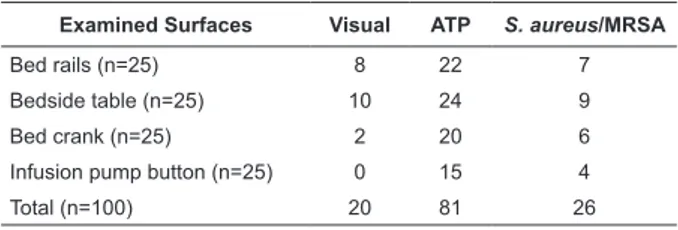
The majority of surfaces were dry and visually free from dirt, dust, stains and smears. Twenty surfaces, 10 bedside tables, 8 bed rails and 2 cranks failed due to sticky deposits.
Failure rates for surface cleanliness, using the different methods, varied considerably (Table 1).
Differences in ATP, visual and microbiological failure
rates (Table 2) were signiicant (p<0.05) and consistent,
and varied from 14% to 18%. The differences between visual and microbiological failure rates were not
signiicantly different. The differences between ATP and Staphylococcus aureus/MRSA failure rates were not
signiicant and varied from 12% to 18%.
Examined Surfaces Visual ATP S. aureus/MRSA
Bed rails (n=25) 8 22 7
Bedside table (n=25) 10 24 9
Bed crank (n=25) 2 20 6
Infusion pump button (n=25) 0 15 4
Total (n=100) 20 81 26
Table 1 – Failure rates (%) after cleaning using different assessment methods for surfaces near patients in Intensive Care Unit
ATP, Adenosine Triphosphate.
MRSA= Meticillin-resistant Staphylococcus aureus.
Table 2 – Differences in failure rates after cleaning, between visual and two other assessment methods for surfaces near patients in Intensive Care Unit
Examined Surfaces ATP (%) S. aureus/MRSA (%)
Bed rails 14 1
Bedside table 14 1
Bed crank 18 4
Infusion pump button 15 4
Total 60 10
ATP, Adenosine Triphosphate.
MRSA= Meticillin-resistant Staphylococcus aureus.
Failure rates provide an indication of cleaning
eficacy in relation to benchmark values but do not
provide an indication of the extent of failure. A summary of the overall ATP data to illustrate mean, median values and the range of data points is provided in Table 3. Wide variations in counts, using ATP, were found between sites. The ATP results, after cleaning, varied from 34 RLUs to 7201.
Surface sample Mean (RLU)
Median (RLU)
Range (RLU)
Bed rails (n=25) 983 160 72-7201
Bedside table (n=25) 830 398 102-2341
Bed crank (n=25) 388 121 54-4654
Infusion pump button (n=25) 509 354 34-3672
Table 3 – Adenosine Triphosphate (ATP) readings of samples obtained from 4 frequently touched surfaces near patients in intensive care unit after daily cleaning
It can be seen that there is no relationship between ATP failures and microbiological failures. It can be seen, too, that there is relationship between microbiological failures and visual failures (p<0.05). However, only one indicator microorganism was used, which may have
inluenced this result.
In the present study, 80% of surfaces in the ward were considered visually clean after cleaning. Using visual assessment, most sites would have been considered acceptably clean, but when benchmark ATP values were applied, only 19% were considered clean after cleaning.
After routine cleaning, S. aureus was most frequently isolated from bedside tables (six times), bed rails (four times), and bed cranks and infusion pump buttons both had positive results three times. Although only a low number of samples were available at each site, there were no apparent differences in isolation frequencies between the surfaces. Only six were
identiied as meticillin resistant.
Discussion
The aim of cleaning should be to keep surfaces visibly clean, to disinfect commonly touched surfaces more frequently than surfaces not commonly touched, and to clean up spills promptly(1-3). Thus, near patient
(e.g. chart tables, bed frames) and frequently touched (bed crank, bed rail, Infusion pump button) environmental surfaces may become contaminated with epidemiologically important microbes and should be cleaned regularly, as well as at patient discharge as per hospital policy.
Although the recommendation of a Brazilian regulatory body(8) is to irst clean the target surface with
soap and detergent, during the accomplishment of this study, cloths soaked in alcohol, applied directly to the surfaces, was observed. This can undermine the process of disinfection.
The results indicate that visual assessment, on its own, was an unreliable indicator of surface cleanliness and as a means for assessing the effectiveness of cleaning protocols. The visual assessment method used in this study, as shown by others, proved the least sensitive method for assessing cleanliness. The disparity is especially clear when compared with such rapid hygiene-testing methods as ATP bioluminescence(19-23).
Visual assessment of cleanliness in isolation can be overestimated. Instead, an integrated approach to monitoring cleanliness is recommended. Previous
studies have identiied poor standards of cleanliness in hospitals, often with normal cleaning resulting in no improvement (i.e. reduction) in ATP or microbiological levels(11,16-17,19).
An early study(11) speciically examined concurrent
visual assessment of hospital environments against chemical (bioluminescence detection) and microbiological methods of measuring organic and microbial soil. While 82% of wards seemed visibly clean (after cleaning), only 30% were microbiologically clean, and only 25% were free from organic soil. Another study(19) has evaluated
the effectiveness and thoroughness of routine cleaning activities in hospitals. It compared 2 standardized, observation-based audit guidelines with a risk-based audit tool used in conjunction with rapid environmental testing via an ATP bioluminescence tool for several observation periods in 4 hospitals. Although 90% of the sites tested appeared visually clean immediately after routine disinfection/cleaning activities, none of the sites were found to be effectively sanitized using the ATP bioluminescence monitor, and only 10% met bacteriologic food-handling standards. In comparison, another study(23) showed that 93.3% of areas were
visibly clean, 92% were microbiologically clean and 71.5% were free from organic soil.
The present study did not show a correlation between ATP and Staphylococcus aureus/MRSA values,
a inding replicated by others(23). However, as the two
techniques measure different parameters, an integrated approach to monitoring cleaning regimens may be the most useful. Indicator organisms such as MRSA indicate contamination and do relate to a potential risk of infection. It has been shown that 1-27% of general ward surfaces harbor MRSA(1).
Microbiological testing may or may not correlate with ATP readings, since the two techniques measure different parameters. Microbiological methods detect residual micro-organisms (usually bacteria), which should decrease as a result of cleaning. The magnitude of any decrease will depend on the method, materials and chemicals used. ATP bioluminescence is a measure of cleanliness that detects organic soiling (microbial and non-microbial ATP)(18).
Staphylococci were found on surfaces after the existing cleaning regimen, of which 16% were presumptive S. aureus/MRSA. Other studies on this topic have reported contamination rates in isolation rooms, such as 27.0%,(24) and 50%(22).
Rev. Latino-Am. Enfermagem 2011 May-Jun;19(3):557-64.
protocols to prevent HAIs, isolation precautions and hand hygiene is recommended in order to control it(25).
Numerous studies illustrate that many different inanimate surfaces in hospitals can become a reservoir for MRSA (1-23). Several studies speciically address environmental
MRSA contamination within isolation units(4-7,11-12,18-23).
However, the various studies of MRSA detection on surfaces in isolation rooms are generally not comparable, since the patient characteristics, the microbiological screening methods, the sampling regimen, as well as the manner, frequency, and effectiveness of cleaning and disinfection methods vary considerably.
Environmental contamination may contribute to the transmission of healthcare pathogens when healthcare workers contaminate their hands or gloves by touching contaminated surfaces, or when patients come into direct contact with contaminated surfaces(24).
Contaminated environmental surfaces that are commonly touched by patients and/or staff may act as sources for hand transfer. In support of this, a study of 12 nurses(12) demonstrated that ive (42%) of the 12
contaminated their gloves with MRSA while performing activities that required no direct patient contact but involved touching objects in the rooms of MRSA patients. In another study, 31% of volunteers who touched bed rails and over-the-bed tables in patient rooms contaminated their hands with S. aureus (35% of which were an MRSA strain)(12). When volunteers touched bed
rails and over-the-bed tables in unoccupied rooms that
had been given a inal cleaning, as opposed to a daily
one, 7% contaminated their hands with S. aureus(13).
The role of contaminated environmental surfaces in the transmission of healthcare-associated pathogens is also supported by the fact that cleaning and/or disinfection of the environment can reduce the incidence of healthcare-associated colonization or infection. However, evidence for the effect of basic cleaning on reducing the acquisition rate of MRSA in hospitals is scant. Studies have demonstrated that an intervention consisting of increased cleaning, Environmental Services staff education, use of a black-light monitoring system and the use of ATP Bioluminescence improved cleaning and decreased the likelihood of positive cultures for either MRSA(16-17,23).
The ATP and microbiological results after cleaning varied greatly; this has been previously reported(11)
and generally indicates inconsistencies in the quality of cleaning.
The present results indicate considerable levels of invisible organic soiling remaining on surfaces
after cleaning. In the present study it is possible that irregularly or infrequently changing cleaning materials was a source of contamination. Results obtained with routine cleaning may in part relate to the use of reusable cleaning materials rather than disposable ones, which were not changed at adequate intervals in the existing routine protocol, and are known to spread contamination(26). It is likely that a number of the
failure rates in ATP/microbiological counts after cleaning were as a result of dirt and/or microorganisms being redistributed rather than removed by cleaning. Simple changes to the cleaning processes used in hospitals can achieve substantial improvements leading to a reduction in the residual surface levels of ATP, indicator organisms, and methicillin-resistant Staphylococcus aureus(22).
If cleaning is intended to remove pathogens from a surface, it is necessary that cleaning be able to reduce residual organic material to a low level. Thus, a cleaning protocol that fails to achieve benchmark values for the removal of organic soil, as determined by a sensitive
ATP test, is unlikely to be it for that purpose. In a
hospital environment, this would necessitate either reassessment of staff adherence to the protocol, or the adoption of new cleaning methods or frequencies.
Microbiological assessment in speciic instances, and a
more general use of sensitive ATP testing in training and process management, may be one way of formulating an integrated and cost effective cleaning assessment strategy(21).
This study has limitations. A convenience sample of only four objects does not represent the ICU as a whole and there may be items that could have been positive for MRSA but were not sampled. Samples before cleaning were not measured, which only allows describing the cleanliness conditions of surfaces close to patients. Financial constraints limited the amount of samples taken.
Further investigations of the clinical signiicance
of hospital environmental contamination and of more effective cleaning methods are required.
Conclusion
Visual assessment alone did not always provide a meaningful measure of surface cleanliness or cleaning
eficacy and should be used only as the irst stage in an
integrated monitoring program.
reconsidered. A well designed cleaning schedule should specify monitoring and the corrective action to be taken if, after cleaning, the site is still not cleaned satisfactorily. In the present study, the cleaning program required neither monitoring nor corrective action. Simple changes to the cleaning processes used in hospitals can achieve substantial improvements leading to a reduction in the residual surface levels of ATP, indicator organisms, and methicillin-resistant Staphylococcus aureus.
References
1. Boyce JM. Environmental contamination makes an important contribution to hospital infection. J Hosp Infect. 2007;65(Suppl. 2):50-4.
2. Dettenkofer M, Spencer RC. Importance of environmental contamination e a critical view. J Hosp Infect. 2007;65(Suppl. 2):55-7.
3. Fraise AP. Decontamination of the environment. J Hosp Infect. 2007;65(Suppl. 2):58-9.
4. Hardy KJ, Oppenheim BA, Gossain S, Gao F, Hawkey PM, A study of the relationship between environmental contamination with meticillin-resistant Staphylococcus aureus (MRSA) and patients’ acquisition of MRSA. Infect Control Hosp Epidemiol. 2006;27:127-32.
5. Sexton T, Clarke P, O’Neill E, Dillane T, Humphreys H. Environmental reservoirs of meticillin-resistant
Staphylococcus aureus in isolation rooms: correlation with patient isolates and implications for hospital hygiene. J Hosp Infect. 2006;62:187-94.
6. Lemmes SW, Hafner H, Zolldann D, Stanzel S, Lutticken R. Distribution of multi-resistant gram-negative versus gram-positive bacteria in the hospital inanimate environment. J Hosp Infect. 2004;56:191-7.
7. Al-Hamad A, Maxwell S. How clean is clean? Proposed methods for hospital cleaning assessment. J Hosp Infect. 2008;70:328-34.
8. Agência Nacional de Vigilância Sanitária Segurança do
paciente em serviços de saúde: limpeza e desinfecção
de superfícies. Agência Nacional de Vigilância Sanitária.
Brasília: Anvisa; 2010. 116 p.
9. Dancer SJ. The role of environmental cleaning in the control of hospital-acquired infection. J Hosp Infect. 2009;73:378-85.
10. Dancer SJ. How do we assess hospital cleaning? A proposal for microbiological standards for surface hygiene in hospitals. J Hosp Infect. 2004;56:10-5. 11. Griffith CJ, Cooper RA, Gilmore J, Davies C, Lewis M. An evaluation of hospital cleaning regimes and standards. J Hosp Infect. 2000;45:19-28.
12. Bhalla A, Pultz NJ, Gries DM, Ray AJ, Eckstein EC, Aron DC, et al. Acquisition of nosocomial pathogens on hands after contact with environmental surfaces near hospitalized patients. Infect Control Hosp Epidemiol. 2004;25:164-7.
13. Dancer SJ. Importance of the environment in meticillin-resistant Staphylococcus aureus acquisition: the case for hospital cleaning. Lancet Infect Dis. 2008;8:101-13.
14. White L, Dancer SJ, Robertson C, McDonald J. Are hygiene standards useful in assessing infection risk? Am J Infect Control. 2008;36:381-4.
15. Blythe D, Keenlyside D, Dawson SJ, Galloway A. Environmental contamination due to methicillin-resistant Staphylococcus aureus (MRSA). J Hosp Infect. 1998;38:67-70.
16. Carling PC, Briggs JL, Perkins J, Highlander D. Improved cleaning of patient rooms using a new targeting method. Clin Infect Dis. 2006;42:385-8. 17. Boyce JM, Havill NL, Dumigan DG, Golebiewski M, Balogun O, Rizvani R. Monitoring the effectiveness of hospital cleaning practices by use of an adenosine triphosphate bioluminescence assay. Infect Control Hosp Epidemiol. 2009;30:678-84.
18. Griffith CJ, Obee P, Cooper RA, Burton NF, Lewis M. The effectiveness of existing and modified cleaning regimens in a Welsh hospital. J Hosp Infect. 2007;66:352-9. 19. Malik R, Cooer R, Griffith C. Use of audit tools to evaluate the efficacy of cleaning systems in hospitals. Am J Infect Control. 2003;31:181-7.
20. Larson EL, Aiello AE, Gomez-Duarte C, Lin SX, Lee L, Della-Latta P, et al. Bioluminescence ATP monitoring as a surrogate marker for microbial load on hands and surfaces in the home. Food Microbiol. 2003;20:735-9. 21. Lewis T, Griffith C, Gallo M, Weinbren M. A modified ATP benchmark for evaluating the cleaning of some hospital environmental surfaces. J Hosp Infect. 2008;69:156-63.
22. Lewis T, Gallo M, Weinbren M, Griffith CJ. An assessment of the effectiveness of modified hospital cleaning protocols using visual, ATP bioluminescence, and microbiological analysis. J Hosp Infect. 2006;64(Suppl. 1):55-6.
Rev. Latino-Am. Enfermagem 2011 May-Jun;19(3):557-64.
24. Boyce JM, Potter-Bynoe G, Chenevert C, King T. Environmental contamination due to meticillin-resistant Staphylococcus aureus: possible infection control implications. Infect Control Hosp Epidemiol. 1997;18:622-7.
25. Fortaleza CR, Melo EC de, Fortaleza CMCB. Nasopharyngeal colonization with methicillin-resistant
staphylococcus aureus and mortality among patients in an intensive care unit. Rev. Latino-Am. Enfermagem. [periódico na Internet]. out 2009 [acesso 15 fev 2010]; 17(5): 677-82. Disponível em: http://www. scielo.br/scielo.php?script=sci_arttext&pid=S0104-1692009000500013&lng=pt. doi: 10.1590/S0104-11692009000500013.
26. Moore G, Griffith CJ. A laboratory evaluation of the decontamination properties of microfiber cloths. J Hosp Infect. 2006;64:379-85.