Evaluation of levels of cortisol in saliva
using electro-chemical luminescence in
low-risk and high-low-risk pregnancies
Avaliação do cortisol salivar por
electroquimioluminecência em gestantes de
baixo e de alto risco
Ana Carla P. Montenegro
1
Viviane Rosado D’ Assunção
2
Monique Gabrielli B. Luna
3
Pollyanna Valente N. Raposo
4
Francisco Bandeira
5
1-5Faculdade de Ciências Médicas. Universidade de Pernambuco. Rua Arnóbio Marques, 310. Recife, PE, Brasil. CEP: 50.100-130. E-mail: [email protected]
Abstract
Objectives: to compare the levels of cortisol (cortisolemia refers to the level of cortisol in blood) in women with a high-risk pregnancy compared with those with a low-risk pregnancy, by way of evaluation of levels of cortisol in saliva, using the electroche-mical luminescence technique (ECL).
Methods: 38 women aged between 17 and 40 years in the third trimester of pregnancy were divided in two groups: 20 low-risk pregnancies and 18 high-risk ones. Cortisol in saliva was collected at midnight and measured using ECL. The mean levels of cortisol in saliva in the two groups were compared using the Kruskal-Wallis test.
Results: the mean systolic and diastolic pressure was normal in both groups. The levels of cortisol in the saliva of women with high-risk pregnancies was significantly higher than those for the low-risk preg-nancy group (20.2 (±21,1) nmol/L vs 11.4(±16.2) nmol/L; p=0.007).
Conclusions: a high risk pregnancy involves higher levels of cortisol than a low-risk one. The levels of cortisol in saliva, as measured using ECL, can be used to identify hypercortisolism in pregnancy.
Key words
Cortisol, Pregnancy, Pregnancy, high-riskResumo
Objetivos: comparar os níveis de cortisol em mulheres com gravidez de alto risco em relação às gestantes de baixo risco, por meio da avaliação do cortisol salivar utilizando a técnica da electroquimio-luminescência (EQL).
Métodos: foram estudadas 38 mulheres de 17a 40 anos de idade, no terceiro trimestre de gestação, divi-didas em dois grupos: 20 gestantes de baixo risco e 18 gestantes de alto risco. O cortisol salivar foi cole-tado à meia-noite e medido através da EQL. As médias do cortisol salivar foram comparadas entre os dois grupos de gestantes através do teste de Kruskal-Wallis.
Resultados: a média das pressões sistólica e diastólica foi normal nos dois grupos. O cortisol salivar das gestantes de alto risco foi significativa-mente mais elevado do que das gestantes baixo risco: (20,2 (±21,1) nmol/L vs 11,4(±16,2) nmol/L; p=0,007).
Conclusões: a gestação de alto risco cursa com níveis mais elevados de cortisol quando comparada à gestação de baixo risco. O cortisol salivar, medido pelo EQL mostrou-se promissor para identificar o hipercortisolismo na gestação.
Introduction
Human gestation brings about changes in the activity
of most endocrine systems in a woman, including the hypthalamic-pituitary-adrenal axis (HPA).1These
alterations in the HPA axis are important for
main-taining an adequate environment for growth and
development of the fetus, since an excess or lack of cortisol results in disruption of maternal-fetal
home-ostasis.2-4
Excess maternal corticoid during pregnancy has been being observed for forty years now, with
elevated levels beginning to be detected at around
the 12thweek of pregnancy.5The difficulty studying alterations in the HPA axis in a pregnant woman is
that of establishing a reliable biological marker and
a practical diagnostic method which is not
influ-enced by the physiological alterations arising from pregnancy itself.5,6
The neuroendocrine changes that occur in every
pregnancy alter the parameters used by laboratory techniques for dealing with hypercortisolism.1
Increased levels of estrogen stimulate the hepatic
production of glycocorticoid carrying globulin (GCG).1 This increase in hepatic GCG, which
continues until the 12thday after birth, causes a rise
in circulating levels of cortisol linked to the protein
and occasions a temporary fall in levels of free cortisol, which, in turn, leads to a reduction in
nega-tive feedback to the HPA axis.7Consequently, levels
of the adrenocorticotrophic hormone (ACTH) rise and this stimulates the production of cortisol. The
levels of free cortisol are initially normal and then
rise during pregnancy and reach maximum levels at the end of the second and third trimesters.5Total
cortisol and free plasmatic cortisol can reach values
that are two or three times higher compared with
women who are not pregnant. These high levels of plasmatic cortisol observed in pregnant women are
equivalent to those found in Cushing’s syndrome.1
The increase in levels of free cortisol during preg-nancy also leads to restriction of the action of
cortisol during this period. Despite the increase in
serum cortisol during pregnancy, the circadian rhythm of the system is preserved,8 but it is not
known what variation in levels of cortisol is
respon-sible for complications occurring during high-risk
pregnancies. Evaluating the levels in normal preg-nant women as well as in the high risk group may
provide information on these questions.
Studies evaluating hypercortisolism using dexamethasone are difficult to interpret during
preg-nancy, owing to the alterations that normally occur
during pregnancy itself.1 Studies have shown a
smaller reduction in levels of plasmatic and urinary cortisol in pregnant women, after a suppression test
with 1 mg of dexamethasone, compared to women
who are not pregnant.1In the post-partum period, this abnormality may persist for two or three weeks
in a significant proportion of women.1-2This
reduc-tion in the suppressive acreduc-tion of dexamethasone
contributes to the GCG effect on cortisol and restric-tion of the acrestric-tion of cortisol,1,5or to the possible
antiglycocorticoid effect of progesterone on tissue.5
It is thus necessary to establish laboratory techniques and paradigms for pregnancy in order to identify
variations in levels of hormones that truly differ
from the values considered to be physiological.7 It is common to measure the levels of free
urinary cortisol when diagnosing hypercortisolism in
pregnancy.1-4The principle underlying this method
is the detection of the free fraction of cortisol, thereby diminishing the influence of pregnancy on
concentrations of cortisol in serum. However, this
diagnostic method has limitations regarding elabora-tion and interpretaelabora-tion, as collecelabora-tion is a laborious
process and the laboratory technique used is
radioimmunoassay (RIA).8At present, the “gold standard” test for measuring free urinary cortinsol
involves structural assays, such as mass
spec-troscopy, which are more accurate than
methodolo-gies using assays based on antibodies.5
Measurement of cortisol in saliva at midnight
has been used as a tracking test to identify
hypercor-tisolism in non-pregnant women.9-13This method measures the free fraction of cortisol in serum and
has sensitivity and specificity similar to that of other
established methods for tracking increases in cortisol leves.14It also has the advantage that it is a
non-invasive method and samples can easily be collected
in the patient’s own home.15The concentration of
cortisol in saliva reflects the levels of free fraction cortisol in plasma and is not affected by the amount
of saliva produced or by variations in the
concentra-tion of glycocorticoid carrying globulins caused by the use of oral contraceptives and pregnancy.12
Measurement of cortisol in pregnancy to evaluate
hormone response to psychological stress has been used as a predictor of adversities in pregnancy.13-14
The aim of this study is to compare levels of
cortisol in high-risk pregnancies by evaluating
cortisol in saliva using electrochemical lumines-cence (ECL) to study the HPA axis during
preg-nancy.
Methods
women with high and low risk pregnancies after the 24thweek of gestation, measured from the last date
of menstruation or using ultrasound parameters. The
participants were recruited at the pre-natal outpa-tients clinic of the Agamenon Magalhães Hospital,
which attends women with high- and low-risk
preg-nancies referred by the Brazilian National Health
System in the city of Recife and its surrounding metropolitan region, in the State of Pernambuco,
Brazil, between August and October 2006. Levels of
cortisol were analyzed in samples of saliva from the two groups of women with high-and low-risk
preg-nancies.
Thirty-eight pregnant women were selected, 20 with low-risk pregnancies and 18 with high-risk. The
inclusion criteria were: a good level of
under-standing on being invited, not having systemic
arte-rial hypertension, diabetes mellitusor heart disease either prior to pregnancy or related to it. The
preg-nant women were selected at the outpatients clinic
and from the wards of the above mentioned hospital and the criteria were: being in the third term of
preg-nancy, being aged between 18 and 35 years, not
using corticoid and not presenting any psychiatric disorders.
After selection the participants signed a free and
informed consent form, answered the questionnaire
to provide information on the woman and her preg-nancy, and were told how to collect the saliva
samples at home from the free flow of saliva,
without expectoration, i.e. being careful to expel only salivary secretion and to avoid mixing it with
tracheal secretion. The saliva was collected in a dry
cylindrical plastic tube (8 mL), the amount of saliva collected was around 2 mL, always at midnight, the
same time established for the investigation of
non-pregnant women and men with Cushing’s, since
sali-vary cortisol is low at this time and alterations in cortisol production are thus easier to identify. The
patients were informed that they could not eat, sleep
or engage in strenuous exercise for at least three hours before collection. This advice was given to
avoid any interference of these activities in the levels
of cortisol in both groups. After collection, the women were told to store the sample in a refrigerator
or a polystyrene container with ice in their own
home (in the case of patients in hospital, the samples
were kept in the high-risk ward refrigerator, at an average temperature of 4ºC. The samples collected
at home were brought to the hospital by the patient
and sent in a polystyrene container to the laboratory researcher, within at most 10 hours after collection.
The technique used measure levels of cortisol
involved electro-chemical luminescence
immuno-assay (EQL). EQL is a very precise method for measuring hormone levels. The test used for the
study, Elecsys Cortisol, is based on the competition
test principle using a polyclonal antibody specifi-cally directed against cortisol. The use of saliva for
measurement of cortisol levels facilitates the
tech-nique, as there is no need to separate the hormone
from its protein carrier. This is a very simple tech-nique, as it requires no catalyst, the reagents used for
the reaction are highly stable, and have maximum
sensitivity. The COBAS apparatus with the Elecsys 1010/2010 analyser and Roche modular analytics
E170 ® were used. The saliva collected was
centrifuged and incubated directly without treat-ment. The sensitivity of the test was <0.5 nmol/L.
The intra-assay variation coefficient of the samples
used to normatize the technique ranged from 1.5% to
6.1% and the corresponding interassay variation coefficients ranged from 4.1% to 37.1%.
The results were expressed as the medians of
their respective interquartile intervals. The statistical comparisons for the quantitative variables describing
the clinical characteristics of the groups were carried
out using Student’s t test. Evaluation of the level of salivary cortisol in the two groups of women was
conducted using the Kruskal-Wallis test. In all the
analyses, a pvalue equal to or lower than 0.05 was considered statistically significant.
The project received prior approval from the
Agamenom Magalhães Hospital’s Ethics Committee,
protocol FR-072669.
Results
In the low-risk pregnancy group, 22 of the 30
women originally selected returned with the sample,
two of whom (9%) were excluded for not having
provided a large enough sample. For this reason, only 20 low-risk pregnant women were finally
included in the study. Of the 25 high-risk pregnant
women, 20 collected the sample. Two of these had to be excluded for not providing a large enough sample
(10%) resulting in a final study sample of 18 in this
group.
Of the 18 pregnant women in the high-risk
group, six (33.3%) were in hospital with pre-term
labor, five (27.7%) had chronic systemic arterial
hypertension, five (27.7%) had pre-eclampsia, one (5.5%) chronic hypertension and diabetes and one
(5.5%) heart disease (heart failure following
valvu-lopathy). Three of the women with chronic hyperten-sion were using methyldopa. No pregnant woman
from either group presented clinical signs of
The main clinical characteristics of the two groups are shown in Table 1. The women in the
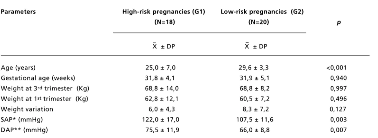
high-risk group were, on average, younger (p<0.001) and had higher systolic (p=0.003) or diastolic (p=0.007) arterial pressure than those in the low-risk group.
Table 2 shows the levels of salivary cortisol in
the two groups. The levels of cortisol in high-risk
pregnant women were significantly higher than those
in women with low-risk pregnancies (p=0.007). There was an asymmetrical distribution of salivary
cortisol in each group. Two extreme values were
identified, one in each group, and, even when these were disregarded, the asymmetry persisted, as can
be seen in Figure 1.
Table 1
Clinical characteristics of the population under study.
Parameters
High-risk pregnancies (G1)
Low-risk pregnancies (G2)
(N=18) (N=20)
p
X ± DP
X ± DP
Age (years)
25,0 ± 7,0
29,6 ± 3,3
<0,001
Gestational age (weeks)
31,8 ± 4,1
31,9 ± 5,1
0,940
Weight at 3
rdtrimester (Kg)
68,8 ± 14,0
68,8 ± 8,2
0,997
Weight at 1
sttrimester (Kg)
62,8 ± 12,1
60,5 ± 7,2
0,496
Weight variation
6,0 ± 4,3
8,3 ± 7,2
0,127
SAP* (mmHg)
122,0 ± 17,0
107,5 ± 11,6
0,003
DAP** (mmHg)
75,5 ± 11,9
66,0 ± 8,8
0,007
* SAP=systolic arterial pressure; ** DAP=diastolic arterial pressure.
Table 2
Levels of cortisol in the saliva of 38 pregnant women, as measured using electro-chemical illuminescence.
High-risk pregnancies
Low-risk pregnancies
p
(N=18) (N=20)
Mean (±SD)
20,23 (21,10)
11,45 (16,23)
0,007
Median
12,64
6,533
0,007
25% percentile
8,757
3,892
75% percentile
21,960
8,896
Interquartile difference
13,205
5,004
Figure 1
Distribution of levels of cortisol in saliva, by group.
Gr
o
u
p
1
Cortisol (Mmol/L)
2
0
14
28
42
56
70
84
98
Group 1 = Low-risk; Group 2 = High-risk. Each point represents an individual.
Discussion
The results of the present study suggest that salivary
cortisol, measured using electrochemical
lumines-cence (ECL), is found at significantly higher levels in women with high-risk pregnancies compared to
the low-risk group. The women with low risk
preg-nancies had mean cortisol levels in their saliva of 11.450 nmol/L, almost four times higher than
stan-dard laboratory reference value for cortisol at
midnight, in individuals without hypercortisoism,
which is 3.058 nmol/L.1,5
The use of ECL to measure cortisol in saliva in
pregnant women has not been the subject of much
study4,15and those that use this technique have not collected their samples at midnight.15In pregnancy,
the evaluation of the neuro-endocrine system should
take into account the alternations that are normal in women in this condition. Physiological
hypercorti-solism in pregnant women affects the measurements
and interpretation of values obtained for salivary
cortisol. As circadian rhythms remain the same, it is important that the sample be collected at midnight to
reduce the interference of high morning levels of
cortisol on the results.15
Many factors influence the level of cortisol in
saliva. The figures produced by this study
demons-trate an asymmetrical distribution. The data were collected in an attempt to establish general
parame-ters for the health of pregnant women, although the
concepts of well-being, stress-levels and family
stability were not clearly defined. The literature reports that these factors affect the dynamic of the
neuro-endocrine system during pregnancy.16
The women who attend prenatal clinics in public hospitals are predominantly from the low-income
sector.17In view of this, many of these women may
be undernourished, with vitamin deficiencies and
anemia, have an insalubrious home environment and low levels of education.17These factors may have
interfered in the measurement of cortisol in both
groups of pregnant women. Another point worth making is the fact that many of the women with
high-risk pregnancies had already been hospitalized,
which may have affected the levels of cortisol. This, along with the number of participants, makes it
diffi-cult to extrapolate references for pregnant women in
general from these figures.
Cortisol levels were measured only once for each participant. One of the methodological limitations of
this study was the difficulty in showing pregnancy
events in chronological order. There were no labora-tory data on the women’s cortisol levels prior to
pregnancy or in the first trimester. Nevertheless, the
women did not have clinical manifestations of hypercortisolism on the initial evaluation, which
supports the idea that the increase in cortisol levels
was a result of pregnancy. It should be pointed out
that the clinical symptoms of Cushing’s syndrome worsen during pregnancy and that the two cases of
extreme levels of cortisol need to be evaluated
subsequent to pregnancy to rule out the presence of a neuro-endocrine disorder. The pregnant woman in
the low-risk group who had a high level of cortisol
was 24 weeks pregnant, had experienced greater than average weight-gain (4 kg) and did not display
any of the clinical manifestations of Cushing’s
syndrome. These figures may be distorted by the
influence of extraneous factors that were not clearly identified in the study, such as, for example, the
degree of stress experienced in everyday life. Other
factors that might have affected the results, such as contamination by bleeding gums, are less likely,
each of the women before the saliva was collected. This study found that the cortisol levels in the
high-risk pregnancy group were on average higher
than they would be during a risk-free pregnancy, which raises a number of questions regarding the
influence of this hormone in the course of gestation.
Cortisol plays an important part in the development
of the fetus, although excessive levels are related to negative metabolic changes. Given the limitations of
the study, it was not possible to establish a
prospec-tive relationship between these events. Identification
of reference values for cortisol in pregnancy or vari-ations in physiological hypercortisolism could lead
the way to the development of new tools for health
professionals managing pregnancy. Hitherto there have been no studies that have analyzed and
compared cortisol levels in the saliva of women with
low- and high-risk pregnancies. Further research
needs to be carried out to clear up the outstanding questions.
References
1. Lindsay JR, Nieman LK. The
hypothalamic-pituitary-adrenal axis in pregnancy: challenges in disease detection
and treatment. Endocr Rev. 2005; 6: 2004-25.
2. Hougaard KS, Andersen MB, Kjaer SL, Hansen AM, Werge
T, Lund SP. Prenatal stress may increase vulnerability to
life events: comparison with the effects of prenatal
dexam-ethasone. Brain Res. 2005; 159: 55-63.
3. Huizink AC, Robles de Medina PG, Mulder EJH, Visser
GHA, Buitelaar JK. Stress during pregnancy is associated
with developmental outcome in infancy. J Child Psychol
Psychiatry Allied Discipl. 2003; 44: 810-8.
4. Maureen KW, Charles W. Pituitary-adrenal physiology
during pregnancy. Endocrinologist. 2001; 11: 159-70.
5. Lindsay JR, Jonklaas J, Oldfield EH, Nieman LK.
Cushing’s syndrome during pregnancy: personal experience
and review of the literature. J Clin Endocrinol Metab. 2005;
90: 3077-83.
6. Jones A, Godfrey KM, Wood P, Osmond C, Goulden P,
Phillips IW. Fetal growth and the adrenocortical response
to pshychological stress. J Clin Endocrinol Metab. 2006;
91: 1868-71.
7. Lindholm J. Plasma and urinary cortisol in pregnancy and
during estrogen-gestagen treatment. Scand J Clin Lab
Invest. 1973; 31: 119-22.
8. Papanicolaou DA, Mullen N, Kyrou I, Nieman LN.
Nighttime salivary cortisol: a useful test for the diagnosis
of cushing’s syndrome. J Clin Endocrinol Metab. 2002; 87:
4515-21.
9. Castro M, Elias PC, Quidute AR, Halah FP, Moreira AC.
Out-pacient screenig for cushing’s syndrome: the sensitivy
of the combination of circadian rhythm and overnight
dexamethasone suppression salivary cortisol tests. J Clin
Endocrinol Metab. 1999; 84: 878-82.
10. Putignano P, Toja P, Dubini A, Giraldi FP, Corsello SM,
Cavagnini F. Midnight salivary cortisol versus urinary free
and midnight serum cortisol as screening tests for cushing’s
syndrome. J Clin Endocrinol Metab. 2003; 88: 4153-67.
11. Raff H, Raff JL, Findling JW. Late-night salivary cortisol
as a screening test for cushing’s syndrome. J Clin
Endocrinol Metab. 1998; 83: 2681-6.
12. Nierop A, Bratsikas A, Klinkenberg A, Nater UM,
Zimmermann R, Ehelert U. Prolonged salivary cortisol
recovery in second-trimester pregnant women and
attenu-ated salivary α-amylase responses to psychosocial stress in
human pregnancy. J Clin Endocrinol Metab. 2006;
91:1329-35.
13. Yaneva M, Mosnier-Pudar H, Dugué MA, Grabar S, Fulla
Y, Bertagna X. Midnight salivary cortisol for the inicial
diagnosis of cushing’s syndrome of various causes. J Clin
Endocrinol Metab. 2004; 89: 3345-51.
14. Viardot A, Huber P, Puder J, Zulewski H, Keller U, Muller
B. Reproducitibility of nighttime salivary cortisol and its
use in the diagnosis of hypercortisolism as compared to
urinary free cortisol and overnight dexamethasone
suppres-sion test. J Clin Endocrinol Metab. 2005; 90: 5730-6.
15. Power C. Association of early growth and adult adiposity
with patterns of salivary cortisol in adulthood. J Clin
Endocrinol Metab. 2006; 91: 4264-70.
16. Egliston KA, McMahon C, Austin MP. Stress in pregnancy
and infant HPA axis function: conceptual and
methodolog-ical issues relating to the use of salivary cortisol as an
outcome measure. Psychoneuroendocrinology. 2006; 10:
2-10.
17. Vanderplaat M, Samson Y, Raven P. The politics and
prac-tice of empowerment evalualtion and social intervation:
lessons from the atlantic comunity actin progrma for
chil-dren regional evaluation. Canadian Journal of Program
Evaluation. 2001; 16: 79-98.
______________
Recebido em 6 de março de 2009
Versão final apresentada em 14 de setembro de 2009