SUMMARY
Objective: he values of bone mineral density (BMD) were compared in postmenopausal
women with and without breast cancer. Methods: A cross-sectional study was conducted,
including 51 breast cancer survivors (BCS) and 71 women without breast cancer, who were non-users of hormone therapy, tamoxifen, or aromatase inhibitors. BMD T-scores and
mea-surements in grams per centimeter squared (g/cm2) were obtained at the femoral neck,
tro-chanter, Ward’s triangle, and lumbar spine. Osteopenia and osteoporosis were grouped and categorized as abnormal BMD. Unconditional logistic regression analysis was used to esti-mate the odds ratios (OR) of abnormal BMD values as measures of association, with 95% con-idence intervals (CIs), adjusting for age, years since menopause, parity, and body mass index
(BMI). Results: he mean age of the women with and without breast cancer was 54.7 ± 5.8
years and 58.2 ± 4.8 years (p < 0.01), respectively. Ater adjusting for age, parity and BMI, abnormal BMD at the femoral neck (adjusted OR: 4.8; 95% CI: 1.5-15.4), trochanter (adjusted OR: 4.6; 95% CI: 1.4-15.5), and Ward’s triangle (adjusted OR: 4.5; 95% CI: 1.5-12.9) were signiicantly more frequent in postmenopausal BCS than in women without breast cancer.
Postmenopausal BCS had a signiicantly lower mean BMD at the trochanter (0.719 vs. 0.809
g/cm2, p < 0.01) and at the Ward’s triangle (0.751 vs. 0.805 g/cm2, p = 0.03). Conclusion: he
prevalence of abnormal BMD was higher in postmenopausal BCS than in postmenopausal women without breast cancer. Bone health requires special vigilance and the adoption of in-terventions should be instituted early to minimize bone loss in BCS.
Keywords: Breast cancer; menopause; bone loss; abnormal bone mineral density.
©2012 Elsevier Editora Ltda. All rights reserved.
RESUMO
Densidade mineral óssea em mulheres na pós-menopausa com e sem câncer de mama
Objetivo: Comparar a densidade mineral óssea (DMO) de mulheres na pós-menopausa com e sem cân-cer de mama. Métodos: Conduziu-se estudo de corte transversal, incluindo 51 mulheres com câncer de mama e 71 mulheres sem câncer de mama, não usuárias de terapia hormonal, tamoxifeno ou de inibido-res da aromatase. Avaliou-se a DMO, em T-score e em gramas por centímetro quadrado (g/cm²), no colo do fêmur, trocânter, triângulo de Wards e na coluna lombar. Osteopenia e osteoporose foram agrupadas e categorizadas como DMO alterada. Utilizou-se a análise de regressão logística não condicional para estimar o odds ratios (OR) de DMO alterada como medida de associação, com intervalo de coniança de 95% (IC 95%), ajustando-se por idade, anos de menopausa, paridade e índice de massa corpórea (IMC).
Resultados: A média de idade de mulheres com e sem câncer de mama foi 54,7 ± 5,8 anos e 58,2 ± 4,8 anos (p < 0,01), respectivamente. Após ajustar por idade, paridade e IMC, DMO alterada no colo do fêmur (OR ajustado: 4,8; IC 95%: 1,5-15,4), trocânter (OR ajustado: 4,6; IC 95%: 1,4-15,5) e no triângulo de Wards (OR ajustado: 4,5; IC 95%: 1,5-12,9) foi mais frequente em mulheres com câncer de mama. Mulheres com câncer de mama apresentaram signiicativamente menor média de DMO no trocânter (0,719 vs. 0,809 g/cm2, p < 0,01) e no triângulo de Wards (0,751 vs. 0,805 g/cm2, p = 0,03). Conclusão: A prevalência de DMO alterada foi maior em mulheres na pós-menopausa com câncer de mama. A saúde óssea requer vigilância especial e a adoção precoce de intervenções para minimizar a perda óssea de mulheres com câncer de mama.
Unitermos: Câncer de mama; menopausa; perda óssea; densidade mineral óssea alterada.
©2012 Elsevier Editora Ltda. Todos os direitos reservados.
Study conducted at the Department of Gynecology and Obstetrics, Universidade Estadual de Campinas (UNICAMP),
Campinas, SP, Brazil
Submitted on: 02/02/2012
Approved on: 07/01/2012
Correspondence to:
Délio Marques Conde Rua R-7 com Av. Perimetral, S/N St. Oeste – Goiânia, GO, Brazil CEP: 74125-120 Phone: +55 62 3209-6151 [email protected]
Conlict of interest: None.
Bone mineral density in postmenopausal women with and without breast
cancer
DÉLIO MARQUES CONDE1,LÚCIA COSTA-PAIVA2, EDSON ZANGIACOMI MARTINEZ3, AARÃO MENDES PINTO-NETO4 1 MD, PhD; Mentor, Medical Residency Training Program in Breast Disorders, Breast Service, Hospital Materno Infantil (HMI), Goiânia, GO, Brazil 2 MD, PhD; Associate Professor, Department of Gynecology and Obstetrics, Universidade Estadual de Campinas (UNICAMP), Campinas, SP, Brazil 3 PhD; Associate Professor, Department of Social Medicine, Universidade de São Paulo (USP), Ribeirão Preto, SP, Brazil
INTRODUCTION
Breast cancer is the most frequently diagnosed cancer in women worldwide, representing 23% (1.38 million) of the total new cancer cases in 20081. Breast cancer is
primarily a disease of postmenopausal women2. In the
postmenopause, there is a reduction in serum estrogen levels leading to bone loss. Estrogen plays an important role in skeletal maintenance due to its osteoprotective action3. In this context, postmenopausal status is a major
risk factor for low bone mineral density (BMD)4.
Previous studies have demonstrated a relationship between a higher BMD and a higher risk for post-menopausal breast cancer5,6. Although the mechanism
of this relationship is not fully understood, cumulative exposure to estrogen may play a role5. BMD may be a
predictor for breast cancer risk, but breast cancer survi-vors (BCS) can also develop bone loss related to cancer treatment7,8. Chemotherapy-induced ovarian failure7,8,
aromatase inhibitor use9,10, and a potential direct toxic
chemotherapy efect on bone tissue11 may induce and
intensify bone loss in BCS. Tamoxifen use is associated with signiicant bone loss in premenopausal BCS. How-ever, tamoxifen exerts a bone-protective action in wom-en with early mwom-enopause induced by chemotherapy12.
Premenopausal women with breast cancer undergo-ing chemotherapy with preserved ovarian function do not have a signiicant reduction in bone mass7,8.
How-ever, those who develop amenorrhea have a rapid and signiicant bone loss7,8. Menopausal hormone therapy
may be indicated for the prevention of bone loss13, and
many women on hormone therapy at the time of breast cancer diagnosis are instructed to discontinue its use2.
Discontinuation of hormone therapy is based on an in-creased risk of breast cancer recurrence14. his group of
factors contributes to increased bone loss in postmeno-pausal BCS.
Previous studies have investigated BMD in post-menopausal survivors of breast cancer15-22. However,
few studies have evaluated a group of women without breast cancer for comparison18,19,22. In one study, mean
BMD values in postmenopausal BCS were similar to those in population-based non-BCS18. In contrast, the
prevalence of osteoporosis in another study was 27.2% in BCS and 19.4% in women without breast cancer19.
Furthermore, it was demonstrated that postmeno-pausal BCS had a higher fracture risk than cancer-free women20.
Despite these findings, a notable proportion of postmenopausal BCS have undiagnosed abnormal BMD15,16,19. Considering these aspects, the present study
was conducted aiming to compare BMD in Brazilian postmenopausal women with and without breast cancer
METHODS
PATIENTS
Participant selection has been previously described in de-tail23. he current sample was derived from a study that
in-vestigated menopause symptoms, sexual activity, quality of life, some cardiovascular risk factors, and BMD in BCS23.
he present study focused on the indings of BMD in post-menopausal women with and without breast cancer.
A cross-sectional study of women undergoing routine follow-up care was conducted in the Women’s Hospital of the School of Medicine of the Universidade Estadual de Campinas, Brazil. Patients who met the inclusion cri-teria were invited to participate in the study during out-patient consultation. Women aged 45-65 years, who were non-users of hormone therapy or tamoxifen in the last six months, and who had no history of other malignancies were invited. Only postmenopausal women who complet-ed oncology treatment were includcomplet-ed in this study. None of the BCS were using aromatase inhibitors. A total of 187 women were invited to participate in the study, 100 BCS and 87 without breast cancer. Among the BCS, three re-fused to participate in the study due to lack of time, 22 were undergoing oncology treatment, 12 were premenopausal, seven had no knowledge of time since menopause, and ive did not have bone density measurements. Among women without breast cancer, two refused to participate in the study due to lack of time, four were premenopausal, and ten had no knowledge of time since menopause. herefore, 122 postmenopausal women, 51 with breast cancer and 71 without breast cancer, constituted the present study sample.
Participants responded to an interview that assessed sociodemographic characteristics, including age, race/ ethnicity, age at menarche, parity, smoking, age at meno-pause, and years since menopause. Clinical characteristics included body mass index (BMI) in kilograms per square meter (kg/m2), time since breast cancer diagnosis, type of
surgery, tumor stage, chemotherapy, and radiotherapy. his study was approved by the institutional review board; all participants signed an informed consent.
BONEMINERALDENSITYMEASUREMENT
BMD tests were ordered in the initial interview with the patient. BMD in grams per centimeter squared (g/cm2) was
measured at the femoral neck, trochanter, Ward’s triangle, and the lumbar spine (L2-L4; anteroposterior plane) us-ing a Lunar DPX device (DXA; Lunar DPX, Madison, WI, USA). BMD values were also expressed as T-scores, using the World Health Organization criteria24: normal: T-score
STATISTICALANALYSIS
he means of descriptive continuous variables were com-pared between women with and without cancer using Student’s t-test for independent samples. he frequencies
of binary variables were compared by Fisher’s exact test. Unconditional logistic regression analysis25 was used to
es-timate the odds ratios (OR) of abnormal BMD as measures of association, with 95% conidence intervals (CIs). In these models, the independent binary variable was breast cancer (with cancer/without cancer), and the dependent binary variable was BMD, classiied as normal or abnor-mal. he ORs were estimated as measures of the associa-tion between cancer and abnormal BMD in the femoral neck, trochanter, Ward’s triangle and lumbar spine with and without adjustment for age, parity, years since meno-pause, and BMI in quartiles. When age and years since menopause were considered, regression models could not be itted because of the strong collinearity between these variables. he ORs adjusted for age, parity, and BMI, and adjusted for years since menopause, parity, and BMI were estimated. When bone density (g/cm2) was regarded as a
continuous variable, linear regression models26 were used
to compare the population means between women with and without cancer adjusting for covariates. All analyses were performed using the SAS version 9.2 sotware. he signiicance level was set at 5%.
RESULTS
he mean age of women with breast cancer was 54.7 ± 5.8 years and 58.2 ± 4.8 years for those without breast can-cer (p < 0.01). he mean BMI was 27.5 ± 5.2 kg/m2 and
30.2 ± 5.7 kg/m2 for women with and without breast
can-cer (p < 0.01), respectively. he mean age at menopause was 47.1 ± 5.0 years for women with breast cancer and 47.4 ± 4.9 years for women without breast cancer (p = 0.98). Bone density measurement was performed from one to two months ater the irst interview. Other characteristics are shown in Table 1. In the breast cancer group, the mean time since breast cancer diagnosis was 67.9 ± 53.1 months.
A little over half of the postmenopausal BCS (52.9%) underwent mastectomy, and 47.1% underwent breast-conserving therapy. Approximately 72% of women under-went chemotherapy, 76.5% underunder-went radiation therapy, and 60.8% underwent chemotherapy and radiation ther-apy. Distribution according to tumor stage was (n, %): 0 (4, 7.8%), I (7, 13.7%), II (31, 60.8%), and III (9, 17.7%).
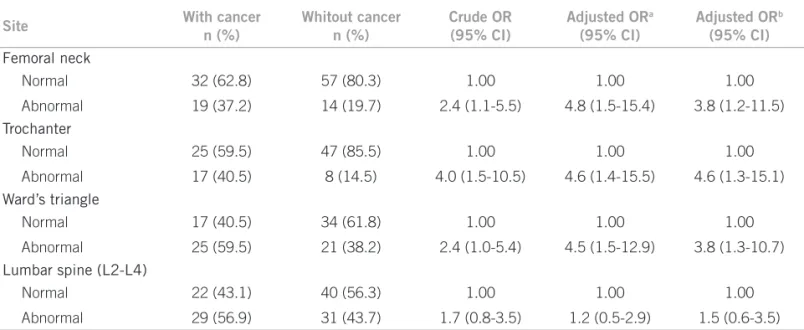
In the unadjusted analysis, the results in Table 2 show that postmenopausal BCS had a signiicantly higher preva-lence of abnormal BMD at the femoral neck, trochanter, and Ward’s triangle than women without breast cancer. Ater adjusting for age, parity, and BMI, abnormal BMD at the femoral neck (adjusted OR: 4.8; 95% CI: 1.5-15.4), trochanter (adjusted OR: 4.6; 95% CI: 1.4-15.5), and Ward’s triangle (adjusted OR: 4.5; 95% CI: 1.5-12.9) was signiicantly more frequent in postmenopausal BCS than in women without breast cancer. Ater adjusting for years since menopause, parity, and BMI, the prevalence of ab-normal BMD at the femoral neck, trochanter, and Ward’s triangle remained signiicantly higher in postmenopausal BCS. In unadjusted and adjusted analysis, there was no signiicant diference in the prevalence of abnormal lum-bar spine (L2-L4) BMD between BCS and women without breast cancer.
Figure 1 displays the mean BMD (g/cm2) of
post-menopausal BCS and women without breast cancer. On analysis adjusted for age, parity, and BMI, BCS showed a signiicantly lower mean BMD at the trochanter (0.719 vs. 0.809 g/cm2, p < 0.01) and Ward’s triangle (0.751
vs. 0.805 g/cm2, p = 0.03). Ater adjusting for years since
menopause, parity, and BMI, BCS had a signiicantly lower mean BMD at the trochanter (p < 0.01), and Ward’s tri-angle (p = 0.03).
DISCUSSION
his study aimed to compare BMD in women with and without breast cancer. Few studies have investigated BMD in postmenopausal BCS, in comparison to a group of post-menopausal women without breast cancer. Based on BMD
Characteristics With cancer (n = 51) Without cancer (n = 71) p-value
Age (years) 54.7 ± 5.8 58.2 ± 4.8 < 0.01a
Age at menarche (years) 13.1 ± 1.8 13.1 ± 1.9 0.85a
Body mass index (kg/m2) 27.5 ± 5.2 30.2 ± 5.7 < 0.01a
Age at menopause (years) 47.1 ± 5.0 47.4 ± 4.9 0.98a
Years since menopause 7.4 ± 5.2 10.6 ± 5.3 < 0.01a
Race (% white women) 21.6 32.4 0.22b
Parity (% nulliparous) 17.7 4.2 0.03b
Smokers (%) 13.7 5.6 0.22b
Data are expressed as mean ± SD unless otherwise speciied. a Student’s t-test for independent samples; b Fisher’s exact test.
T-scores, it was observed that abnormal BMD at the femo-ral neck, trochanter, and Ward’s triangle was signiicant-ly more frequent in BCS than in women without breast cancer. Furthermore, BCS had a signiicantly lower mean BMD (g/cm2) at the trochanter and Ward’s triangle.
In this case study, it was observed that the prevalence of abnormal BMD in postmenopausal BCS was higher at the Ward’s triangle (59.5%), and lumbar spine (56.9%). In another study, including only postmenopausal BCS with a mean age of 54 years and 36.6% of tamoxifen users, it was observed that 56.3% of the participants had abnormal
BMD at the lumbar spine15. he indings of these authors15
were similar to the present results regarding the high prev-alence rate of abnormal BMD at the lumbar spine (56.9%), although the participants of the present cohort were non-users of tamoxifen. Other studies have also reported a high frequency of abnormal BMD at the lumbar spine19,21,22.
he lumbar spine and Ward’s triangle have a high con-tent of trabecular bone. A higher frequency of abnormal BMD at these bone sites may be related to a greater sensi-tivity of trabecular bone to estrogen deiciency27. his is a
characteristic of postmenopausal women who do not use hormone therapy.
Few studies comparing BMD between postmenopaus-al BCS and women without breast cancer have been iden-tiied18,19,22. Based on cross-sectional data18, BMD Z-scores
of BCS were investigated. his study found that postmeno-pausal patients who had not received adjuvant therapy had higher whole body BMD compared to age-matched women. Furthermore, the authors reported that hip and spine BMD measurements in postmenopausal BCS were similar to those in a population-based sample without breast cancer, suggesting that the treatment had caused no major deleterious efect on the BMD of BCS18. However,
a retrospective study found that women who were post-menopausal at the time of breast cancer diagnosis and re-ceiving adjuvant chemotherapy had lower BMD Z-scores at the total hip, femoral neck, trochanter, and lumbar spine than those who had not received chemotherapy17,
indicat-ing a potential detrimental efect of adjuvant therapy on the BMD of postmenopausal women.
In another study22, BMD was compared between a
BCS group with chemotherapy-induced amenorrhea and a premenopausal control group without breast cancer.
Site With cancer
n (%)
Whitout cancer n (%)
Crude OR (95% CI)
Adjusted ORa
(95% CI)
Adjusted ORb
(95% CI)
Femoral neck
Normal 32 (62.8) 57 (80.3) 1.00 1.00 1.00
Abnormal 19 (37.2) 14 (19.7) 2.4 (1.1-5.5) 4.8 (1.5-15.4) 3.8 (1.2-11.5)
Trochanter
Normal 25 (59.5) 47 (85.5) 1.00 1.00 1.00
Abnormal 17 (40.5) 8 (14.5) 4.0 (1.5-10.5) 4.6 (1.4-15.5) 4.6 (1.3-15.1)
Ward’s triangle
Normal 17 (40.5) 34 (61.8) 1.00 1.00 1.00
Abnormal 25 (59.5) 21 (38.2) 2.4 (1.0-5.4) 4.5 (1.5-12.9) 3.8 (1.3-10.7)
Lumbar spine (L2-L4)
Normal 22 (43.1) 40 (56.3) 1.00 1.00 1.00
Abnormal 29 (56.9) 31 (43.7) 1.7 (0.8-3.5) 1.2 (0.5-2.9) 1.5 (0.6-3.5)
SD, standard deviation; OR, odds ratio; CI, conidence interval, a OR estimates adjusted for age, parity and body mass index in quartiles; b OR estimates adjusted for years since menopause, parity and body mass index in quartiles.
Table 2 – Comparison of prevalence of abnormal bone mineral density (T-score < - 1 SD) of postmenopausal women with and without breast cancer
Figure 1 – Comparison of mean bone mineral density (g/cm2) in postmenopausal women with and without breast
cancer.
a Adjusted for age, parity and body mass index in quartiles. b Adjusted for years since menopause, parity and body mass index in quartiles.
n 71 51
Mean 0.943 0.900 SD 0.103 0.123 p = 0.08 (a), p = 0.05 (b)
with cancer without cancer
55 42
0.809 0.719 0.112 0.117 p < 0.01 (a), p < 0.01 (b)
with cancer without cancer
55 42
0.805 0.751 0.124 0.148 p = 0.03 (a), p = 0.03 (b)
with cancer without cancer
71 51
1.090 1.060 0.162 0.128 p = 0.58 (a), p = 0.64 (b) with cancer Femoral neck Trochanter Ward’s triangle Lumbar spine (L2-L4)
Bone mineral density (g/cm
2) 1.4
1.2
1.0
0.8
0.6
0.4
he authors of this study observed that signiicantly more BCS had low spine BMD based on T-scores22. In the
Wom-en’s Health Initiative Observational Study (WHI-OS), data on the prevalence of low bone mass was reported in a large cohort of postmenopausal BCS, compared to cancer-free women19. In the WHI-OS, the frequency of osteoporosis
was 27.2% in postmenopausal BCS and 19.4% in cancer-free women. A comparison of BMD T-scores in post-menopausal BCS and in women with no history of breast cancer, adjusting for covariables, except menopausal hor-mone therapy, showed BCS with a signiicantly higher prevalence of osteoporosis in the total hip, total body, and at any site19. In the present study, the prevalence of
abnor-mal BMD T-scores at the femoral neck, trochanter, and Ward’s triangle was higher in BCS in adjusted and unad-justed analyses. he mean BMD (g/cm2) at the trochanter
and Ward’s triangle was higher among postmenopausal non-BCS, adjusting for potential confounding variables (age, years since menopause, parity, BMI). In the WHI-OS, when menopausal hormone therapy was added as a covariate, the diference in the prevalence of osteoporosis between women with and without breast cancer was no longer signiicant19. Participants in the present study were
not using hormone therapy, thus it was not possible to ad-just for this variable.
Diferences observed between the present indings and results of other studies may be related to the age difer-ence between the participants, bone sites investigated, and variables evaluated. In the WHI-OS, a low BMD and high prevalence of osteoporosis in BCS occurred, especially be-cause of lower hormone therapy use by these women19. he
present study included women who did not take tamoxi-fen or aromatase inhibitors, since the aim at the time of study design was to gain knowledge about BMD in BCS who were not receiving any hormone therapy.
From the present indings and previous studies, it can be inferred that abnormal BMD is a concern for post-menopausal BCS15-19,21,22. Despite these indings, 77.8% of
the WHI-OS postmenopausal BCS had undiagnosed os-teoporosis at baseline. hus, a considerable proportion of BCS women had no diagnosis of abnormal BMD and con-sequently did not receive therapy, as previously reported16.
he indings should be interpreted considering some limitations, such as the cross-sectional study design and a lack of information about chemotherapy regimen. he immunohistochemical proile of tumors in BCS, which could have allowed the investigation of a relationship between hormone receptor status and BMD, was not in-cluded. Physical activity, and calcium, cafeine, and alco-hol consumption were not investigated. However, there was inconsistent evidence of an association between cur-rent physical activity or calcium intake and BMD in a
systematic review4. In this review, there was fair evidence
that current cafeine intake was not associated with BMD, and that moderate alcohol consumption was not associ-ated with lower BMD4.
To the authors’ knowledge, this was the irst study con-ducted in Latin America investigating BMD in postmeno-pausal BCS. he existence of a control group of women with similar menopausal status, i.e., postmenopausal women with no history of breast cancer, is highlighted. Despite some diferences between both groups, the com-parisons between BMD were adjusted for potential con-founding variables.
he current guidelines of the American Society of Clinical Oncology recommend the use of aromatase in-hibitors in postmenopausal BCS with hormone receptor-positive tumors during adjuvant therapy as an initial treat-ment, or sequential or extended treatment ater tamoxifen use28. Although further studies are still required, a recent
randomized, placebo-controlled, double-blinded trial in healthy postmenopausal women has demonstrated that exemestane reduced the relative incidence of invasive breast cancers by 65%29. his group of data suggests that
the use of aromatase inhibitors will become increasingly more frequent. In this context, the prevalence of abnormal BMD in BCS may be higher in the future than in the pres-ent study. In BCS, there is a need for increased vigilance of bone health, especially in postmenopausal women.
CONCLUSION
In conclusion, the authors believe that these results are of interest to healthcare professionals involved in the man-agement of women with breast cancer. he prevalence of abnormal BMD was higher in postmenopausal BCS than in postmenopausal women without breast cancer. Bone health requires special vigilance and the adoption of inter-ventions should be instituted early to minimize bone loss in BCS.
REFERENCES
1. International Agency for Research on Cancer. GLOBOCAN 2008. World-female. Estimated incidence by age. [cited 2012 jan 12]. Available from: http:// globocan.iarc.fr/age-speciic_table_r_PDF.asp?selection=221900&title=Worl d&sex=2&type=0&stat=0&PDF=1&window=1&sort=0&submit=%A0Execut e%A0).
2. Ganz PA, Greendale GA, Petersen L, Zibecchi L, Kahn B, Belin TR. Manag-ing menopausal symptoms in breast cancer survivors: results of a randomized controlled trial. J Natl Cancer Inst. 2000;92:1054-64.
3. Imai Y, Kondoh S, Kouzmenko A, Kato S. Minireview: osteoprotective action of estrogens is mediated by osteoclastic estrogen receptor-alpha. Mol Endocri-nol. 2010;24:877-85.
4. Waugh EJ, Lam MA, Hawker GA, McGowan J, Papaioannou A, Cheung AM, et al. Risk factors for low bone mass in healthy 40-60 year old women: a sys-tematic review of the literature. Osteoporos Int. 2009;20:1-21.
5. Zhang Y, Kiel DP, Kreger BE, Cupples LA, Ellison RC, Dorgan JF, et al. Bone mass and the risk of breast cancer among postmenopausal women. N Engl J Med. 1997;336:611-7.
7. Saarto T, Blomqvist C, Välimäki M, Mäkelä P, Sarna S, Elomaa I. Chemical castration induced by adjuvant cyclophosphamide, methotrexate, and luo-rouracil chemotherapy causes rapid bone loss that is reduced by clodronate: a randomized study in premenopausal breast cancer patients. J Clin Oncol. 1997;15:1341-7.
8. Shapiro CL, Manola J, Lebof M. Ovarian failure ater adjuvant chemotherapy is associated with rapid bone loss in women with early-stage breast cancer. J Clin Oncol. 2001;19:3306-11.
9. Coleman RE, Banks LM, Girgis SI, Kilburn LS, Vrdoljak E, Fox J, et al. Skeletal efects of exemestane on bone-mineral density, bone biomarkers, and fracture incidence in postmenopausal women with early breast cancer participating in the Intergroup Exemestane Study (IES): a randomised controlled study. Lancet Oncol. 2007;8:119-27.
10. Eastell R, Adams JE, Coleman RE, Howell A, Hannon RA, Cuzick J, et al. Efect of anastrozole on bone mineral density: 5-year results from the an-astrozole, tamoxifen, alone or in combination trial 18233230. J Clin Oncol. 2008;26:1051-7.
11. Bani A, Podestà M, Fazzuoli L, Sertoli MR, Venturini M, Santini G, et al. High-dose chemotherapy shows a dose-dependent toxicity to bone marrow osteoprogenitors: a mechanism for post-bone marrow transplantation osteo-penia. Cancer. 2001;92:2419-28.
12. Vehmanen L, Elomaa I, Blomqvist C, Saarto T. Tamoxifen treatment ater adju-vant chemotherapy has opposite efects on bone mineral density in premeno-pausal patients depending on menstrual status. J Clin Oncol. 2006;24:675-80. 13. he North American Menopause Society. Management of osteoporosis in
postmenopausal women: 2010 position statement of he North American Menopause Society. Menopause. 2010;17:25-54.
14. Kenemans P, Bundred NJ, Foidart JM, Kubista E, von Schoultz B, Sismondi P, et al. Safety and eicacy of tibolone in breast-cancer patients with vasomotor symptoms: a double-blind, randomised, non-inferiority trial. Lancet Oncol. 2009;10:135-46.
15. Twiss JJ, Waltman N, Ott CD, Gross GJ, Lindsey AM, Moore TE. Bone mineral density in postmenopausal breast cancer survivors. J Am Acad Nurse Pract. 2001;13:276-84.
16. Lindsey AM, Gross G, Twiss J, Waltman N, Ott C, Moore TE. Postmenopausal survivors of breast cancer at risk for osteoporosis: nutritional intake and body size. Cancer Nurs. 2002;25:50-6.
17. Greep NC, Giuliano AE, Hansen NM, Taketani T, Wang HJ, Singer FR. he efects of adjuvant chemotherapy on bone density in postmenopausal women with early breast cancer. Am J Med. 2003;114:653-9.
18. Crandall C, Petersen L, Ganz PA, Greendale GA. Bone mineral density and adjuvant therapy in breast cancer survivors. Breast Cancer Res Treat. 2004;88:257-61.
19. Chen Z, Maricic M, Pettinger M, Ritenbaugh C, Lopez AM, Barad DH, et al. Osteoporosis and rate of bone loss among postmenopausal survivors of breast cancer. Cancer. 2005;104:1520-30.
20. Chen Z, Maricic M, Bassford TL, Pettinger M, Ritenbaugh C, Lopez AM, et al. Fracture risk among breast cancer survivors: results from the Women’s Health Initiative Observational Study. Arch Intern Med. 2005;165:552-8.
21. Twiss JJ, Gross GJ, Waltman NL, Ott CD, Lindsey AM. Health behaviors in breast cancer survivors experiencing bone loss. J Am Acad Nurse. Pract. 2006;18:471-81.
22. Winters-Stone KM, Nail L, Bennett JA, Schwartz A. Bone health and falls: frac-ture risk in breast cancer survivors with chemotherapy-induced amenorrhea. Oncol Nurs Forum. 2009;36:315-25.
23. Conde DM, Pinto-Neto AM, Cabello C, Sá DS, Costa-Paiva L, Martinez EZ. Menopause symptoms and quality of life in women aged 45 to 65 years with and without breast cancer. Menopause. 2005;12:436-43.
24. Kanis JA. Assessment of fracture risk and its application to screening for post-menopausal osteoporosis: synopsis of a WHO report. WHO Study Group. Os-teoporos Int. 1994;4:368-81.
25. Hosmer DW, Lemeshow S. Applied logistic models. 2nd ed. New York: John Wiley& Sons; 2000.
26. Montgomery DC, Peck EA, Vining GG. Introduction to linear regression anal-ysis. 4th ed. New York: John Wiley & Sons; 2007.
27. Beerthuizen R, van Beek A, Massai R, Mäkäräinen L, Hout J, Bennink HC. Bone mineral density during long-term use of the progestagen contraceptive implant Implanon compared to a non-hormonal method of contraception. Hum Reprod. 2000;15:118-22.
28. Burstein HJ, Prestrud AA, Seidenfeld J, Anderson H, Buchholz TA, Davidson NE, et al. American Society of Clinical Oncology clinical practice guideline: update on adjuvant endocrine therapy for women with hormone receptor-positive breast cancer. J Clin Oncol. 2010;28:3784-96.