1. PhD. Faculdade de Ciências Médicas, Universidade de Pernambuco (UPE).
2. Titular Professor of Cardio-thoracic surgery at Faculdade de Ciências Médicas/UPE.
3. MD. Pediatric cardiologist at Hospital Universitário Oswaldo Cruz/ UPE.
4. MD. Pediatric cardiologist at Unitórax/ RHP. 5. Adjunt Professor of Thoracic surgery at UFPE.
Work done at Hospital Universitário Oswaldo Cruz, Faculdade de Ciências Médicas/UPE, Recife - Pernambuco, Brasil UNITORAX/ RHP - Real Hospital Português, Recife - Pernambuco, Brasil. This work was suported by Fundo de Desenvolvimento à Pesquisa e Extensão, FDPE/UPE.
Correspondence address: Rua dos Navegantes 1515/101. Boa Viagem. Recife, PE. CEP: 51021-010.
E-mail: [email protected]
Alline de Souza Alves OLIVEIRA1,Breno Barbosa de Siqueira CARNEIRO1,Ricardo de Carvalho LIMA2,Catarina CAVALCANTI3,Roberta VILLACHAN3,Nadja ARRAES4,Ricardo de Albuquerque LINS5,Mozart ESCOBAR5
Artigo received in Novembro 15th, 2006 Article accepted in August 8th, 2007
RBCCV 44205-907
Tratamento cirúrgico da coarctação da aorta: experiência de três décadas
Surgical treatment of the aortic coarctation:
three-decade experience
Abstract
Objective: To make a 30-year review of the immediate
results of coarctation of the aorta (CoAo) operation, between 1974 and 2004. All the patients underwent CoAo whether in isolation or associated with other congenital defects.
Methods: The following data was investigated: age at the
time of surgery, gender, associated lesions, and type of surgical technique, and immediate surgical outcome, particularly focusing the presence of systemic arterial hypertension.
Results: One hundred and four patients underwent CoAo.
Of the 104 enrolled patients, 75 (72%) were pediatric patients and 29 (28%) adults patients. In the pediatric group, 23 (22%) were considered neonates, 17 (16%) infants, and 35 (34%) children. The associated defects were present in 66 (63.5%) patients, 54 (51.9%) in the pediatric group and
12 (11.3%) in the adult group. Seven (6.7%) deaths were observed in the immediate postoperative period. Among the various surgical techniques employed, aortoplasty was used in 80 patients (76.9%); end-to-end anastomosis in 15 (14.4%); Teles technique in seven (6.7%), Waldhausen technique in one (1%), and it was not possible to identify the technique in one patient (1%).
Conclusions: Despite the limitations of the present study,
it may be noted that the results were good with the corrective surgery being performed safely and with a low mortality rate. Medium- and long-term follow-up was impaired by the well-known structural deficiencies in Brazil.
Descriptors: Aortic coarctation, surgery. Cardiac surgical
hypertension. The aortic coarctation was classified from the surgeon’s point of view such as pre-, juxta-, and postductal.
In the present study two groups of patients were considered: the pediatric group included the newborns and the patients’ age d” 15 yrs; the adult group included patients older than 15 yrs. The pediatric group was subdivided into three groups: newborns (age d” one month); infants (age ranging from on month to 12 months); and children (age > 12 months to 15 yrs).
This study was approved by the Research and Ethics Committee of the University of Pernambuco, Recife, PE, Brazil
RESULTS
During the study period, 104 patients (45 [43.3%] HUOC, 59 [56.7%] RHP) underwent aortic coarctation repair surgery. The study population comprised 63 men (61%) and 37 women (39%). The pediatric group included 75 (72.1%) patients divided as follows: 23 (22%) newborns, 17 (16%) infants, and 35 (34%) children. The adult group included 29 (27.9%) patients. At the time of the surgery, the minimum age was 24 days and the maximum age was 54 years. The age of the patients was 11.8 ± 12.3 years.
In our study, most patients with coarctation have juxtaductal coarctation 54 (52%); 27 (26%) preductal (infantile-type); and 23 (22.1%) postductal (adult-type). The association with other diseases have been present in 66 (63.5%) of the patients. Among the associated defects, the most frequent ones were: patency of ductus arteriosus 46 (48.9%); related to the aortic valve 12 (12.8%), interventricular communication 9 (9.6%), to the mitral valve 9 (9.6%), and to interatrial communication 2 (2.1%); INTRODUCTION
The aortic coarctation is congenital cardiopathy of easy diagnosis. However, frequently, the diagnosis is made relatively late leading to additional complications to the patients with this congenital defect. The causes for the late diagnosis range from the generalists lack of habit to palpate the lower limbs, check both upper and lower limbs pressure, and even the own unfamiliarity of the disease [1].
The present study aims at to perform a retrospective study of the patients who had underwent surgical treatment at Hospital Universitário Oswaldo Cruz (HUOC) UPE and at Unitórax/Real Hospital Português (RHP), with the purpose of analyzing the short-term outcomes of surgical treatment performed in two regional reference institutions, in order to provide better information regarding the associated diseases, the techniques used, and the immediate complications in the treatment of this congenital cardiopathy.
METHOD
Exclusion criteria included the following: presence of aortic coarctation either isolated or associated with other congenital defects, and surgeries performed between March 1974 and January 2004. Patients of all ages were eligible. The medical charts of both hospitals HUOC and RHP were used as sources of research data.
All participating patients in the study underwent conventional thoracic radiography, electrocardiogram, echocardiogram, and aortogrphy. Data collected from each patient included gender, age at the time of surgery, associated lesions, technique used, and the postoperative immediate outcomes focusing the systemic arterial
Resumo
Objetivo: Revisar os resultados imediatos de 30 anos da
cirurgia de coarctação da aorta (CoAo), no período entre 1974 e 2004. Foram incluídos todos pacientes operados de CoAo, isolada ou associada a outros defeitos congênitos.
Método: Foram pesquisados os seguintes dados: idade no
momento da cirurgia, sexo, lesões associadas, tipo de técnica cirúrgica utilizada, resultado cirúrgico imediato, com ênfase à presença de hipertensão arterial sistêmica.
Resultados: Foram operados 104 pacientes, dos quais 75
(72%) eram pacientes pediátricos e 29 (28%), adultos. No grupo pediátrico, 23 (22%) foram considerados neonatos, 17 (16%), lactentes, 35 (34%), crianças. Os defeitos associados estiveram presentes em 66 (63,5%) pacientes, sendo 54 (51,9%) no grupo pediátrico e 12 (11,3%) no grupo dos adultos. Foram observados sete (6,7%) óbitos no
pós-operatório imediato (POI). Dentre as diversas técnicas cirúrgicas utilizadas a aortoplastia foi usada em 80 pacientes (76,9%); anastomose término-terminal em 15 (14,4%); técnica de Teles em sete (6,7%); Waldhausen em um (1%), e não foi possível identificar a técnica em um (1%) paciente.
Conclusões: Apesar das limitações do presente estudo,
pode-se observar que os resultados foram bons, sendo a correção cirúrgica realizada de maneira segura e com baixa mortalidade. O seguimento a médio e longo prazo foi prejudicado pelas deficiências estruturais conhecidas em nosso meio.
Descritores: Coartação aórtica, cirurgia. Procedimentos
transposition of the great vessels 2 (2.1%); regarding to a single atrium 1 (1.1%); tricuspid atresia 1 (1.1%); dextrocardia 1 (1.1%); complete atrioventricular canal 1 (1.1%); and single ventricle 1 (1.1%). Endocardial fibroelastosis was associated with three patients (3.2%), two patients (2.1%) had Shone syndrome, one (1.1%) had descending aorta aneurysm, and one had dilated cardiomyopathy (Table 1).
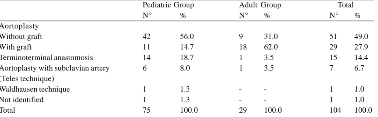
Among the surgical technique used, aortoplasty was performed in 80 (76.9%) patients; aortoplasty without patch-graft in 51 (49%), aortoplasty by patch-graft in 7 (6.7%) using Teles technique, and aortoplasty in 1 (1%) using Waldhausen technique.
Both Bovine pericardium and Dracon synthetic grafts were used. Terminoterminal anastomosis (end-to-end
anastomosis) was performed in 15 (14.4%) patients and it was not possible to identify the technique in one patient only (Table 2).
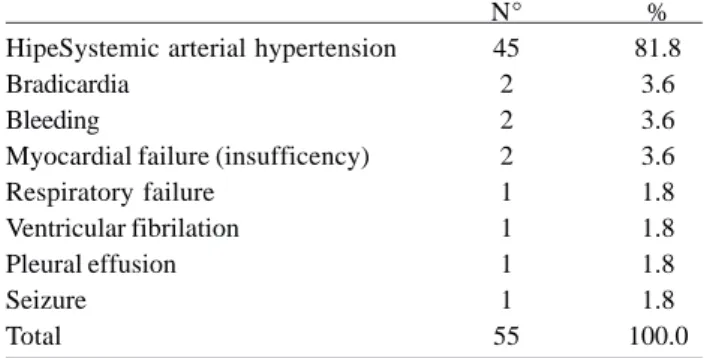
The most common postoperative complication was systemic arterial hypertension. It was observed in 45 (81.8%) patients. The remaining complications observed were as follows: bradycardia, bleeding, respiratory failure, myocardial failure, ventricular fibrillation, pleural effusion, and seizure (Table 3).
In the immediate postoperative, seven (6.7%) patients died; six (5.7%) in the pediatric group and one (0.9%) in the adult group. The causes were as follows: cardiac arrest in two (1.9%) patients, respiratory failure in two (1.9%) patients, myocardial failure in one (o.9%) patient, and two (1.9%) patients died from unknown causes.
Table 1. Disease associated with the coarctation of the aorta.
Patency of the ductus arteriosus Aortic valve injuries
Interventricular communication Mitral valve injuries
Endocardial fibroelastosis Descending aorta aneurysm Shone Syndrome
Transposition of the great vessels Interatrial communication Tricuspid atresia Single atrium Dextrocardia
Single atrioventricular valve Single ventricle
Dilated cardiomyopathy Total
Pediatric Group
N° %
38 48.1
8 10.1
9 11.4
7 8.9
3 3.8
2 2.5
2 2.5
2 2.5
2 2.5
1 1.3
1 1.3
1 1.3
1 1.3
1 1.3
1 1.3
79 100.0
Adult Group
N° %
8 53.3
4 26.7
-
-2 13.3
-
-1 6.7
-
--
--
--
--
--
--
--
--
-15 100.0
Total
N° %
46 48.9
12 12.8
9 9.6
9 9.6
3 3.2
3 3.2
2 2.1
2 2.1
2 2.1
1 1.1
1 1.1
1 1.1
1 1.1
1 1.1
1 1.1
94 100.0
Table 2. Surgical techniques performed in the correctionof aortic coarctation.
Aortoplasty Without graft With graft
Terminoterminal anastomosis Aortoplasty with subclavian artery (Teles technique)
Waldhausen technique Not identified Total
Pediatric Group
N° %
42 56.0
11 14.7
14 18.7
6 8.0
1 1.3
1 1.3
75 100.0
Adult Group
N° %
9 31.0
18 62.0
1 3.5
1 3.5
-
--
-29 100.0
Total
N° %
51 49.0
29 27.9
15 14.4
7 6.7
1 1.0
1 1.0
DISCUSSION
The benefits acquired from the surgical option to treat aortic coarctation are unquestionable. Since this type of intervention was first used half century ago, the subsequent publications have demonstrated satisfactory outcomes regarding the increase of survival, symptomatic improvement, and reduced systemic arterial hypertension [2-4]. In our study, among the congenital defects observed, the patency of ductus arteriosus was the most frequent one, affecting 48.9% of the patients. A similar patency of ductus arteriosus distribution in both groups pediatric (48.1%) and adult corroborates the findings found in the literature [2].
The second most observed defect in patients with aortic coarctation was the changes in the aortic valve with an incidence of 12.8% compared to values described by Brouwer et al. (8.3%) [5], and lower than the ones described by Corno et al. (20.3%) [2]. However, when the adult group is observed this incidence is much higher than that of the pediatric group (26.7% vs 10.1%, respectively) [5].
The third most associated defect in this study was the interventricular communication (IVC) in 11.4% of the pediatric patients. Nevertheless, it was not seen in any patient of the adult group. The incidence of IVC and aortic coarctation in adult patients is low reaching as low as 3% [5]. This may be justifiable by the fact that the aortic coarctation alone causes less hemodynamic repercussion, while those patients with associated IVC must be submitted to earliest surgical intervention. Of the pediatric patients, the newborn subgroup showed the highest incidence, ranging from 44.1% to 76% [2].
The incidence of the mitral valve injury was observed in 8.9% and 13.3% of the patients in both groups the pediatric and the adult, respectively. In the pediatric group all the changes were caused by the congenital origin, while in the adult group one patient had mitral valve prolapse and another had rheumatic mitral insufficiency.
Table 3. Postoperative surgical complications in the correction of the coarctation of the aorta.
HipeSystemic arterial hypertension Bradicardia
Bleeding
Myocardial failure (insufficency) Respiratory failure
Ventricular fibrilation Pleural effusion Seizure Total
N° 45 2 2 2 1 1 1 1 55
% 81.8
3.6 3.6 3.6 1.8 1.8 1.8 1.8 100.0
The aortic coarctation had been associated to several different diseases, most significantly with lower incidence to endocardial fibroelastosis, Shone syndrome, transposition of the great vessels, interatrial communication, tricuspid atresia, single atrium, dextrocardia, complete atrioventricular canal, single ventricle, and dilated cardiomyopathy.
The descending aorta aneurysm was most frequent in the adult group, affecting 6.7% of the patients, while in the pediatric group it affected 2.5% only. Possibly, in the adult group, it is correlated to a greater time without correcting the disease with a persistence of the hemodynamic change. This is in accordance to the literature findings 91.6%) [5].
Several technique were used such as aortoplasty without graft, graft aortoplasty, aortoplasty using Teles technique, aortoplasty using Waldhausen technique, resection and anastomosis terminoterminal (end-to-end anastomosis). The aortoplasty without graft was performed in the majority of the patients (49%) and the graft aortoplasty was performed in 27.9% of the patients, totaling 76.9% of the patients submitted to these two techniques. Although in the literature it has not been observed the use of aortoplasty without graft, this technique has been preferred in our group, especially in pediatric patients, and has yet the advantage to rule out the application of foreign material. The resection had been performed in 14.4% of the patients, a lower incidence than that observed by several authors. This has been the technique of choice, mainly by the pediatric patients [6, 7]. foreign , especially in pediatric patoi4ntshausen technique The Teles technique was performed in seven (6.7%) patients, being performed in six in the pediatric group (8%) and in one patient (3.5%) in the adult group.
Despite the excellent outcomes, the Teles technique has not been performed by our group in usual practice, maybe due to the need of a greater technical requirement [8]. The Waldhausen technique was performed only in one (1%) patient. This technique has frequently been used by other authors in newborns and infants, especially in children with aortic arch distal hypoplasia [9, 10]. However, it has not been the procedure of choice in our medical services.
REFERENCES
1. Yee ES, Soifer SJ, Turley K, Verrier ED, Fishman NH, Ebert PA. Infant coarctation: a spectrum in clinical presentation and treatment. Ann Thorac Surg. 1986;42(5):488-93.
2. Corno AF, Botta U, Hurni M, Payot M, Sekarski N, Tozzi P, et al. Surgery for aortic coarctation: a 30 years experience. Eur J Cardiothorac Surg. 2001;20(6):1202-6.
We recognize that the study has limitations. The retrospective nature of this study making a 30-year review of the literature where the data collection was difficult was mainly based on the surgical medical charts of the time of the surgery and on the immediate postoperative outcomes.
CONCLUSION
The aortic coarctation has been over the past few years a highly surgical disease, with both good immediate and delayed outcomes. Basically, at the present study, 70% of the patients had as associated diseases the patency of the ductus arteriosus, aortic valve injuries, and interventricular communication. Regarding the techniques, aortoplasty with and without graft, was the most performed technique by the authors. Systemic arterial hypertension was the most frequent complication in the immediate postoperative. The overall surgical morbimortality could be considered equivalent to other studies in the literature. Until new ways of treatment can present better outcomes, we believe that up to the present, the aortic coarctation surgical treatment is the considered the “gold standard” procedure.
3. Lerberg DB, Hardesty RL, Siewers RD, Zuberbuhler JR, Bahnson HT. Coarctation of the aorta in infants and children: 25 years of experience. Ann Thorac Surg. 1982;33(2):159-70.
4. Harrison DA, McLaughlin PR, Lazzam C, Connelly M, Benson LN. Endovascular stents in the management of coarctation of the aorta in the adolescent and adult: one year follow up. Heart. 2001;85(5):561-6.
5. Brouwer RM, Erasmus ME, Ebels T, Eijgelaar A. Influence of age on survival, late hypertension, and recoarctation in elective aortic coarctation repair. Including long-term results after elective aortic coarctation repair with a follow-up from 25 to 44 years. J Thorac Cardiovasc Surg. 1994;108(3):525-31.
6. Van Heurn LW, Wong CM, Spiegelhalter DJ, Sorensen K, de Leval MR, Stark J, et al. Surgical treatment of aortic coarctation in infants younger than three months: 1985 to 1990. Success of extended end-to-end arch aortoplasty. J Thorac Cardiovasc Surg. 1994;107(1):74-86.
7. Crafoord C, Nylin G. Congenital coarctation of the aorta and its surgical treatment. J Thorac Surg. 1945;14:347-61.
8. Mendonça JT, Carvalho MR, Costa RK, Franco Filho E. Coarctation of the aorta: a new surgical technique. J Thorac Cardiovasc Surg. 1985;90(3):445-7.
9. Waldhausen JA, Nahrwold DL. Repair of coarctation of the aorta with a subclavian flap. J Thorac Cardiovasc Surg. 1966;51(4):532-3.