SUMMARY
Objective: Human anti-tumor necrosis factor (TNF-α) monoclonal antibody (inliximab) is used to treat au-toimmune diseases such as rheumatoid arthritis (RA). Although the risk of worsening heart failure has been described in patients under chronic treatment, the acute cardiovascular efects of this drug are unknown in RA patients without heart failure. Methods: 14 RA patients with normal echocardiography and no history of heart failure were evaluated during the 2-hour inliximab (3-5 mg/kg) infusion period, using a noninvasive hemody-namic beat-to-beat system (Portapres). Stroke volume (SV); systolic, diastolic and mean blood pressures (SBP, DBP and MBP, respectively); cardiac output (CO); heart rate (HR); and total peripheral vascular resistance (PVR) were recorded. All patients also received saline infusion instead of inliximab as a control. Signiicant diferences in hemodynamic parameters were determined using Tuckey’s test. All values were expressed as mean ± standard deviation (SD). Results: Fourteen RA patients (6M/8F) with mean age of 47.2 ± 8.8 years were evalu-ated. A signiicant decrease was found in cardiac output and stroke volume (7.04 ± 2.3 to 6.12 ± 2.1 l/min and 91 ± 29.0 to 83 ± 28.8 mL/beat, respectively) ater inliximab infusion. Although not statistically signiicant, a progressive increase was detected in SBP, DBP and total PVR during infusion. Saline infusion did not cause signiicant hemodynamic changes in the same group of RA patients. No adverse efects were observed during the infusion period. Conclusion: Acute inliximab administration decreased cardiac output due to low stroke volume in RA patients without heart disease. he results also demonstrated that, in spite of its negative inotropic efect, inliximab enhanced BP, probably by increasing PVR.
Keywords: Inliximab; TNF-α inhibitors; autoimmune diseases; rheumatoid arthritis; heart failure.
©2012 Elsevier Editora Ltda. All rights reserved.
RESUMO
Inliximabe reduz débito cardíaco em pacientes com artrite reumatoide sem insuiciência cardíaca
Objetivo: O inibidor de fator de necrose tumoral (TNF-α) inliximabe é usado no tratamento de doenças au-toimunes como a artrite reumatoide (AR). Embora o risco de piora de insuiciência cardíaca em pacientes sub-metidos a tratamento crônico tenha sido descrito, os efeitos cardiovasculares agudos da infusão desta droga em pacientes com AR sem insuiciência cardíaca são desconhecidos. Métodos: Pacientes com AR e ecocar-diogramas normais e sem antecedentes de insuiciência cardíaca foram avaliados durante o período de infusão de inliximabe (3-5mg/kg), de 2 horas, utilizando um sistema de monitoramento hemodinâmico não invasivo batimento-a-batimento (Portapres). As variáveis avaliadas foram: volume sistólico (VS), pressão arterial sistó-lica, diastólica e média (PAS, PAD e PAM, respectivamente), débito cardíaco (DC), frequência cardíaca (FC) e resistência vascular periférica total (RVPT). Todos os voluntários também receberam infusão de soro isiológico (SF) como estudo controle. Estatísticas foram avaliadas usando o teste de Tuckey. Os valores estão expressos em média ± desvio-padrão. Resultados: Catorze pacientes (6M/8F), com idade média de 47,2 ± 8,8 anos, foram avaliados. Reduções signiicativas no débito cardíaco e volume sistólico foram encontradas após a infusão do inliximabe (7,04 ± 2,3 a 6,12 ± 2,1 L/min e 91 ± 29,0 a 83 ± 28,8 mL/batimento, respectivamente). Embora não estatisticamente signiicante, detectaram-se aumentos progressivos na PAS, PAD e RVPT durante a infusão. A infusão controle de SF não causou mudanças hemodinâmicas signiicativas nos pacientes estudados. Não foram observados efeitos adversos no período de infusão. Conclusão: A administração de inliximabe reduz aguda-mente o débito cardíaco devido a redução no volume sistólico em pacientes com AR sem insuiciência cardíaca. Nossos resultados mostram que, apesar do efeito inotrópico negativo, o inliximabe elevou a pressão arterial, provavelmente devido ao aumento na RVPT.
Unitermos: Inliximabe; inibidores de TNF-α; doenças autoimunes; artrite reumatoide; insuiciência cardíaca.
©2012 Elsevier Editora Ltda. Todos os direitos reservados.
Study conducted at Universidade Estadual de Campinas,
Campinas, SP, Brazil
Submitted on: 01/20/2011 Approved on: 07/24/2012
Correspondence to:
Heitor Moreno Junior Cardiovascular Pharmacology
Laboratory Universidade Estadual de
Campinas FCM 10 Building, 1st 30 Floor Campinas – SP, Brazil CEP: 13083-970 Phone: +55 19 3521 9538 Fax: +55 19 3289 2968 [email protected]
Conlict of interest: Leandro Boer-Martins is an employee
of Novartis Biociências S.A. (Brazil). The other authors declare to have no conlict of interest.
Infliximab reduces cardiac output in rheumatoid arthritis patients
without heart failure
RODRIGO CARDOSO SANTOS1, VALÉRIA NASSER FIGUEIREDO1, LUIZ CLÁUDIO MARTINS1, CAROLINADE HARO MORAES2, THIAGO QUINAGLIA1, LEANDRO BOER-MARTINS3, SÍLVIA ELAINE FERREIRA-MELO4, MICHEL ALEXANDRE YAZBEK5, MANOEL BERTOLO1, HEITOR MORENO JUNIOR1
1 MD, PhD; Department of Medicine, Faculty of Medical Sciences, Universidade Estadual de Campinas (UNICAMP), Campinas, SP, Brazil 2 Pharm D, MSc; Department of Medicine, Faculty of Medical Sciences, UNICAMP, Campinas, SP, Brazil
3 MD, PhD; Department of Medicine, Faculty of Medical Sciences, UNICAMP, Campinas, SP, and Cardiovascular and Metabolism Unit, Pharma Sector, Novartis Biociências SA,
São Paulo, SP, Brazil
INTRODUCTION
Afecting men and women, rheumatoid arthritis (RA) is an autoimmune disease of unknown etiology, whose prev-alence increases with age. It primarily afects distal joints, causing destruction and deformation due to bone and car-tilage erosion. In addition to joints, it afects other sites, such as the lungs and heart.
Over the last decade, the use of some antirheumatic drugs (DMARDs) has changed the disease course; partic-ularly, methotrexate (MTX) and corticosteroids have dra-matically enhanced the success of RA management1,2. In
addition, the use of tumor necrosis factor-alpha (TNF-α) inhibitors was a major breakthrough in RA treatment.
Cardiovascular diseases are associated with increased inlammatory activity in RA, and this fact may be related to increased risk of heart failure3. TNF-α is an
inlamma-tory cytokine present in RA and is also related to cardiac injury through a variety of biological mechanisms, thus contributing to the progression of heart failure3. Although
TNF-α inhibitors represent a major advance in the treat-ment of rheumatic disease, their impact on cardiovascular risk in RA is unknown. hese observations led to several large randomized controlled trials designed to assess the eicacy of TNF-α inhibitor therapy in the treatment of heart failure. Unfortunately, these eforts were unsuccess-ful, since such trials were stopped prematurely due to lack of eicacy and worsening of heart failure in the groups treated with anti-TNF-α3-5.
hus, although some useful insights were ofered, there are still several unanswered questions regarding the safety of anti-TNF-α use6. For example, it is still unknown
whether TNF-α inhibitor infusion causes acute cardiovas-cular efects in RA patients without cardiac diseases.
METHODS
PATIENTPOPULATION
RA patients followed in the Rheumatology Outpatient Clinic at the Teaching Hospital of the Universidade de Campinas with RA refractory to the usual treatment, ac-cording to the Brazilian Consensus for the Diagnosis and Treatment of Rheumatoid Arthritis7 as well as the
Ameri-can College of Rheumatology/European League Against Rheumatism classiication criteria8, participated in a
pro-tocol using the TNF-α inhibitor inliximab (Remicade
®
, Merck & Co – United States). Fourteen patients following treatment with methotrexate 12.5mg to 20mg/week and at least one conventional DMARD were included in the pres-ent study. Eight of them were also using corticosteroids. At the time of the study, each patient had received at least four cycles of inliximab infusion.All individuals completed a medical history ques-tionnaire, and were submitted to physical examinations, electrocardiography, echocardiography, and biochemical
tests. Patients with signs and symptoms of heart failure and abnormal echocardiogram9 were excluded. In
addi-tion, patients with impaired renal funcaddi-tion, ischemic heart disease, liver disease, stroke, peripheral vascular disease, dyslipidemia, diabetes, or any other major diseases, as well as smokers, were also excluded. All subjects signed an informed consent, and the study was approved by the university’s ethics committee.
STUDYDESIGN
his study comprised 14 patients with RA. Data collection was performed before, during, and ater a 3-5 mg/kg inlix-imab dose was intravenously administered to each patient over a two-hour period. During this time, the calm, com-fortably seated patients were monitored by a noninvasive system for hemodynamic evaluation, 15 minutes before and 15 minutes ater infusion, and by oice blood pressure (BP) measurements. Ater infusion, patients underwent a two-hour observation period, and were then discharged.
Two weeks ater this irst protocol, all 14 patients re-ceived a saline intravenous infusion (500 mL) for two hours, and hemodynamic measurements were obtained as a control.
OFFICEBLOODPRESSURE
Clinical values of BP were obtained three times from each patient, using a digital monitor (HEM-907 XL OMRON)10.
While measuring BP, the participant remained seated with the arm comfortably placed at heart level11. BP was
con-sidered the mean of the two readings. Pulse pressure (PP) was calculated as: systolic BP (SBP) - diastolic BP (DBP).
HEMODYNAMICASSESSMENT (PORTAPRESSYSTEM)
he Portapres system (Finapres Medical Systems B.V. – Amsterdam, the Netherlands) was used for hemody-namic monitoring4,12-14. Two cufs were positioned on
DISEASEACTIVITY
Disease activity was measured using two parameters: a functional disability questionnaire known as Health As-sessment Questionnaire (HAQ)15,16 and the disease activity
score (DAS 28), as well as certain routine inlammatory markers such as c-reactive protein (CRP) and erythrocyte sedimentation rate (ESR). he anti-cyclic citrullinated peptide usually used for RA diagnosis was not performed in this study8.
STATISTICALANALYSIS
he Statistical Analysis System, version 8.02 (SAS Institute Inc. – Cary, NC, USA), was used for all statistical analy-ses. Normal distribution was assessed by the Kolmogorov-Smirnov test. Signiicant hemodynamic diferences during the protocol were determined using Tuckey’s test. A p-val-ue < 0.05 indicated signiicance. All valp-val-ues were expressed as mean ± standard deviation (SD).
RESULTS
he general characteristics of the patients are listed in Ta-ble 1. Baseline echocardiographic indings are shown in Table 2. he mean age of RA was 47.2 ± 8.8 years; women comprised 57% of the patients, and biochemical test re-sults were normal. he mean let ventricle ejection frac-tion was 70.3 ± 4.3%.
Disease activity variables are also shown in Table 1, in-cluding rheumatoid factor levels at the time of treatment evaluation.
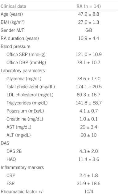
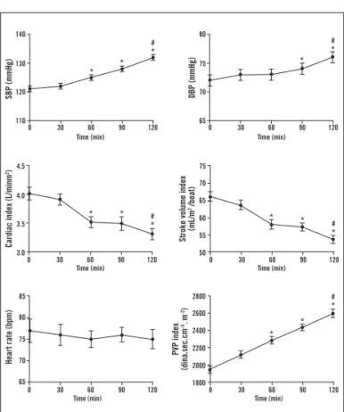
A signiicant decrease in cardiac output and stroke volume was observed ater two-hour inliximab infusion (7.04 ± 2.3 to 6.12 ± 2.1 L/min and 91± 29.0 to 83 ± 28.8 mL/beat, respectively). Conversely, SBP, DBP, and PVR progressively increased during inliximab infusion. hese hemodynamic indings were normalized to each individu-al body surface area and are expressed in Graphic 1.
No symptoms were reported during the TNF-α inhibi-tor administration.
No hemodynamic changes were observed during sa-line infusion protocol in the same RA patients. Results are shown in Table 3.
DISCUSSION
he present study demonstrated that cardiac index and stroke volume index progressively decreased during inf-liximab infusion in individuals not presenting clinical and echocardiographic evidences of heart failure and follow-ing chronic treatment for rheumatoid arthritis. In healthy subjects, but not in RA patients, the normal cardiac re-sponse for saline infusion should be an increase in stroke volume and cardiac output of approximately 10%, without signiicant change in heart rate or blood pressure17. hese
results found could not be due to other drugs taken by
Clinical data RA (n = 14)
Age (years) 47.2 ± 8.8
BMI (kg/m2) 27.6 ± 1.3
Gender M/F 6/8
RA duration (years) 10.9 ± 4.4
Blood pressure
Ofice SBP (mmHg) 121.0 ± 10.9
Ofice DBP (mmHg) 78.1 ± 10.7
Laboratory parameters
Glycemia (mg/dL) 78.6 ± 17.0
Total cholesterol (mg/dL) 174.1 ± 20.5
LDL cholesterol (mg/dL) 89.3 ± 16.7
Triglycerides (mg/dL) 141.8 ± 58.7
Potassium (mEq/L) 4.1 ± 0.7
Creatinine (mg/dL) 1.0 ± 0.1
AST (mg/dL) 20 ± 3.4
ALT (mg/dL) 20 ± 10
DAS
DAS 28 4.3 ± 2.0
HAQ 11.4 ± 3.6
Inlammatory markers
CRP 2.4 ± 1.8
ESR 31.9 ± 18.6
Rheumatoid factor +/- 10/4
RA, rheumatoid arthritis; n, number of patients; BMI, body mass index; M/F, male/female; SBP, systolic blood pressure; DBP, diastolic blood pressure; LDL, low-density lipoproteins; AST, aspartate transaminase; ALT, alanine transaminase; DAS, Disease Activity Score; HAQ, Health Assessment Questionnaire; CRP, c-reactive protein; ESR, erythrocyte sedimentation rate. Values are expressed as means ± SD.
Table 1 – General characteristics of patients with rheumatoid arthritis
RA (n = 14)
Aorta (mm) 28.1 ± 2.0
LA (mm) 36.6 ± 2.7
LV septal thickness (mm) 9.5 ± 0.5
LV posterior wall thickness (mm) 9.1 ± 1.1
LV end-diastolic diameter (mm) 50.8 ± 3.0
LV end-systolic diameter (mm) 31.9 ± 2.8
Ejection fraction (%) 70.3 ± 4.3
LV mass/BSA (g/m2) 96.0 ± 9.2
RA, rheumatoid arthritis; LA, left atrium; LV, left ventricle; BSA, body surface area. Values are expressed as means ± SD.
patients, since inliximab was the only new treatment ad-ministrated during the study. Also, there are no reports of cardiac injury caused by inliximab synergism with other drugs. herefore, only a direct efect of inliximab on the heart could be responsible for the impairment in cardiac output in these patients. Inversely to healthy individuals17,
SPB and total PVR increased during the TNF-α inhibitor administration. To the authors’ knowledge, this is the irst time that hemodynamic parameters have been evaluated in RA patients during acute infusion of inliximab in order
to better evaluate its impact on cardiovascular function. It is noteworthy that hemodynamic measurements were obtained by using the Portapres system, which has a near-zero bias (7% error) when compared with gold standard invasive methods such as triplicate thermodilution. It is probable that the two large randomized trials that stud-ied anti-TNF-α infusion in heart failure patients did not detect cardiovascular efects because low doses of inlix-imab were administrated, and hemodynamic parameters were not monitored by an accurate method such as the Portapres system. Using this system, the present ind-ings demonstrated that, even using moderate inliximab doses, its acute infusion may cause asymptomatic cardiac dysfunction.
It is known that the main cause of death in RA is car-diovascular disease (CVD)18, and the overall increase
in heart failure among RA patients may be related to in-creased inlammatory activity, perhaps leading to prema-ture atherosclerosis. In fact, clinical markers of inlamma-tion have been associated with cardiovascular mortality and morbidity in RA patients5. Contrarily to the healthy
cardiac tissue, heart failure is of special interest because the failing heart produces TNF19. Inversely to the lack of
data regarding the efects of circulating TNF-α on cardio-vascular function in humans19-22, studies in mice using
heart failure models suggest improvement in ventricular dysfunction by circulating TNF blockade23,24. However,
these results could not be reproduced in patients, and on the contrary, clinical trials showed that patients with heart failure had no beneit from the anti-TNF-α drug use, and it could even make their disease worse. hese large ran-domized trials conirmed that patients with heart failure NYHA class III-IV had disease progression associated with infusion of high doses of anti-TNF-α (10mg/kg). Nevertheless, the safety of patients without heart failure is still uncertain21,25.
Several pathophysiologic mechanisms contribute to cardiac injury, especially elevated oxidative stress and in-lammation, which correlate with a variety of conditions such as hypertension, coronary artery disease, cardiomy-opathy, atherosclerosis, and heart failure26. TNF-α is the
most important cytokine related to these mechanisms. Increased TNF-α levels in inlammatory states lead to a high concentration of reactive oxygen species (ROS), in a vicious cycle, causing elevation of pro-oxidants. he im-balance between pro-oxidants and antioxidants causes systemic injury, including heart failure by DNA damage, protein nitration, lipid peroxidation, and activation of ma-trix metalloproteinases27,28.
Conversely, despite the fact that TNF-α inhibition can both preserve cardiac function and partially reverse pathological changes in congestive heart failure24 in
ani-mal models, this could not be reproduced in humans. Such
RA (n=14) Before saline After saline
SBP (mmHg) 163 ± 13 161 ± 15
DBP (mmHg) 86.7 ± 8.2 88.3 ± 3.1
Heart rate (bpm) 74.3 ± 17.0 74.67 ± 11.7
Stroke volume (mL) 80.5 ± 16.9 80.9 ± 24.4
Cardiac output (L/min) 6.1 ± 2.7 6.2 ± 2.9
Peripheral vascular resistance (dina.sec.cm-5)
1,701 ± 708.9 1,771 ± 718.4
RA, rheumatoid arthritis; n, number of patients; SBP, systolic blood pressure; DBP, diastolic blood pressure. Values are expressed as means ± standard deviation.
Table 3 – Control group hemodynamic parameters
Graphic 1 – Hemodynamic changes during 2-hour inliximab infusion (3-5 mg/kg) in RA patients (n = 14). *p < 0.001
versus before infusion. #p < 0.05 versus 60 minutes. SBP,
systolic blood pressure; DBP, diastolic blood pressure, PVR, peripheral vascular resistance.
SBP (mmHg) DBP (mmHg)
Cardiac index (L/minm
2)
Heart rate (bpm)
PVP index
(dina.sec.cm
-5. m -2)
Stroke volume index
(mL/m
2 /beat)
0 30 60 90 120 0 30 60 90 120
0 30 60 90 120 0 30 60 90 120
0 30 60 90 120 0 30 60 90 120
120
110 130 140
3.5
3.0 4.0 4.5
70
65 75 80
Time (min) Time (min)
Time (min) Time (min)
Time (min) Time (min)
55 60
50 70 65 75
2000 2200
1800 2600 2400 2800
75
65 80 85
70
* *
*
#
* *
#
* * *
#
* *
* #
*
* *
diiculty has probably occurred because mice heart failure models were transgenic (TNF1.6) with cardiac-speciic overexpression of TNF-α, and heart failure in humans has several enrolled cytokines and ROS due to inlamma-tion. he question “why is TNF-α inhibition harmful?” remains answered. According to some authors, inliximab can cause cell lyses in the presence of a complement29, as
well as a rebound efect of TNF-α toxicity on inliximab infusion22. Since the actual cause is not known, there is no
evidence regarding the safe dose of inliximab.
Some relevant aspects should be highlighted when ana-lyzing these results, because a number of potential limita-tions must be considered. First, a small number of RA pa-tients were enrolled in the study. However, even studying a small sample of subjects, the Portapres system allows for continuous recording (beat-to-beat) of hemodynamic vari-ables; the standard errors of means were very small and a test power of 0.72 was achieved. Second, patients did not have a long-term follow-up, so the accumulative dose efects of inliximab on cardiac function could not be analyzed. And inally, due to the small sample of patients studied, there was no standardization of RA duration and severity.
he present indings suggest that a two-hour inliximab infusion decreases cardiac output and stroke volume, even in RA patients without clinical and echocardiographic evidences of previous cardiac dysfunction. he results also demonstrate that, in spite of its possible negative inotropic efect, inliximab may enhance BP, probably by increasing PVR. However, considering the limitations of this study and of others, further investigations on acute and long-term administration of this anti-TNF-α drug, involving a higher number of subjects, should be performed in order to assess the safety of this RA treatment.
ACKNOWLEDGEMENTS
his study was supported by the Fundação de Amparo à Pesquisa do Estado de São Paulo (Fapesp), by the Conse-lho Nacional de Desenvolvimento Cientíico e Tecnológi-co (CNPq), and by the Coordenação de Aperfeiçoamento de Pessoal de Nível Superior (Capes), Brazil.
REFERENCES
1. Smolen JS, Aletaha D, Koeller M, Weisman MH, Emery P. New therapies for treatment of rheumatoid arthritis. Lancet. 2007;370:1861-74.
2. Doan T, Massarotti E. Rheumatoid arthritis: an overview of new and emerging therapies. J Clin Pharmacol. 2005;45:751-62.
3. Mann DL. Inlammatory mediators and the failing heart: past, present, and the foreseeable future. Circ Res. 2002;91:988-98.
4. Lysander WJ, Van Lieshout JJ. Non-invasive pulsatile arterial pressure and stroke volume changes from the human inger. Exp Physiol. 2005;90:437-46. 5. Wallberg-Jonsson S, Johansson H, Ohman ML, Rantapaa-Dahlqvist S. Extent
of inlammation predicts cardiovascular disease and overall mortality in sero-positive rheumatoid arthritis. A retrospective cohort study from disease onset. J Rheumatol. 1999;26:2562-71.
6. Wolfe F, Flowers N, Burke TA, Arguelles LM, Pettitt D. Increase in lifetime adverse drug reactions, service utilization, and disease severity among patients who will start COX-2 speciic inhibitors: quantitative assessment of channeling bias and confounding by indication in 6689 patients with rheumatoid arthritis and osteoarthritis. J Rheumatol. 2002;29:1015-22.
7. Bértolo MB, Brenol CV, Schainberg CG, Neubarth F, Lima FAC, Laurindo IM, et al. Atualização do consenso brasileiro no diagnóstico e tratamento da artrite reumatóide. Rev Bras Reumatol. 2007;47:151-9.
8. Aletaha D, Neogi T, Silman AJ, Funovits J, Felson DT, Bingham CO, 3rd, et al. 2010 rheumatoid arthritis classiication criteria: an American College of Rheumatology/European League Against Rheumatism collaborative initiative. Arthritis Rheum. 2010;62:2569-81.
9. Lang EA. Recommendations for chamber quantiication: a report from the American Society of Echocardiography’s Guidelines and Standards Commit-tee and the Chamber Quantiication Writing Group. J Am Soc Echocardiogr. 2005;18:1440-63.
10. El Assaad MA, Topouchian JA, Darne BM, Asmar RG. Validation of the Om-ron HEM-907 device for blood pressure measurement. Blood Press Monit. 2002;7:237-41.
11. Chobanian AV, Bakris GL, Black HR, Cushman WC, Green LA, Izzo JL, Jr., et al. he Seventh report of the Joint National Committee on Prevention, Detec-tion, EvaluaDetec-tion, and Treatment of High Blood Pressure: the JNC 7 report. JAMA. 2003;289:2560-72.
12. Langewouters GJ, Settels JJ, Roelandt R, Wesseling KH. Why use Finapres or Portapres rather than intra-arterial or intermittent non-invasive techniques of blood pressure measuremant?. J Med Eng Technol. 1998;22:37-43.
13. Jansen JRC, Schreuder JJ, Mulier JP, Smith NT, Settels JJ, Wesseling KH. A comparison of cardiac output derived from the arterial pressure wave against thermodilution in cardiac surgery patients. Br J Anaesth. 2001;87:212-22. 14. Leonetti P, Audat F, Girard A, Laude D, Lefrère F, Elghozi J. Stroke volume
monitored by modeling low from inger arterial pressure waves mirrors blood volume withdrawn by phlebotomy. Clin Auton Res. 2004;14:176-81. 15. Ramey DR, Raynauld JP, Fries JF. he health assessment questionnaire 1992:
status and review. Arthritis Care Res. 1992;5:119-29.
16. Wolfe F. A reappraisal of HAQ disability in rheumatoid arthritis. Arthritis Rheum. 2000;43:2751-61.
17. Kumar A, Anel R, Bunnell E, Habet K, Neumann A, Wolf D, et al. Efect of large volume infusion on let ventricular volumes, performance and contractil-ity parameters in normal volunteers. Intensive Care Med. 2004;30:1361-9. 18. Ku IA, Imboden JB, Hsue PY, Ganz P. Rheumatoid arthritis: model of systemic
inlammation driving atherosclerosis. Circ J. 2009;73:977-85.
19. van de Putte LB, Atkins C, Malaise M, Sany J, Russell AS, van Riel PL, et al. Ef-icacy and safety of adalimumab as monotherapy in patients with rheumatoid arthritis for whom previous disease modifying antirheumatic drug treatment has failed. Ann Rheum Dis. 2004;63:508-16.
20. Weinblatt ME, Keystone EC, Furst DE, Moreland LW, Weisman MH, Bir-bara CA, et al. Adalimumab, a fully human anti-tumor necrosis factor alpha monoclonal antibody, for the treatment of rheumatoid arthritis in patients taking concomitant methotrexate: the ARMADA trial. Arthritis Rheum. 2003;48:35-45.
21. Mann DL, McMurray JJ, Packer M, Swedberg K, Borer JS, Colucci WS, et al. Targeted anticytokine therapy in patients with chronic heart failure: results of the Randomized Etanercept Worldwide Evaluation (RENEWAL). Circulation. 2004;109:1594-602.
22. Chung ES, Packer M, Lo KH, Fasanmade AA, Willerson JT. Randomized, dou-ble-blind, placebo-controlled, pilot trial of inliximab, a chimeric monoclonal antibody to tumor necrosis factor-alpha, in patients with moderate-to-severe heart failure: results of the anti-TNF herapy Against Congestive Heart Failure (ATTACH) trial. Circulation. 2003;107:3133-40.
23. Kubota T, Bounoutas GS, Miyagishima M, Kadokami T, Sanders VJ, Bruton C, et al. Soluble tumor necrosis factor receptor abrogates myocardial inlamma-tion but not hypertrophy in cytokine-induced cardiomyopathy. Circulainlamma-tion. 2000;101:2518-25.
24. Kadokami T, Frye C, Lemster B, Wagner CL, Feldman AM, McTiernan CF. Anti-tumor necrosis factor-alpha antibody limits heart failure in a transgenic model. Circulation. 2001;104:1094-7.
25. Zink A, Strangfeld A, Schneider M, Herzer P, Hierse F, Stoyanova-Scholz M, et al. Efectiveness of tumor necrosis factor inhibitors in rheumatoid arthritis in an observational cohort study: comparison of patients according to their eligi-bility for major randomized clinical trials. Arthritis Rheum. 2006;54:3399-407. 26. Khaper N, Bryan S, Dhingra S, Singal R, Bajaj A, Pathak CM, et al. Targeting
the vicious inlammation-oxidative stress cycle for the management of heart failure. Antioxid Redox Signal. 2010;13:1033-49.
27. Giordano FJ. Oxygen, oxidative stress, hypoxia, and heart failure. J Clin Invest. 2005;115:500-8.
28. Matsuzawa A, Ichijo H. Redox control of cell fate by MAP kinase: physiological roles of ASK1-MAP kinase pathway in stress signaling. Biochim Biophys Acta. 2008;1780:1325-36.