55
Tumor of orbit
Tumor orbitário
Mirjana A. Janicijevic-Petrovic
1, Tatjana S. Sarenac-Vulovic
1, Katarina M. Janicijevic
2, Dejan D. Vulovic
2, Dragan I. Vujic
31Clinic of Ophthalmology, Clinical Centre in Kragujevac, Serbia. 2Faculty Medical Sciences, University of Kragujevac, Serbia. ³State University of Novi Pazar, Serbia.
ABSTRACT
The cavernous hemangiomas are the most common intra orbital tumors found in adults of the middle age. Although histological benign, they can encroach on intra orbital or the adjacent structures (optic nerve) and be considered anatomically or positional malignant. We present a case report of orbital cavernous hemangioma of right orbit in young women after pregnancy, from Topola near Kragujevac (Central Serbia) with visual compromise and it’s by trans-nasal endoscopic surgical management. Our patient was controlled and treated with the symptomatic therapy, topical therapy with artificial tears and surgical treatment. Our patient has optimal visual acuity of affected right eye after surgical treatment of orbital tumor. Surgical treatment of symptomatic orbital cavernous hemangioma is safe and effective, so that the cosmetic results are the important parameter to evaluate the clinical outcome.
Keywords: Cavernous hemangioma/diagnosis; Cavernous hemangioma/surgery; Orbit/pathology; Case reports
RESUMO
Os hemangiomas cavernosos são os tumores intra-orbitais mais comuns encontrados em adultos de meia-idade. Embora histológico benigno, eles podem invadir orbital intra ou as estruturas adjacentes (nervo óptico) e ser considerado anatomicamente ou posicional maligno. Apresentamos um relato de caso de hemangioma cavernoso orbital da órbita direita em mulheres jovens após a gravidez, a partir de Topola perto Kragujevac (Central Sérvia) com comprometimento visual e é por tratamento cirúrgico endoscópico trans-nasal. A paciente foi controlado e tratado com a terapia, terapia tópica sintomático de lágrimas artificiais e tratamento cirúrgico. Nosso paciente tem acuidade visual ideal do olho direito afetada após o tratamento cirúrgico de tumor orbital. O tratamento cirúrgico do sintomático hemangioma cavernoso orbital é segura e eficaz, de modo que os resultados cosméticos são o parâmetro importante para avaliar o resultado clínico.
Descritores: Hemagnioma cavernoso/diagnostic; Hemangioma cavernoso/cirurgia; Orbita/patoogia; Relatos de casos
C
ASER
EPORTThe authors declare no conflicts of interest
Received for publication: 4/4/2013 - Accepted for publication: 27/11/2013
Rev Bras Oftalmol. 2014; 73 (1): 55-8
56
INTRODUCTION
O
rbit tumor occurs in middle-aged women, and has its peak incidence in early middle age. The capillary and cavernous hemangiomas are the most common vascu-lar tumors and are classified separately because of distinct clini-cal and pathologic differences1.Visual disability results from a high degree of relative hy-peropia or from optic nerve compression. Relative hyhy-peropia may persist, in spite of complete removal of tumor. The pattern of choroidal folds generally reflected the location of the tumor within the orbit2. B-ultrasound and MRI in combination with
clini-cal manifestations facilitated the diagnosis and orientation of orbital cavernous hemangiomas3.
The most patients underwent B-ultrasound and surgical exploratory procedures. The most of tumors are unilateral and can increase intra orbital volume with a resultant the mass ef-fect. Complete excision is usually possible and surgical morbid-ity is low. The morbidmorbid-ity associated with orbital cavernous he-mangioma is the threat of compressive optic neuropathy, extra ocular muscle dysfunction and cosmetic disfigurement4.
Case report
A 28 years old female presented with two months history of a progressive right visual loss, that had worsened during the last pregnant in 2012.
She presents with ptosis and eye movement limitation of right eye, about two years ago.
After a complete ophthalmologic evaluation, she performed the perimetry that showed severe, peripheral decrease of visual field sensitivity, as incomplete hemianopsy.
Ultrasonography and MRI showed intra orbital mass that filled up homogeneously at the right orbital apex, compressing the orbital nerve and with close relationship with extra ocular muscles.
Visual acuity of the right eye was 0.1 with correction, and 1.0 with correction of the left eye (hypermetropio/anisometropio) by Snellen’s test. On examination, ptosis of 4mm was noted in
the right eye. Intraocular pressures (Goldman’s tonometry) were regular (pre and post operative). Hertel’s measurement (by egzoophthalmometry) was 24mm on the right eye and 22mm on the left eye (both-preoperative) and the less-postoperative was 22mm on the right eye. The test of double vision was regular always. Slit examination of the both eye was regular. Fundus examination of right eye indicated for the choroidal folds with-out clear margin of the right optic nerve. Indirect ophthalmos-copy of left eye showed correct clinical state. Ultrasonography showed retro bulbar marked regular and oval formation 20x19mm, near of the right optic nerve, figure 1.
Perymetry (Humphrey-perimetry tests) showed the spe-cific scotoma - hemianopsy with transitory field compromise, with the best results after surgical treatment, figure 2.
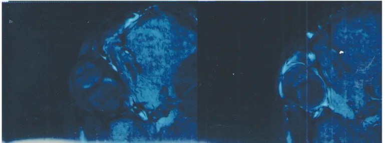
MRI showed the hyperdense, retro bulbar, oval, intraconal
Figure 1. Ultrasonography of the right eye (standardized B-scan) showed well circumscribed intraconal mass in the right orbit, as vascular tumor and R=19 mm
Figure 2. The perimetry tests of the right eye with orbit tumor and the scotoma – incomplete hemianopsy (before and after surgical treatment - as the best results) Janicijevic-Petrovic MA, Sarenac-Vulovic TS, Janicijevic KM, Vulovic DD, Vujic DI
57
mass in the right orbit, located in the inferior temporal quadrant. This formation (21x20mm) was as tissue, clear margin, and with compression of the neurovascular elements of the right orbit, figure 3.
Clinical indication for MRI was act to consultation of neu-rosurgical and plastic doctors.
Operation was done in general anesthesia in Belgrade as a low-risk procedure.
Hystopathological test was showed the benign lesion of cavernous subtype (large anastomosing vascular spaces observed filled of blood, separated by fibrous stroma) and the pathologi-cal diagnosis was the cavernous hemangioma (from the Belgrade).
The adjuvant therapy during hospitalization was the symp-tomatic therapy (steroid drugs, antibiotics) and topical therapy with artificial tears.
DISCUSSION
The most orbital hemangiomas require no intervention, but especially when there is visual compromise, orbit surgery is needed as treatment, but MRI evidence of intralesional hemor-rhage was confirmed by histopathology in diagnosis, as authors known1.
Indications for surgical therapy were visual impairment, progressive unilateral ptosis, diplopia and orbital pain clearly related to orbital cavernoma. The hemangiomas are found be-tween extra ocular muscles and optic nerve within the intraconal place, classically within medial aspect of the orbit and it leads to axial exophthalmia, was in our case5.
They are approached through an upper eyelid or a transcaruncular based medial orbitotomy. Gentle dissection al-lows for en-bloc removal after all vessels have been identified and cauterized with bipolar cautery, that the resection via these approaches can be considered as a low-risk procedure6.
Friberg et al., speak that comparison with uninvolved fel-low eyes showed that preoperative refractive errors were usu-ally shifted toward hyperopia with intraconal tumor, whereas extraconal tumors were typically associated with higher astig-matic errors on involved side, as authors showed2.
The vascular tumors of the orbit include capillary
heman-gioma, cavernous hemanheman-gioma, hemolymphanheman-gioma, hemangiopericytoma (the rare tumor), and we had the cavern-ous hemangioma7.
Transnasal endoscopic resection of intra orbital tumors is feasible and may offer advantages when compared to traditional approaches. It is paramount to have specialized instruments such as long hand piece drills, good camera systems, and long bipolar forceps, as well as an experienced endoscopic surgeon, to con-trol vascular lesions and potential life threatening complications, as was the surgical treatment in our patient8,9. The endoscopic
transnasal approach is safe, effective and less invasive thera-peutic modality for removal of lesions extending from inferomedial part of the left orbital apex to pterygopalatine fossa. With appropriate patient selection, this approach improves ac-cess and visualization, and it enables performance of operative procedures with less risk than conventional microscopic transcranial or transfacial approaches8,9.
Wiegand et al., say that the histopathologic slides were evaluated and additional immunohistochemical stains were done, if necessary to get the correct diagnosis, as we got from the Belgrade3.
Visual function and cosmetic result are main parameters to evaluate clinical outcome. Surgical approach and dissection technique are crucial in determining visual outcome, as in our case report10.
Rootman et al., reported on use of fractionated stereotac-tic radiotherapy (SFRT) for treatment of surgically complicated cavernous. For symptomatic cavernous malformations demon-strating anatomical position that may increase risk of surgical excision, so that SFRT is effective and safe method to control lesion size and improve visual function, but authors had not re-sults of this therapy4.
CONCLUSION
The transnasal endoscopic resection of intra orbital tumors is feasible and may offer some advantages when compared to traditional approaches. This surgical treatment of orbital cavern-ous hemangioma is safe and effective.
Our young patient has optimal visual acuity of the affected right eye, after this surgical treatment of orbital hemangioma.
Figure 3. MRI examination of the right orbit with cavernous hemangioma Tumor of orbit
58
Surgical treatment of symptomatic orbital cavernomas is revers-ible, so that the cosmetic results are the important parameters to evaluate the clinical outcome.
REFERENCES
1. Arora V, Prat MC, Kazim M. Acute presentation of cavernous heman-gioma of the orbit. Orbit. 2011;30(4):195-7.
2. Friberg TR, Grove AS Jr. Choroidal folds and refractive errors associ-ated with orbital tumors. An analysis. Arch Ophthalmol. 1983;101(4):598-603.
3. Wiegand S, Zimmermann AP, Eivazi B, Sesterhenn AM, Sekundo W, Bien S, et al. Analysis of clinically suspected orbital cavernomas. Br J Ophthalmol.
4. Rootman DB, Rootman J, Gregory S, Feldman KA, Ma R. Stereotactic fractionated radiotherapy for cavernous venous malformations (he-mangioma) of the orbit. Ophthal Plast Reconstr Surg. 2012;28(2):96-102.
5. Bouguila J, Yacoub K, Bouguila H, Neji NB, Sahtout S, Besbes G. [In-traorbital cavernous hemangioma]. Rev Stomatol Chir Maxillofac. 2008;109(5):312-5. French.
Corresponding author: Mirjana A. Janicijevic-Petrovic
Clinic of Ophthalmology, Clinical Centre in Kragujevac, Zmaj Jovina 30, 34000 Kragujevac, Serbia
Mob: +38166013691 - Fax: +38134370073 E-mail:[email protected]
6. Bertelmann E, Hartmann C, Minko N. [Intraorbital cavernous heman-giomas: symptoms, diagnostics and surgical approaches]. Klin Monbl Augenheilkd. 2011;228(1):49-53. German.
7. Cophignon J, d’Hermies F, Civit T. [Vascular tumors of the orbit]. Neurochirurgie. 2010;56(2-3):197-212. French.
8. Yoshimura K, Kubo S, Yoneda H, Hasegawa H, Tominaga S, Yoshimine T. Removal of a cavernous hemangioma in the orbital apex via the endoscopic transnasal approach: a case report. Minim Invasive Neurosurg. 2010;53(2):77-9.
9. Stamm A, Nogueira JF. Orbital cavernous hemangioma: transnasal endoscopic management. Otolaryngol Head Neck Surg. 2009;141(6):794-5. 2010;94(12):1653-6.
10. Boari N, Gagliardi F, Castellazzi P, Mortini P. Surgical treatment of orbital cavernomas: clinical and functional outcome in a series of 20 patients. Acta Neurochir (Wien). 2011;153(3):491-8. Review.