Subcutaneous Cavernous Hemangioma in
the Nasal Dorsum: Report of Case Treated
with Endoscopic Rhinoplasty
Jan Alessandro Socher
1Maurício F. de Sá Marchi
2Jeniffer C. Kozechen Rickli
21Department of Otorhinolaryngology, Regional University of
Blumenau Foundation (Fundação Universidade Regional de Bumenau–FURB), Blumenau/SC, Brazil
2Department of Medicine, Regional University of Blumenau
Foundation (Fundação Universidade Regional de Bumenau–FURB),
Blumenau/SC, Brazil
Int Arch Otorhinolaryngol 2014;18:213–216.
Address for correspondence Prof. Dr. Jan Alessandro Socher, PhD, Alameda Duque de Caxias, 145 sala 306, Bairro Centro, CEP 89015-010, Blumenau/SC, Brazil (e-mail: [email protected]).
Introduction
Subcutaneous hemangioma is a rare variant of slow-flow venous malformations.1It occurs in both adults and children and is more prevalent in females.2It shows an aggressive growth pattern, can occur in any part of the body, and
sometimes recurs after excision.1,3,4 The clinical features include local whitening of the skin, followed often by the formation of thin telangiectasias progressing to a cherry-red stain. They are usually papular lesions of a variable thickness that may encompass both superficial and deep layers of the dermis, including subcutaneous regions, giving the lesion a Keywords
►
hemangioma
►
cavernous
►
rhinoplasty
►
natural ori
fi
ce
endoscopic surgery
Abstract
Introduction
Hemangiomas are vascular malformations, with slow blood
fl
ow, that can
occur in any part on the body. They are more common in women and, predominantly, are
isolated lesions. The malformation does not spontaneously regress. Subcutaneous
hemangi-oma is a rare variant with an aggressive growth pattern that sometimes recurs after excision.
Objective
Case report of a subcutaneous cavernous hemangioma in the nasal dorsum
treated with endoscopic rhinoplasty.
Resumed Report
A 27-year-old woman had a
fi
broelastic tumor mass in the midline of
the nasal dorsum, which was pulsatile; she had obstruction and nasal congestion with
associated rhinorrhea, with evolution and worsening over the previous 2 years.
Computed tomography showed a tumor demarcated in the nasal dorsum without
evidence of intracranial communication. Endoscopic rhinoplasty with septoplasty and
associated paranasal sinus sinusectomy was performed without arteriography
emboli-zation, sclerotherapy, or laser. Pathologic diagnosis showed cavernous hemangioma.
Postoperative follow-up shows no recurrence at 3 years.
Discussion
This case presented with atypical features, thus making the diagnosis a
challenge. Imaging studies were required to con
fi
rm the vascular nature of the tumor.
Excisional biopsy is the procedure of choice for pathologic examination. Subcutaneous
hemangiomas never involute and always need treatment. The surgical approach is
exceptional because there was no preoperative diagnosis. In addition, the closed
technique provided best aesthetic results in this case.
Conclusion
Endoscopic rhinoplasty is suitable for nasal dorsum tumor resection and
has superior aesthetic result to open techniques.
received
September 30, 2012 accepted
April 15, 2013
DOI http://dx.doi.org/ 10.1055/s-0033-1351675. ISSN 1809-9777.
Copyright © 2014 by Thieme Publicações Ltda, Rio de Janeiro, Brazil
THIEME
bluish aspect.5,6Nasal hemangiomas account for 15.8% of all the facial hemangiomas. The complications caused by the tumor include uni- or bilateral nasal obstruction, changes in valve and nasal septum, ulcerations, amblyopia, heart and respiratory failure, feeding difficulties, bleeding, and infec-tions, along other psychosocial factors.7,8 The differential diagnosis includes lymphatic malformations, pyogenic gran-uloma, gliomas, and other benign and malignant tumors.9
Objective
We report a case of localized subcutaneous cavernous hem-angioma of the nasal dorsum treated through endoscopic rhinoplasty.
Case Report
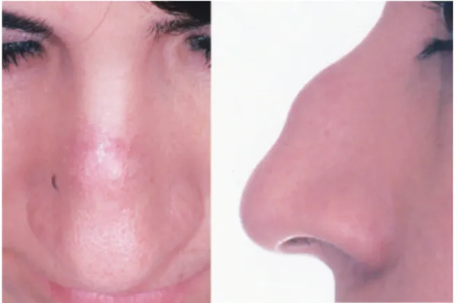
A 27-year-old woman was sent to an otolaryngology service in Blumenau/SC for evaluation of a tumor mass in the midline of the nasal dorsum, with a history of worsening over the past 2 years. The patient complained of nasal congestion associated with obstruction and rhinorrhea. Physical examination re-vealed a tumor in the midline of the nasal dorsum, with a fibroelastic, pulsatile, and motionless aspect, presenting hy-peremia of the skin color and no sign of ulceration (►Fig. 1).
The videoendoscopic exam showed a deviated nasal septum, hypertrophy of the inferior turbinates, and bilateral hyaline rhinorrhea in the middle nasal meatus. The computed tomog-raphy (CT) scan and nuclear magnetic resonance (NMR) of the paranasal sinuses and nasal cavity, with and without contrast, in the axial, coronal, and sagittal planes revealed a tumor restricted to the nasal dorsum, without evidence of intracra-nial communicationto the nasal cavities, with little contrast impregnation (►Figs. 2 and3). Endoscopic rhinoplasty was
planned for resection of the tumor, along with septoplasty surgery and sinusotomy of the paranasal sinuses. The patient was treated in the hospital under general anesthesia and orotracheal intubation. Closed rhinoplasty was performed, with an intercartilaginous incision to approach the nasal dorsum, using an endoscopic technique for complete resection of the subcutaneous tumor with a margin of safety and
preservation of the cutaneous tissue of the nasal dorsum (►Fig. 4). Arteriography with superselective embolization,
sclerotherapy, and laser were not used beforehand or even during the surgical procedure. The pathologic diagnosis was confirmed postoperatively as cavernous hemangioma of the nasal dorsum (►Fig. 5). The patient was followed weekly for
thefirst postoperative month, biweekly during the second postoperative month, then monthly until the sixth month.
Fig. 1 Front and profile photographs showing the detail of the tumor mass in the midline of the nasal dorsum, along withfibroelastic, pulsatile, and motionless aspect, presenting hyperemia of the skin.
Fig. 2 Computed tomography scan in sagittal plane identifying discrete area of contrast enhancement of soft tissue in the nasal dorsum.
Fig. 3 Nuclear magnetic resonance in sagittal plane identifying discrete area of contrast enhancement in the nasal dorsum.
Fig. 4 Intraoperative endoscopic visualization with 30-degree optic demonstrating complete tumor resection of the nasal dorsum.
International Archives of Otorhinolaryngology Vol. 18 No. 2/2014
While monitoring every 6 months until postoperative year 3, there were no detectable signs of tumor recurrence and the patient was satisfied with cosmetic and functional results.
Discussion
Cavernous hemangiomas are tumors formed by vascular ectasia. They can be located deeper in the skin and mucous membranes, but also can involve deeper structures such as subcutaneous tissue, muscle, bone. Hemangiomas may be localized or diffuse. In this case, we identified a case of subcutaneous cavernous hemangioma with atypical clinical features on the nasal dorsum; differential diagnosis should be done with all midline nasal tumors, such as nasal gliomas, meningocele or meningoence-phaloceles, dermoid cysts, teratomas, sebaceous cysts, papillo-mas, lipopapillo-mas,fibromas, and others.10,11
Although the diagnosis may be exclusively clinical on the superficial lesions, cavernous hemangiomas can still be iden-tified through the patient’s history and by the lesion charac-teristics on clinical examination. Imaging tests such as ultrasound, CT, or magnetic resonance imaging (MRI) are needed to confirm the vascular nature and identify the venous, arterial, or lymphatic components and involvement of deeper structures. Regardless, in atypical cases the diagno-sis can be difficult.12
Hemangiomas are congenital malformations that are pres-ent since birth, when they are still incipipres-ent. They evolve with progression proportional to the child’s growth or with an abrupt increase occurring from hormonal changes or local pressure or as a result of injuries, which may explain the behavior in this case with such late growth.4
Radiologic studies are important in the investigation of lesions of the nasal dorsum midline. Pensler cites that CT examination is essential to detect defects at the foramen cecum and helps rule out intracranial communication in some cases.13 According to Lusk et al, MRI may be more useful to assess soft tissue and intracranial communication;
furthermore, no radiation exposure is reported.14To Barko-vich et al, MRI should be the test of choice for the“screening” of initial patient with midline nasal mass. In this case, neither CT nor MRI elucidated connection with the mass of the CNS.15 Negative results on imaging studies, even with contrast, do not exclude the intracranial communication.
The diagnosis and confirmation of nasal dorsum midline tumors were obtained through pathologic examination; ex-cisional biopsy is the gold standard procedure and an inci-sional biopsy is never indicated, because it may lead to meningitis and cerebrospinalfluid leak due to tumor com-munication and common intracranial bleeding of hemangio-mas.10,11In this case, we obtained the diagnosis of cavernous hemangioma postoperatively, after the tumor resection.
Cavernous hemangiomas never involute and should al-ways be treated. Treatment modalities commonly employed in these cases are sclerotherapy and use of laser. Embolization by superselective arteriography is restricted to cases with an arterial component or arteriovenousfistula. The feasibility of removing a cavernous hemangioma depends of the character-istics and where the lesion is located and must be indicated only when it would not cause functional or aesthetic prob-lems. The surgical approach was considered exceptional in this circumstance because there was no preoperative diagno-sis and the case was treated as a midline tumor of the nasal dorsum. Yokoyama et al advocate for endoscopic resection of tumor of the nasal midline when it has no intracranial extension, because the external accesses are associated with aesthetic problems postoperatively.11Closed technique by endoscopic resection was chosen to obtain better aesthetic result and provide greater safety of tumor resection and margins, without need of an external incision and/or skin tissue resection of the nasal dorsum.
Conclusion
Endoscopic rhinoplasty has shown to be an appropriate tech-nique for the resection of tumors of the nasal dorsum, includ-ing cavernous hemangioma, presentinclud-ing no signs of recurrence and with aesthetic results superior to open techniques.
References
1 Enjolras O. Malformações vasculares. In: Bolognia JL, Jorizzo JL, Rapini RP, eds. 2nd ed. Rio de Janeiro, Brazil: Elsevier; 2011:1581–1595
2 Gorlin RJ, Kantaputra P, Aughton DJ, Mulliken JB. Marked female predilection in some syndromes associated with facial hemangio-mas. Am J Med Genet 1994;52:130–135
3 Casanova D, Norat F, Bardot J, Magalon G. [Cutaneous hemangio-ma: clinical aspects]. Ann Chir Plast Esthet 2006;51:287–292
4 Boon LM, Mulliken JB, Enjolras O, Vikkula M. Glomuvenous malfor-mation (glomangioma) and venous malformalfor-mation: distinct clinico-pathologic and genetic entities. Arch Dermatol 2004;140:971–976
5 Hochman M, Adams DM, Reeves TD. Current knowledge and management of vascular anomalies: I. Hemangiomas. Arch Facial Plast Surg 2011;13:145–151
6 Antaya RJ, Ortonne JP, Wells MJ, Perry V, Gelfand JM, James WD. Infantile hemangioma. In: Emedicine. Updated May 20, 2013. Available at: http://emedicine.medscape.com/article/1083849-over-view. Accessed August 20, 2013
Fig. 5 Cavernous hemangioma: photomicrograph showing blood vessels juxtaposed with ample light andfibrous wall lined by a single layer of endothelium. (Hematoxylin staining, 100.)
7 Simic R, Vlahovic A, Subarevic V. Treatment of nasal hemangiomas. Int J Pediatr Otorhinolaryngol 2009;73:1402–1406
8 Waner M, Kastenbaum J, Scherer K. Hemangiomas of the nose: surgical management using a modified subunit approach. Arch Facial Plast Surg 2008;10:329–334
9 Cohen M, Caputy G, Ben-Amitai D, et al. Plastic surgery for hemangi-oma workup. In: Emedicine. Updated August 7, 2013. Available at: http://emedicine.medscape.com/article/1296001-workup. Accessed August 20, 2013
10 Harley EH. Pediatric congenital nasal masses. Ear Nose Throat J 1991;70:28–32
11 Yokoyama M, Inouye N, Mizuno F. Endoscopic management of nasal glioma in infancy. Int J Pediatr Otorhinolaryngol 1999;51:51–54
12 Reilly JR, Koopman CF, Cotton R. Nasal mass in a pediatric patient. Head Neck 1992;14:415–418
13 Pensler JM, Ivescu AS, Ciletti SJ, Yokoo KM, Byrd SE. Craniofacial gliomas. Plast Reconstr Surg 1996;98:27–30
14 Lusk RP, Lee PC. Magnetic resonance imaging of congenital midline nasal masses. Otolaryngol Head Neck Surg 1986;95(3 Pt 1):303–306
15 Barkovich AJ, Vandermarck P, Edwards MS, Cogen PH. Congenital nasal masses: CT and MR imaging features in 16 cases. AJNR Am J Neuroradiol 1991;12:105–116
International Archives of Otorhinolaryngology Vol. 18 No. 2/2014