151
Copyright © 2015 by Academic Publishing House
Researcher
Published in the Russian Federation
Nanotechnology Research and Practice
Has been issued since 2014.ISSN: 2312-7856 E-ISSN: 2413-7227
Vol. 8, Is. 4, pp. 151-164, 2015
DOI: 10.13187/nrp.2015.8.151
www.ejournal13.com
UDC 577.37 + 537.86
The Improved Method for Isolation of Photochrome Trans-membrane Protein Bacteriorhodopsin from Purple Membranes of Halobacterium Halobacterium
Halobium ET 1001
1 Oleg Mosin 2 Ignat Ignatov
1 Moscow State University of Applied Biotechnology, Russian Federation Senior research Fellow of Biotechnology Department, Ph. D. (Chemistry) 103316, Moscow, Talalihina ulitza, 33
E-mail: [email protected]
2 T h e Scientific Research Center of Medical Biophysics (SRCMB), Bulgaria Professor, D. Sc., director of SRCMB
1111, Sofia, N. Kopernik street, 32 E-mail: [email protected]
Abstract
It was developed the improved method for isolation of photochrome trans-membraine protein bacteriorhodopsin (output – 5 mg from 100 g of wet biomass) capable to transform light energy to electrochemical energy of generated protons H+ and P. The protein was isolated from purple membranes of photo-organotrophic halobacterium Halobacterium halobium ET 1001 by cellular autolysis by distilled water, processing of bacterial biomass by ultrasound at 22 KHz, alcohol extraction of low and high-weight molecular impurities, cellular RNA, carotenoids and lipids, solubilization with 0.5% (w/v) SDS-Na, fractionation by MeOH and column gel permeation chromatography (GPC) of the final protein on Sephadex G-200 with 0.1% (w/v) SDS-Na and 2.5 mM ETDA. The homogeneity of the isolated bacteriorhodopsin was proved by combination of preparative and analytical methods, including elecrtophoresis in 12.5% (w/v) PAAG with 0.1% (w/v) SDS-Na and regeneration of apomembranes with 13-trans-retinal.
Keywords: Halobacterium halobium ET 1001, purple membranes, bacteriorhodopsin, biosynthesis, biomolecular electronics
Introduction
152
In halobacteria the BR functions as a light-driven transmembrane proton pump pumping a proton across the membrane. Along with the BR the cell membrane of halobacteria contains a small amount of other related carotenoid pigments, the main of which bakterioruberin determining the stability of halobacteria to solar radiation [2].
The BR is the focus of bio-and nanotechnology mainly because of its high sensitivity and resolution, and is used in molecular bioelectronics as natural photochromic material for light-controlled electrical regulated computer modules and optical systems [3, 4]. In addition, the BR is very attractive as a model for studies of functional activity and structural properties of photo-transforming membrane proteins in the composition of native and photo-converting membranes [5, 6].
The BR-containing nano-films produced using the BR-containing purple membranes (PM) of halobacteria were first obtained and studied in this country in the framework of the project “Photochrome”з when it was demonstrated the effectiveness and prospects for the use of the BR as a photochromic material for holographic recording. These nanomaterials can reversibly change their structure in response to the physical impact and generate two discrete states, calculable by measurement with using spectral methods. This fact determines their usage as logical computing systems in biomolecular electronics [7–9]. Thus, based on the BR was constructed a photoreceptor with a microelectrode of SnO2, consisting of 64 cells (pixels) having the size of 2.5×2.5 mm and electric voltage of 0.3–0.7 V [10]. For the signal conversion by this photoreceptor, low electric current (3–10 nA) was enhanced to voltage from 1 to 10 V and then was supplied to the light emitting diodes. This construction suggests the possibility of effective integration of the BR into the modern microelectronic systems.
The main task for the manufacture of BR-containing nanofilms is the orientation of purple membranes between the hydrophobic and hydrophilic media, such as between water and air, as is common in nature. Typically, to improve the characteristics of the BR-containing films the multiple layers were applied to the surface of the polymeric carrier and dried up, preserving their natural structure. The best results are achieved in the manufacture of films based on gelatin matrix [11, 12]. This allows for a high concentration of the BR (up to 50%) in nanofilms to avoid an aggregation of magenta membrane fragments and destruction of the BR in the manufacturing process [13]. Being embedded onto a gelatin matrix the PM fragments are durable (104 h) and are resistant to many external factors as temperature variations, the intense light exposure, laser radiation, etc. [14]. The dried PM are stacked on the top of each other, focusing in the plane of the matrix. A layer of dried PM with thickness of 1 m contains approx. 200 monolayers. When illuminated by light in such nanofilms is recorded the electric potential approximately 100200 mV, which coincides with the membrane potential of living cells [15].
The structure and mechanism of function of BR have been widely studied in recent years to develop many various approaches of isolation of the protein in native biologically active state that requires the solubilization of BR in a stable, highly purified state free from native lipids [16, 17]. The large scientific and practical interest for obtaining the BR samples for the reconstruction of nanofilms, defined the aim of the present research, related to the development of simple effective method for isolation of the BR from PM in semi-scale quantities.
Material and methods
Bacterial strain
As a BR producer was used a carotenoid strain of extreme photo-organotrophic halobacterium Halobacterium halobium ET 1001, obtained from Moscow State University (Russia). The strain was modified by selection to individual colonies on solid 2% (w/v) agarose media with peptone and 4.3 M NaCl.
Chemicals
For preparation of growth media were used D,L-amino acids г“Reanal”з Hungaryдз ьMPз
153
were of analytical reagent grade. Filtrated water was provided by the Milli-Q-Plus water filtration system (Millipore, Bedford, MA, USA).
Growth conditions
The bacterial growth was carried out on a synthetic medium (SM) containing (g/l): D,L -alanine 0.43; L-arginine 0.4; D,L-aspartic acid 0.45; L-cysteine 0.05; L-glutamic acid 1.3; L-lycine 0.06; D,L-histidine 0.3; D,L-isoleucine 0.44; L-leucine 0.8; L-lysine 0.85; D,L -methionine 0.37; D,L-phenylalanine 0.26; L-proline 0.05; D,L-serine 0.61; D,L-threonine
0.5; L-tyrosine 0.2; D,L-tryptophan 0.5; D,L-valine 1.0, AMP 0.1; UMP 0.1; NaCl 250;
MgSO4.7H2O 20; KCl 2; NH4Cl 0.5; KNO3 0.1; KH2PO4 0.05; K2HPO4 0.05; Na+-citrate 0.5; MnSO4.2H2O – 3.10-4; CaCl2.6H2O 0.065; ZnSO4.7H2O – 4.10-5; FeSO4.7H2O – 5.10-4; CuSO4.5H2O – 5.10-5; Na-citrate 0.5; glycerol 1.0, biotin – 1.10-4; folic acid 1.5.10-4, vitamin B12 – 2.10-5. The growth medium was autoclaved for 30 min at 0.5 atm, the pH value was adjusted to 6.56.7 with 0.5 M KOH. Bacterial growth was performed in 500 ml Erlenmeyer flasks (volume of the reaction mixture 100 ml) for 45 days at t = +35 0ю on a shaker г“эirad Labs”з Hungaryд under intense aeration and monochromatic illumination by light fluorescent lamps LDS-40-2 (40 W) г“ьlfa-Electro”з Russia д го lamps 1.5 lx). Bacterial growth was studied by optical density of the bacterial suspension measured at a wavelength λ = 620 nm on a spectrophotometer Beckman DU-6 г“эeckman юoulter”з USьдй All further manipulations with BR were carried out with the use of a photomask lamp equipped with an orange light filter PCM -мX гтрЖрл cmд г“Marbel”з Germanyдй
Isolation of purple membranes (PM)
The row biomass (1 g) was washed with distilled water and pelleted by centrifugation on a T-нп centrifuge г“юarl Zeiss”з Germanyд (1500 g, 20 min). The precipitate was suspended in 100 ml of dist. H2O and kept for 3 h at 4 0C. The reaction mixture was centrifuged (1500 g, 15 min), the pellet was re-suspended in 20 ml dist. H2O and disintegrated by infrasound sonication (22 kHz, 1 min) in an ice bath (0 0C). The cell homogenate after washing with dist. H
2O was re-suspended in 10 ml of a buffer containing 125 mM NaCl, 20 mM MgCl2, and 4 mM Tris-HCl (pH = 8.0), then 5 mg of RNA-ase (23 units of activity) was added. The mixture was incubated in the dark for 2 h at t = +37 0C. Then 10 ml of the same buffer was added and kept for 1012 h at 4 0C. The aqueous fraction was separated by centrifugation on T-нп centrifuge г“юarl Zeiss”з Germanyд (1500 g, 20 min), the PM precipitate was treated with 50% (v/v) EtOH (5 times 5 ml) at 4 0C followed by separation of the solvent. This procedure was repeated 5 times to give colorless washings. The protein content in the samples was determined spectrophotometrically on a Beckman DU-6 spectrophotometer г“эeckman юoulter”з USьд by the ratio D280/D568 (280 = 1.1.105 M-1. m-1; 568 = 6.3.104 M-1. m-1)[18]. Regeneration of PM was performed as described in [19]. The output of PM fraction makes up 120 mg (8085%).
Isolation of BR
Fraction of the PM (in H2O) (1 mg/ml) was dissolved in 10 ml of 0.5% (w/v) SDS-Na, and incubated for 57 h at t = +37 0C followed by centrifugation (1200 g, 15 min). The precipitate was separated, after that MeOH was added to the supernatant in divided portions (3 times 2 ml) at 0 0C. The reaction mixture was kept for 1415 h in ice bath at t = +4 0C and then centrifuged (1200 g, 15 min). The fractionation procedure was performed three times, reducing the concentration of 0.5% SDS-Na to 0.2 and 0.1% (w/v). The crystal protein (output, 810 mg) was washed with cold distilled 2H
2O (2 times 1 ml) and centrifuged (1200 g, 15 min). Purification of BR
154
protein fraction was subjected to freeze-drying, sealed in glass ampoules (1050 mm) and stored in frost camera at t = -10 0C.
Electrophoresis of BR
The procedure was performed in 12.5% (w/v) polyacrylamide gel (PAAG) containing 0.1% (w/v) SDS. The samples were prepared for electrophoresis by standard procedures (LKB protocol, Sweden). The electrophoretic gel stained with Coomassie blue R-250 was scanned on a CDS-200 laser densitometer г“эeckman”з USьд for quantitative analysis of the protein level.
Proton translocation
The proton translocation was recorded in visicles with a gel-filled pH electrode (Sensorex No. SG900C) combined with PHM85 Radiometer pH meter. The data were collected (0.8 s per time interval) and analyzed using Lotus Measure (Lotus Development). Initial proton pumping rates were determined over 10 s interval. Changes in pH were calibrated using 1m of 10 mM HCl.
Preparation of apomembranes (AP)
50 mg of PM was suspended in 50 ml of 1 M NH2OH (pH = 6.0). The reaxion mixure was kept for 10 h with stirring in ice bath (4 0C) under illumination with a xenon lamp. The precipitate was separated by centrifugation (1000 g, 10 min), washed twice with distilled water and centrifuged. AP fraction was resuspended in 2 ml of 5 mM 2-(N-morpholino)-ethane sulfonyl amide in 100 mM NaCl.
Regeneration of AP with 13-trans-retinal
To 2 ml suspension of AP (2.10-5 mol) in a quartz cuvette was added with stirring 0.1 ml of 2 mM solution of 13-trans-retinal in methanol and kept for 68 h in the dark at t = +40 0C. The degree of regeneration of PM was determined by spectrophotometry on CDS-200 laser densitometer г“эeckman”з USьд by the ratio: Dnat.280.Dnat..568/Dreg..280.Dreg.568, where D280 and D568 the absorbance of a suspension of native and regenerated PM at λ ш нул and λ ш рсу nmй
Results and discussion
Structure and mechanism of action of BR
155
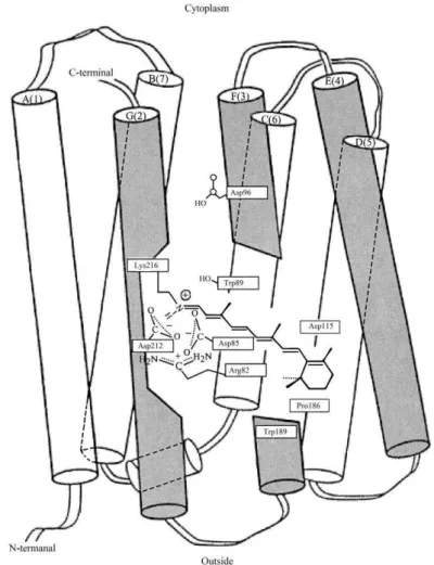
Figure 1: The location of protein and the retinal residue in the BR molecule of halobacterium
H. halobium according to computer modeling: Latin numerals indicate protein fragments of the BR molecule as 7 -helical segments (A, B, C, D, E, G, F) with exposed amino acid residues; gray
color represents the segments responsible for binding of retinal residue to the G-helical segment of BR molecule.
156
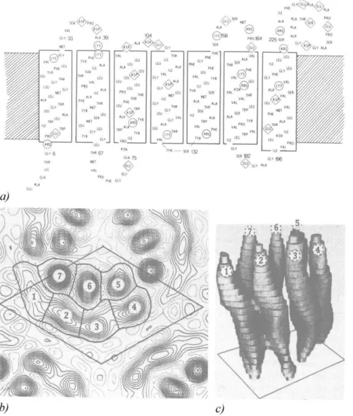
Figure 2: The structure of the BR molecule according to X-ray diffraction analysis:
a) – the primary structure of the BR molecule: amino acids indicated in Latin characters, circles and rhombs show the functionally important amino acids responsible for spatial orientation of α-helical segments of the protein moiety of the BR molecule and the formation of channels for the
transfer of protons H+ across the cell membrane;
b) – the electron density map of PM (a single molecule of the protein is encircled in the center).
Numbers 1–7 are designated α-helical segments of the BR molecule: – 1– A-segment; 2 –B-segment; 3 – C-segment; 4 – D-segment; 5 – E-segment; 6 – F-segment; 7 – G-segment;
c) – the spatial structure of the BR molecule: 1 – A-segment; 2 – B-segment; 3 – C-segment; 4 – D-segment; 5 – E-segment; 6 – F-segment; 7 – G-segment.
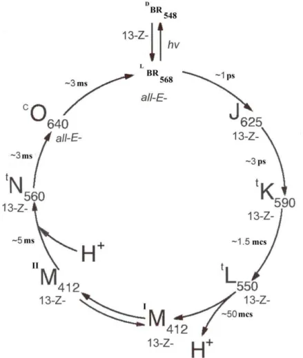
Owing to the unique structure, the BR molecule acts as a light-dependent proton pump, pumping protons across the cell membrane and generates an electrochemical gradient of H+ on the surface of the cell membrane, which energy is used by the cell for the synthesis of ATP in the anaerobic photosynthetic phosphorylation. The mechanism of ATP synthesis is denoted as “ non-chlorophyll photosynthesis”, in contrast to the plant photosynthesis implemented with the participation of chlorophyll. In this mechanism, at absorption of a light photon BR molecule became decolorized by entering into the cycle of photochemical reactions, resulting in the release of a proton to the outside of the membrane, and the absorption of a proton from intracellular space. By the absorption of a light photon is occurred a reversible isomerization of all 13-tras-BR (max = 548 nm) (the quantum yield 0.03 at t = +20 0C) into 13-cis-BR [(
157
intermediates J, К, L, , N, and O, followed by separation of H+ from the retinal residue of BR and its connection from the side of cytoplasm, and finally returns to its 13-trans-conformation while remaining bonded to the protein throughout the photo-cycle (Fig. 3). In this process a proton originating at the Schiff base of the retinal residue is passed across by being transferred to the hydrophilic Asp-85 residue lying in sterically favorable positions, to the other side of the cellular membrane; right after that the vacancy is filled up with a proton transferred from Asp-96 residue [24]. As a result, between the internal and external surface of the membrane forms a concentration gradient of H+, resulting that illuminated by light the halobacteria cells begin to synthesize ATP, i.e. convert light energy into the energy of chemical bonds. This process is reversible and in the dark flows back in the opposite direction, allowing to halobacteria developing in the dark by means of switching of photosynthetic metabolism to the heterotrophic metabolism. Thus, the BR molecule behaves as a photochromic carrier with a short relaxation time the transition from the excited state to the ground state. The optical characteristics of BR vary depending on the method of preparation of PM and the properties of embedded polymer matrix.
The PM are resistant to solar light, the effects of oxygen, the temperatures greater than +80 0C (in water) and up to +140 0C (in air), pH = 112, a high concentration of NaCl (1520% (w/w)) and action of most proteases [25, 26]. Additionally, the PM are resistant to non-polar solvents such as hexane, but are sensitive to mixtures of polar organic solvents with water. These factors are of great practical importance for integration of the PM into a polymeric nanomatrix with keeping photochemical properties.
Figure 3: Photocycle scheme of BR (aqueous solution, pH = 7.2; t = +20±25 0C). Latin numbers J, K, L, M, N, O denote spectral intermediates of BR. IM and IIM represent spectral intermediants of
meta- bacteriorhodopsin with the protonated (IM) and deprotonated (IIM) aldimine bond. L and D denote dark and light forms of pigments. The subscripts correspond to the position of the
158
3.2. Biosynthesis of BRThe technology for preparation of BR consists in growing of halobacteria on liquid synthetic growth media (with 15–20 % (w/w) NaCl) with amino acids, or on natural growth media with peptons – the mixtures of polypeptides and amino acids derived from the partial hydrolysis product or powdered milk, animal meat by proteolytic enzymes (pepsin, trypsin, chymotrypsin), or protein-vitamin concentrate of yeast. The subsequent isolation of BR from purple membranes is carried out by a combination of physical and chemical methods.
For synthesis of the BR used the carotenoid-containing mutant strain of extreme photo-organotrophic halobacterium H. halobium ET 1001. The initial strain was modified by selection of BR synthesizing individual colonies with the purplish red color on the solid (2% agarose) growth SM-medium with peptone and 4.3 M NaCl.
For biosynthesis of the BR the resulting cells were grown aerobically under the monochromatic light illumination on a specifically designed SM-medium containing (g/l):
18 amino acids: D,L-alanine 0.43; L-arginine 0.4; D,L-aspartic acid 0.45; L-cysteine
0.05; L-glutamic acid 1.3; L-lycine 0.06; D,L-histidine 0.3; D,L-isoleucine 0.44; L-leucine
0.8; L-lysine 0.85; D,L-methionine 0.37; D,L-phenylalanine 0.26; L-proline 0.05; D,L -serine 0.61; D,L-threonine 0.5; L-tyrosine 0.2; D,L-tryptophan 0.5; D,L-valine 1.0;
Nucleotides: adenosine-5-monophosphate – 0.1; uridine-5-monophosphate – 0.1;
Inorganic salts: NaCl 250; MgSO4.7H2O 20; KCl 2; NH4Cl 0.5; KNO3 0.1; KH2PO4 0.05; K2HPO4 0.05; Na+-citrate 0.5; MnSO4.2H2O – 3.10-4; CaCl2.6H2O 0.065; ZnSO4.7H2O – 4.10-5; FeSO
4.7H2O – 5.10-4; CuSO4.5H2O – 5.10-5; Glycerol 1.0;
Na-citrate 0.5;
Growth factors: biotin – 1.10-4; folic acid 1.5.10-4, vitamin B
12 – 2.10-5.
159
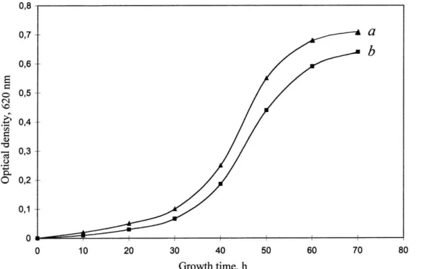
Figure 4: Growth dynamics of H. halobium ET 1001 under various experimental conditions:
a) – SM-medium; b) – peptone medium. Growing conditions: the incubation period: 4–5 days,
temperature: +35 0C, the illumination under monochrome light at λ = 560 nm 3.3. Isolation and purification of BR
The main stages of isolation of the BR were:
Growth of halobacteria H. halobium ET 1001 on the SM-medium; Cell disintegration and autolysis;
Separation of purple membrane (PM) fraction;
Purification of PM from the low and high-molecular weight impurities, cellular RNA, carotenoids and phospholipids;
Solubilization of PM in 0.5% (w/v) solution of ionic detergent SDS–Na to form a micro-emulsion;
Fractionation of the solubilized BR by MeOH;
Gel permeation chromatography (GPC) on Sephadex G-200; Electrophoresis in 12.5% (w/v) PAAG with 0.1% (w/v) SDS-Na.
160
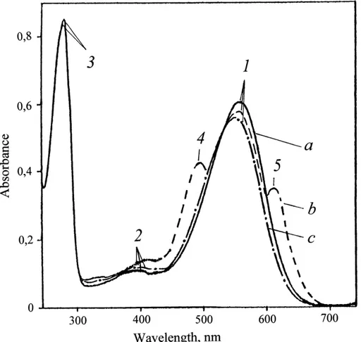
Figure 5: The absorption spectra of PM (50% (v/v) EtOH) at various stages of processing: (a) – natural BR; (b) – PM after intermediate treatment; (c) – PM purified from carotenoids.
The bandwith (1) is the spectral form of BR568, (2) – impurity of spectral form of
meta-bacteriorhodopsin, M412, (3) – the total absorption bandwith of aromatic amino acids, (4) and (5) – extraneous carotenoids. The native BR was used as a control.
161
spectrophotometer г“эeckman юoulter”з USьд for the detergent-solubilized BR or the purified BR-solubilized in detergent.
The fractionation and chromatographic purification of the protein was a necessary next step of the BR purification. For obtaining protein from a biological material in purified, homogeneous state using various detergents assisted to the cleavage of protein-lipid complexes and the rupture of protein-protein bonds. In particular, for the release of proteins (enzymes) which are firmly connected with biomembranes or other subcellular structures used Triton X-100, sodium dodecyl sulfate (SDS) and sodium deoxycholate as detergents.
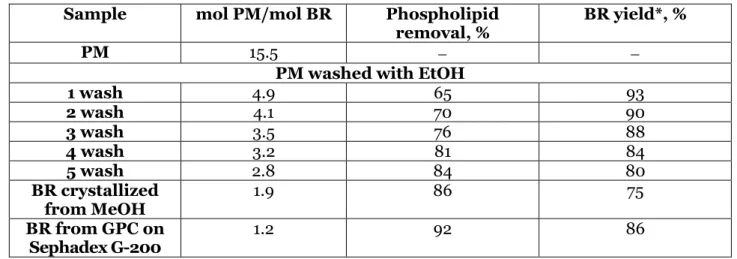
As the BR, being an integral membrane protein intricately penetrates bilipid layer in form of seven -helices, the use of ammonium sulfate and other conventional agents to salting out did not give a positive result for isolation of the protein. The resolving was in the translation of the protein to a soluble form by the colloidal dissolution (solubilization) in appropriate ionic detergent. Using as the ionic detergent SDS-Na was dictated by the need of carrying out the more accomplished solubilization of the protein in a native, biologically active form in complex with 13-trans-retinal, because BR solubilized in 0.5% (w/v) SDS-Na retains a native -helical configuration [27]. However SDS-Na is a strong detergent and its usage for protein fractionation is justified by a limited range of concentration (from 0.1 to 0.5% (w/v)). In addition SDS-Na seems to be more effective for lipid removal that other conventional detergents. Therefore, there was no need the use organic solvents as acetone, methanol and chloroform for purification of phospholipids, while precipitation and delipidization stages being combined in one single step, which significantly simplifies the further fractionation of the protein and reduces its losses during the precipitation procedure. A significant advantage of this method is that the isolated protein in complex with phospholipids and detergent was distributed in the supernatant, while other high molecular weight impurities in the precipitate, which can be easily separated by centrifugation. Fractionation of the solubilized in 0.5% (w/v) SDS-Na protein and its subsequent isolation in crystalline form was performed at 0 0C in three steps precipitating procedure with MeOH, slowly reducing the concentration of SDS-Na from 0.5, 0.25 and 0.1% (w/v). The final stage of the BR purification involved the separation of the protein from low-molecular-weight impurities by GPC. For this purpose the fractions containing BR were passed twice through a chromatography column with dextran Sephadex G-200 balanced with buffer (pH = 8.35) containing 0.1% (w/v) SDS-Na and 2.5 mM EDTA. The data on purification of BR are shown in Table 1.
Table 1: The data on purification stages of the BR
Sample mol PM/mol BR Phospholipid removal, %
BR yield*, %
PM 15.5
PM washed with EtOH
1 wash 4.9 65 93
2 wash 4.1 70 90
3 wash 3.5 76 88
4 wash 3.2 81 84
5 wash 2.8 84 80
BR crystallized from MeOH
1.9 86 75
BR from GPC on Sephadex G-200
1.2 92 86
* Note:
Percentage yield relative to the BR solubilized in SDS-Na before the purification procedure
162
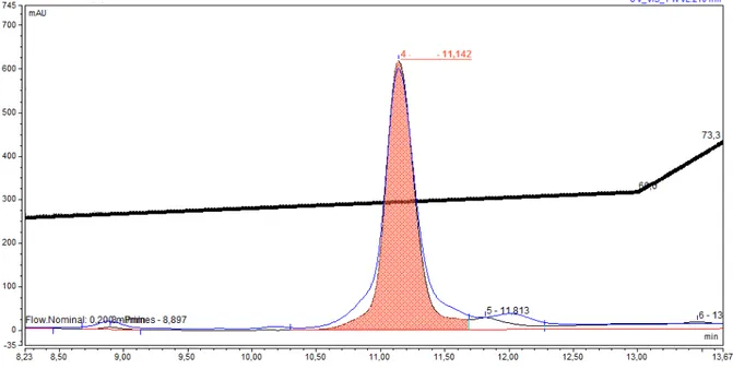
PM. The absorbance profile shows the separated protein. For smaller BR loads (0.1 mg), the analytical column (size) worked equally well.
Figure 6: The GPC-chromatogram of the BR sample after purification (shaded as orange shows the separated protein): GPC (15010 mm) column; stationary phase: Sephadex G-нлл г“Pharmasia”з
USA) equilibrated with buffer with 0.1% (w/v) SDS-Na and 2.5 mM ETDA; specific volume: 3040 units per 1 g dry Sephadex G-200; t = 2025 0C; eluent: 1 mM Tris-HCl buffer (pH = 7.4);
flow rate: 10 ml/cm2.h; detection UV/VIS at λ = 280 and λ = 568 nm
The homogeneity of the isolated BR satisfies to the requirements for reconstruction of native membranes, and was confirmed by electrophoresis in 12.5% (w/v) PAAG with 0.1% (w/v) SDS-Na and in vitro regeneration of AP with 13-trans-retinal. The degree of regeneration of PM was determined by the ratio: Dnat.280.Dnat..568/Dreg..280.Dreg.568 (D280 and D568 the absorbance of a suspension of native and regenerated PM at λ ш нул and λ ш рсу nmд was ср molйай The output of the crystalline protein makes up approximately 5 mg. The isolated protein was washed with cold dist. 2H
2O, centrifuged (1200 g, 15 min), subjected to freeze-drying, sealed into glass ampoules and stored in frost camera at t = -10 0C.
Conclusion
The technology for isolation of BR indicates the high yields of the protein, as a result of which it was isolated 5 mg of BR from 100 g of wet bacterial biomass. To ensure higher yields of BR it needs to accumulate more row biomass feedstock that can be easily achieved in the mass-scale laboratory conditions. The main advantage of this method is that the isolated BR retains its native configuration in combination with 13-trans-retinal, and the ability to photochemical reactions in vitro that is important for further use of the BR for the construction of photo-transforming nanofilms and artificial membranes containing BR.
References:
1. Oesterhelt D., Stoeckenius W. Rhodopsin – like protein from the purple membrane of
Halobacterium halobium кк Natureй мфтмй Vй нооз № уфй Pй мпф–160.
2. Oesterhelt D. The Structure and Mechanism of the Family of Retinal Proteins from Halophilic Archaea // Curr. Op. Struct. Biol. 1988. V. 8. P. 489–500.
163
4. Hampp N., Oesterhelt D. Bacteriorhodopsin and its Potential in Technical Applications / In: Nanobiotechnology (Ch. Niemeyer and C. Mirkin, eds.). – Weinheim: Wiley-VCH-Verlag, 2004. P. 146–167.
5. Mosin O.V., Karnaukhova E.N., Pshenichnikova A.B. Electron impact mass-spectrometry in bioanalysis of stable isotope labeled bacteriorhodopsin / 6th Intern. Conf. on Retinal proteins. – Leiden: Leiden University Press, 1994. p. 115.
6. Mosin O.V., Skladnev D.A., Shvets V.I. The inclusion of deuterated aromatic amino acids in the molecule of bacteriorhodopsin Halobacterium halobium // Applied Biochemistry and Microbiology. 1999. V35, № 1. P. 34–42.
7. Hampp N. Bacteriorhodopsin as a photochromic retinal protein for optical memories //
Chem. Rev. 2000. V. 100. P. 1755–1776.
8. Ignatov I., Mosin O.V. Photoreceptors in visual perception and additive color mixing. Bacteriorhodopsin in nano- and biotechnologies // Advances in Physics Theories and Applications, 2014. V. 27. P. 20–37.
9. Mohammadi S. Bacteriorhodopsin based films in a nanomemory // Advanced Studies in Biology. 2012. V. 4, № 5. P. 207–216.
10. Gui-Ying Ch., Chun-Ping Zh., Zong-Xia G., Jian-Guo T., Guang-Yin Zh., Qi-Wang S. All-optical logic-gates based on bacteriorhodopsin film // Advanced Studies in Biology. 2012. V. 4, № 5. P. 207–216.
11. Shuguang W.U., Ellerby L.M., Cohan J.S. et al. Bacteriorhodopsin rncapsulated in transparent sol-gel glass: a new biomaterial // Chem. Mater. 1993. V. 5. P. 115–120.
12. Wang W.W., Knopf G.K., Bassi A.S. Bioelectronic imaging array based on bacteriorhodopsin film // IEEE Transactions on Nanobioscience. 2008. V. 7, № 4. P. 249–56.
13. Weetall H. Retention of bacteriorhodopsin activity in dried sol-gel glass // Biosensors & Bioelectronics. 1996. V. 11. P. 325–333.
14. Downie J., Timucin D.A., Smithey D.T., Crew M. Long holographic lifetimes in bacteriorhodopsin films // Optics Letters. 1998. V. ноз № фй Pй тол–732.
15. Korposh S.O., Sichka M.Y., Trikur I.I. et al. Films based on bacteriorhodopsin in sol-gel matrices // Proc. of SPIE, 5956, Paper Number 595616. 2005.
16. Mosin O.V., Shvets V.I., Skladnev D.A., Ignatov I. Synthesis of [2 ]bacteriorhodopsin labeled by deuterium on residues of aromatic amino acids // Khimicheskaya Technologiya
(Chemical Engineering), Publishing House “Nauka & Technology” Moscow. 2012. V. 9.
P. 553–564.
17. Mosin O.V., Shvets V.I., Skladnev D.A., Ignatov I. Biosynthesis of trans-membrain photo-transforming protein [2 ]bacteriorhodopsin, labeled with deuterium on residues of aromatic amino acids [2,3,4,5,6-2H
5]Phe, [3,5-2H2]Tyr and [2,4,5,6,7-2H5]Trp // Problems of Biological,
Medical and Pharmaceutical Chemistry. 2013. V. 8. P. 29–39.
18. Nonella M., Windemuth A., Schulten K. Structure of bacteriorhodopsin and in situ
isomerization of retinal: a molecular dynamics study // Journal Photochem. Photobiol. 1991. V. 54, № 6. P. 937–948.
19. Rudiger M., Tittor J., Gerwert K., Oesterhelt D. Reconstitution of bacteriorhodopsin from the apoprotein and retinal studied by Fourier-transformed infrared spectroscopy // Biochemistry.
1997. V. 36. P. 4867-4874.
20. Grigorieff N. Electron-crystallographic refinement of the structure of bacteriorhodopsin // Journal of Molecular Biology. 1996. V. 259. P. 393–421.
21. Jap B.K., Maestre M.F., Hayward S.B., Glaeser R.M. Peptide-chain secondary structure of bacteriorhodopsin // Biophys J., 1983. V. поз № 1. P. 81–89.
22. Mak-Jurkauskas M.L., Baiaj V.S., Hornstein M.K. et al. Energy transformations early in the bacteriorhodopsin photocycle revealed by DNP-enhanced solid-state NMR // Proc. Natl. Acad. Sci. USAй нллуй Vй млрз № ой Pй 883–888.
23. Haupts U., Tittor J., Bamberg E., Oesterhelt D. General concept for ion translocation by halobacterial retinal proteins: the isomerization/switch/transfer model // Biochemistry. 1997. V. осз № н–7. P. 78–85.
164
25. Seitz A., Hampp N. Kinetic optimization of bacteriorhodopsin films for holographic interferometry // J. Phys. Chem. B. 2000. V. млпз № 30. P. 7183–7192.
26. Shamansky L.M., Minh Luong K., Han D., Chronister E.L. Photoinduced kinetics of bacteriorhodopsin in a dried xerogel glass. Biosensors & Bioelectronics. 2002. V. 17. P. 227–231.
27. Mosin O.V., Ignatov I. Biosynthesis of photochrome transmembrane protein bacteriorhodopsin of Halobacterium halobium labeled with deuterium at aromatic amino acids residues of [2,3,4,5,6-2H
5]Phe, [3,5-2H2]Tyr and [2,4,5,6,7-2H5]Trp // Chemistry and material research. нлмпй Vй сй № ой Pй м–15.
К рттйот + 537.86
Halobacterium
halobium ET 1001
1 2
1 з
з й й
млоомсз з й з оо
E-mail: [email protected]
2 - г дз
з г дз
ммммз з й й К з онкс
E-mail: [email protected]
А й
г у–мл дз
й
Halobacterium halobium ET 1001
й з
нн з - - з
Кз з лйра г йк йд -Na,
MeOH - г д
Sephadex G-нлл лйма г йк йд -Na нйр Э й
з мнйра г йк йд лйма г йк йд
-Na - мо- ан - й
х Halobacterium halobium ET 1001з з
![Introduction of Deuterated Aromatic Amino Acids [2,3,4,5,6-2H5]phenylalanine, [3,5-2H2]tyrosine and [2,4,5,6,7-2H5]tryptophan into a Molecule of Photochrome Trans-membrane Protein Bacteriorhodopsin](data:image/gif;base64,R0lGODlhAQABAIAAAP///wAAACH5BAEAAAAALAAAAAABAAEAAAICRAEAOw==)