Characte rizatio n o f a m e thio nine -rich
pro te in fro m the se e ds o f
Cereus
jam acaru
Mill. (Cactace ae )
1Departamento de Biologia, Universidade Estadual do Ceará,
Fortaleza, CE, Brasil
2Departamento de Bioquímica e Biologia Molecular,
Universidade Federal do Ceará, Fortaleza, CE, Brasil
3Instituto de Q uímica, Universidade de Brasília, Brasília, DF, Brasil 4Embrapa/Cenargen, Brasília, DF, Brasil
T.C.F.R. Aragão1, P.A.S. Souza2, A.F. Uchôa2, I.R. Costa2, C. Bloch Jr.3,4 and F.A.P. Campos1
Abstract
We describe here the isolation and characterization of a major albumin from the seeds of Cereus jamacaru (Cactaceae), to which we gave the trivial name of cactin. This protein has a molecular mass of 11.3 kDa and is formed by a light chain (3.67 kDa) and a heavy chain (7.63 kDa). This protein was isolated using a combination of gel filtration chroma-tography and reverse-phase HPLC. The amino acid composition of cactin was determined and found to resemble that of the 2S seed reserve protein from the Brazil nut, a protein remarkable for its high methionine content. The usefulness of cactin as a molecular marker in the taxonomy of the Cactaceae is discussed.
Co rre spo nde nce
F.A.P. Campos
Departamento de Bioquímica e Biologia Molecular
Universidade Federal do Ceará 60001-970 Fortaleza, CE Brasil
E-mail: bioplant@ ufc.br
Research supported by CAPES, CNPq and Fundação Cearense de Apoio à Pesquisa (FUNCAP).
Received July 12, 1999 Accepted April 10, 2000
Ke y wo rds
·Cactaceae ·Mandacaru
·Cereus jam acaru
·Cactin ·2S Albumins
·Methionine-rich protein
Intro ductio n
In the semi-arid regions of Northeastern Brazil, the succulent stems of the manda-caru (Cereus jamacaru Mill.) are used as feed for cattle (1). The plant itself, with its beautiful flowers and its fleshy, red, deli-cious fruits, is almost a symbol of the Brazil-ian Northeast. Despite the potential of the mandacaru as a forage crop and as a fruit crop, very few studies have been conducted in order to help exploit these potentials.
One of the main constraints of the wide acceptance of mandacaru as a forage crop is the low level of protein throughout the plant. The genetic manipulation of this trait
is being hampered by a lack of understanding of the patterns of protein deposition and mobilization in stems and seeds. Here we present data on the characterization of a re-serve protein from the seeds of mandacaru and show that this protein is similar to the 2S methionine-rich seed storage protein from the Brazil nut (2).
Mate rial and Me tho ds
Plant mate rial and se e d ge rminatio n co nditio ns
Aiuába, Ceará, Brazil. Seeds were surface sterilized with 2.5% NaOCl for 5 min, rinsed with sterile water, and sown on wetted, ster-ile filter paper on Petri dishes, under dim light at 28 ± 1oC (3). After germination,
seeds were transferred to the dark. Groups of 20 seedlings were collected at 0, 3, 6, 9 and 12 days of germination and kept at -20o
C until needed.
Pro te in e xtractio n and purificatio n
Salt-soluble proteins were extracted from flour in 0.1 M Tris/HCl and 0.5 M NaCl, pH 8.0, for 2 h. After centrifugation (10,000 g, 20 min, 4o
C), ammonium sulfate was added to the supernatant to a concentration of 90% and the precipitated proteins were recovered by centrifugation. After dialysis against dis-tilled water at 4oC and centrifugation, the
albumin and globulin fractions were obtained. The albumins were fractionated on a Sephacryl S-100-HR column (80 x 2.6 cm) equilibrated and eluted in 0.05 M ammo-nium bicarbonate at a flow rate of 30 ml/h and 4.3-ml fractions were collected. This column was previously calibrated over the range from 6.5 to 66 kDa using the MW-GF-70 kit (Sigma Chemical Co., St. Louis, MO, USA). The effluents containing the 11.3-kDa protein were pooled, freeze-dried and fractionated by molecular exclusion-HPLC on a Superdex Peptide HR 10/30 column from Pharmacia Biotech (Pharmacia Biotech, Uppsala, Sweden), linked to an HPLC sys-tem from Waters Corporation (Milford, MA, USA). The column was equilibrated and eluted in 0.05 M ammonium bicarbonate at a flow rate of 0.5 ml/min. This fractionation yielded two protein peaks.
The peak containing the 11.3-kDa pro-tein was further fractionated by reverse phase-HPLC (RP-phase-HPLC) on a 3.9 x 300 mm µ-Bondapak C-18 column (Waters) linked to a HPLC system from Waters Corporation. The column was equilibrated in 0.1% trifluoro-acetic acid (TFA) and proteins were eluted
in a linear gradient of 0-60% acetonitrile in 0.1% TFA, run in 60 min. In all of the chromatographic steps the effluents were monitored at 280 nm. Protein concentration was determined by the protein-dye binding method of Bradford (4). The homogeneity of the purified protein was ascertained by SDS-PAGE and by isoelectric focusing. For chain separation, the purified protein was reduced and carboxymethylated (5) and the reaction mixture subjected to gel permeation chro-matography in a Superdex Peptide HR 10/30 column, equilibrated and eluted in 0.05 M ammonium bicarbonate at a flow rate of 0.5 ml/min.
Antibo dy pre paratio n and immuno blo tting analysis
alka-line phosphatase-conjugated anti-goat IgG.
SD S-PAGE and iso e le ctric fo cusing
Gel electrophoresis was performed using the tricine-SDS polyacrylamide gel electro-phoresis method for the separation of low molecular mass proteins described by Schag-ger and Jagow (8). The proteins contained in the MW-SDS-17S kit (Sigma) for molecular mass 3.46 to 16.95 kDa were used as molec-ular mass markers. Isoelectric focusing was performed using a method for rapid isoelec-tric focusing in a vertical polyacrylamide minigel system (9).
MALD I-TO F analysis
Freeze-dried samples of the native cactin were prepared for matrix-assisted laser de-sorption time of flight analysis (MALDI-TOF) with a Voyager-DE STR Bioworksta-tion (PerSeptive Biosystems, Framingham, MA, USA) as follows: a 0.5 mg/ml solution of cactin was divided into 2 groups of 3-ml aliquots. To the first group, 9 ml of the matrix a-cyano-4-hydroxycinnamic acid (saturated solution in acetonitrile/0.1% TFA 1:1, v/v) from Sigma Chemicals was added and vortex mixed and 1 ml of the final mix-ture was applied onto the Voyager Biowork-station sample plate. For the second group, prior to the addition of the matrix solution, the 3-ml samples were incubated for 10 min at room temperature with 1 ml of a 1.0 M DTT in 150 mM ammonium bicarbonate solution. Ten ml of the a -cyano-4-hydroxy-cinnamic acid solution was then added to the samples and 1 ml of this mixture was applied onto the sample plate. Both groups of samples were air-dried at room temperature. The spec-trometer equipped with delayed-extraction system was operated in the linear mode. Sample ions were evaporated by irradiation with an N2 laser at a wavelength of 337 nm,
and accelerated at 23 kV potential in the ion source with a delay of 150 ns. Samples were
ionized with 100 to 200 shots of a 3-ns pulse width laser light. The signal was digitized at a rate of 500 MHz and averaged data were presented to a standard Voyager data system for manipulation.
Amino acid analysis
Amino acid compositions of protein samples were analyzed as described previ-ously (10). Lysozyme was also analyzed and the results were compared with published values to ensure the accuracy of the tech-nique. Three independent determinations were performed for each sample. Selected replicates were analyzed in duplicate to en-sure repeatability.
Re sults and D iscussio n
When the albumins from C. jamacaru were analyzed by SDS-PAGE, a major pro-tein band was observed at a position corre-sponding to a molecular mass of 9.5. This protein, to which we gave the trivial name of cactin, was purified by size exclusion chro-matography and by RP-HPLC on a µ-Bondapak C-18 column. The last purifica-tion step yielded cactin in a homogeneous form, as indicated by SDS-PAGE (Figure 1A) and by isoelectric focusing (Figure 1B). This protein accounts for approximately 9% of the total salt-soluble protein.
display-gest that four disulfide bonds must have been reduced and eight hydrogen atoms in-corporated into the molecule. The two low resolution peaks detected in the first experi-ment now are interpreted as possible disso-ciation and/or cleavage products of the na-tive protein yielding the two polypeptide chains of cactin. This might have occurred somewhere during sample manipulation pro-cedures.
The molecular mass calculated from the SDS-PAGE experiments was 9.5 kDa, while MALDI-TOF analysis indicated a molecular mass of 11.3 kDa for the native cactin. This discrepancy between the molecular mass val-ues obtained from SDS-PAGE and MALDI-TOF data probably arises from the anoma-lous electrophoretic behavior which is com-monly presented by basic, low molecular weight proteins.
Polyclonal antibodies were raised against purified cactin and the purified antibodies were highly monospecific. These antibodies were used to show that cactin is mobilized during germination, whereas after 12 days of germination it could no longer be detected (Figure 1C).
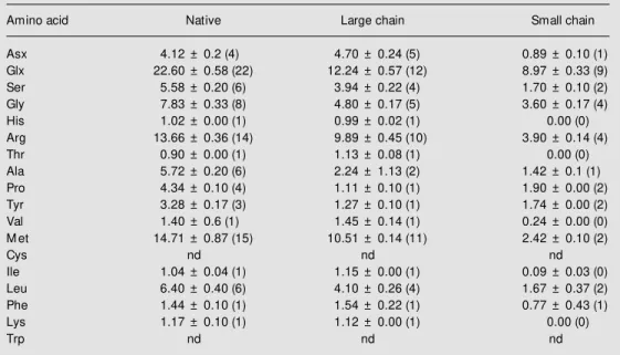
The production of large amounts of the small and large polypeptide chains was made possible by reduction and carboxymethyla-tion of the purified protein and fraccarboxymethyla-tionacarboxymethyla-tion of the reaction mixture on a Superdex Pep-tide HR 10/30 column. This yielded two protein peaks corresponding to the large chain (peak 1, Figure 3) and small chain (peak 2, Figure 3) and two peaks at the end of the run, corresponding to the low molecular weight components of the reduction and carboxy-methylation reaction mixture. The amino acid compositions of the native protein and of its large and small chains were determined by Pico Tag technology (Table 1). The overall amino acid composition of cactin resembles closely that of the 2S seed reserve protein from the Brazil nut (Bertholletia excelsa, Lecythidaceae), a protein remarkable for its high content of methionine (2). Another prop-Figure 1 - A, Assessment of the homogeneity of the methionine-rich protein
isolated from the seeds of C. jamacaru by SDS-PAGE. Purified protein (4 µg) w as added to lane 2 and molecular w eight markers w ere applied to lane 1. B, Assessment of the homogeneity of the methionine-rich protein isolated from the seeds of C. jamacaru by isoelectric focusing. C, Western blot analysis of the mobilization during germination of the methionine-rich protein isolated from the seeds of C. jamacaru. The samples analyzed w ere prepared from seeds 0, 3, 6, 9 and 12 days after germination.
kDa
16.95
14.40
10.16
8.16
6.20
1 2
0 3 6 9 12
A B
C
sug-erty that cactin shares with proteins belong-ing to the 2S seed storage protein family is its high content of amino acids with a high nitrogen to carbon ratio (Arg, Gln and Asn) which are particularly suited for storage of nitrogen (11). The presence of two polypep-tide chains kept together by disulfide bridges is another property that cactin shares with the Brazil nut seed reserve protein and with many members of the 2S seed storage pro-tein family.
The 2S albumins have only become rec-ognized as a major group of seed storage proteins over the last decade (12). These proteins are widely distributed in the seeds of dicots and have been most widely studied in the Cruciferae, notably oil-seed rape and Arabidopsis (12). The presence of two disul-fide-linked polypeptide chains has been dem-onstrated in a number of 2S albumins, al-though some 2S albumins such as those from sunflower (13), prickly-pear (10) and pea (14) have a single polypeptide chain. We are currently exploring the possibilities of using cactin as a molecular marker in the tax-onomy of the Cactaceae. Our preliminary results indicate that proteins cross-reacting with cactin antibodies are present in all spe-cies of the three subfamilies of the Cactaceae (Cactoideae, Opuntioideae and Pereskio-ideae) that we have analyzed (data not shown).
The chief interest in the 2S albumin pro-tein family stems from the fact that many of its members are very rich in the sulfur-con-taining amino acids methionine and cys-teine, which normally are present in very low concentration in plant-derived foodstuffs (15). Within the past decade several groups have focused their attention on using genes encoding these proteins to enhance the nutri-tional quality of plants (16). In the North-eastern states of Brazil, some members of the Cactaceae, notably C. jamacaru and Opuntia ficus-indica, are used as feed for farm animals. It is interesting that both O. ficus-indica (10) and C. jamacaru have these
Figure 2 - A, M ALDI-TOF mass spectrum of native cactin. B, M ALDI-TOF mass spectrum of cactin after reduction w ith DTT.
A2
8
0
0.05 0.10
0
0 30 60
Elution time (min)
Figure 3 - Fractionation of the reduction and carboxymethyla-tion reaccarboxymethyla-tion mixture of cactin on a Superdex Peptide HR 10/ 30 column. 1 and 2, Protein peaks corresponding to large and small chains, respectively.
1 2
R
e
la
ti
v
e
i
n
te
n
s
it
y
(
%
)
100
4000 5000 6000 7000 8000 9000 10000 11000 12000
M ass (m/z)
2000 4000 6000 8000 10000 12000
M ass (m/z) 50
0
5652.8311
11304.8736
R
e
la
ti
v
e
i
n
te
n
s
it
y
(
%
)
50 100
0
3677.3323
5652.5078
7635.4838
11304.7802
A
genes. If their expression can be enhanced, particularly in the stems, then these engi-neered plants would be more nutritious for
farm animals. Work towards this goal is in progress in our laboratory (17,18).
Table 1 - Amino acid composition of the methionine-rich protein isolated from the seeds of Cereus jamacaru, as determined by the Pico Tag method.
Composition is reported as number of residues per mol of protein based on molecular masses of 11300, 7630 and 3670 kDa for the native, large and small chains, respectively. The values for cysteine and tryptophan w ere not determined (nd). Numbers w ithin parentheses represent the assigned integral values.
Amino acid Native Large chain Small chain
Asx 4.12 ± 0.2 (4) 4.70 ± 0.24 (5) 0.89 ± 0.10 (1)
Glx 22.60 ± 0.58 (22) 12.24 ± 0.57 (12) 8.97 ± 0.33 (9)
Ser 5.58 ± 0.20 (6) 3.94 ± 0.22 (4) 1.70 ± 0.10 (2)
Gly 7.83 ± 0.33 (8) 4.80 ± 0.17 (5) 3.60 ± 0.17 (4)
His 1.02 ± 0.00 (1) 0.99 ± 0.02 (1) 0.00 (0)
Arg 13.66 ± 0.36 (14) 9.89 ± 0.45 (10) 3.90 ± 0.14 (4)
Thr 0.90 ± 0.00 (1) 1.13 ± 0.08 (1) 0.00 (0)
Ala 5.72 ± 0.20 (6) 2.24 ± 1.13 (2) 1.42 ± 0.1 (1)
Pro 4.34 ± 0.10 (4) 1.11 ± 0.10 (1) 1.90 ± 0.00 (2)
Tyr 3.28 ± 0.17 (3) 1.27 ± 0.10 (1) 1.74 ± 0.00 (2)
Val 1.40 ± 0.6 (1) 1.45 ± 0.14 (1) 0.24 ± 0.00 (0)
M et 14.71 ± 0.87 (15) 10.51 ± 0.14 (11) 2.42 ± 0.10 (2)
Cys nd nd nd
Ile 1.04 ± 0.04 (1) 1.15 ± 0.00 (1) 0.09 ± 0.03 (0)
Leu 6.40 ± 0.40 (6) 4.10 ± 0.26 (4) 1.67 ± 0.37 (2)
Phe 1.44 ± 0.10 (1) 1.54 ± 0.22 (1) 0.77 ± 0.43 (1)
Lys 1.17 ± 0.10 (1) 1.12 ± 0.00 (1) 0.00 (0)
Trp nd nd nd
Re fe re nce s
1. Braga R (1976). Plantas do Nordeste, Especialmente do Ceará. Coleção M osso-roense. Vol. XLII. Escola Superior de Agricultura de M ossoró, RN, Brazil. 2. Ampe C, Van Damme J, de Castro L,
Sampaio M J, Van M ontagu M & Vande-kerckhove J (1986). The amino acid se-quence of the 2S sulfur-rich protein from seeds of Brazil nut (Bertholletia excelsa H.B.K.). European Journal of Biochemis-try, 159: 597-604.
3. Prisco JT (1966). Action of light on man-dacaru-seed germination. Revista Brasi-leira de Biologia, 26: 261-262.
4. Bradford M (1976). A rapid and sensitive method for the quantitation of microgram quantities of proteins utilizing the prin-ciple of protein-dye binding. Analytical Bio-chemistry, 72: 248-254.
5. Campos FAP & Richardson M (1984). The complete amino acid sequence of the a -amylase inhibitor I-2 from seeds of ragi
(Indian finger millet, Eleusine coracana Gaertn.). FEBSLetters, 167: 221-225. 6. Harlow E & Lane D (1988). Antibodies: A
Laboratory M anual. Cold Spring Harbor Laboratory Press, Cold Spring Harbor, NY. 7. Tow bin H, Staehelin T & Gordon J (1979). Electrophoretic transfer of proteins from polyacrylam ide gels to nitro-cellulose sheets: Procedure and some applications. Proceedings of the National Academy of Sciences, USA, 76: 4350-4354.
8. Schagger H & Jagow G (1987). Tricine-sodium dodecyl sulfate-polyacrylamide gel electrophoresis for the separation of proteins in the range of 1 to 100 kDa. Analytical Biochemistry, 166: 368-379. 9. Robertson EF, Dannelly HK, M alloy PJ &
Reeves HC (1987). Rapid isoelectric fo-cusing in a vertical polyacrylamide minigel system. Analytical Biochemistry, 167: 290-294.
10. Uchoa AF, Souza PAS, Zarat e RM L,
Gomes-Filho E & Campos FAP (1998). The isolation and characterization of a reserve protein from the seeds of Opuntia ficus-indica (Cactaceae). Brazilian Journal of M edical and Biological Research, 31: 757-761.
11. King JE & Gifford DJ (1997). Amino acid utilisation in seeds of loblolly pine during germination and early seedling grow th. Plant Physiology, 113: 1125-1135. 12. Shew ry PR, Napier JA & Tatham AS
(1995). Seed storage proteins: structures and biosynthesis. PlantCell, 7: 945-956. 13. Kortt AA, Caldw ell JB, Lilley GG & Higgins
TJ (1991). Amino acid and cDNA se-quences of a methionine-rich 2S protein from sunflow er seed (Helianthus anuus L.). European Journal of Biochemistry, 195: 329-334.
protein structure, and regulation of the synthesis of a sulfur-rich protein in pea seeds. Journal of Biological Chemistry, 261: 11124-11130.
15. Shotw ell M A & Larkins BA (1989). The biochemistry and molecular biology of seed storage proteins. In: M arcus A (Edi-tor), The Biochemistry of Plants. Vol. 15. Academic Press, New York, 297-345. 16. M olvig L, Tabe LM , Egum BO, M oore AE,
Craig S, Spencer D & Higgins TJ (1997). Enhanced m et hionine levels and in-creased nutritive value of seeds of trans-genic lupins (Lupinus angustifolius L.) ex-pressing a sunflow er seed albumin gene. Proceedings of the National Academy of Sciences, USA, 94: 8393-8398.
17. Llam oca-Zárat e RM , Cam pos FAP & Landsmann J (1999). Establishment and transformation of callus and cell
suspen-sion cultures of the prickly-pear (Opuntia ficus-indica). Journal of the Professional Association for Cactus Development, 3: 27-33.