Rogério Lacerda dos Santos1, Matheus Melo Pithon2, Júlia Barbosa Pereira Leonardo3, Edna Lúcia Couto Oberosler4, Delmo Santiago Vaitsman5, Antônio Carlos de Oliveira Ruellas6
Orthodontic cements: Immediate protection and fluoride release
original article
Objectives: The objective of the authors was to evaluate fluoride release of 3 glass ionomer cements with im-mediate protection of fluoride varnish (Cavitine, SS White), divided into 3 groups: Group M (Meron, VOCO), Group V (Vidrion C, SS White) and Group KC (Ketac-Cem, 3M ESPE).
Methods: Fluoride release was measured during 60 days by means of an ion-selective electrode connected to an ion analyzer. After 4 weeks, the test specimens were exposed to a solution of 0.221% sodium fluoride (1000 ppm of fluoride).
Results: Results showed that the cements reached a maximum peak of fluoride release in a period of 24 h. There was a statistically significant difference between the amount of fluoride released after the applications of fluoride among the groups from the 31st to 60th day (p> 0.05).
Conclusion: The Vidrion C and Meron cements showed better performance to uptake and release fluoride when compared with Ketac-Cem cement.
Keywords: Glass ionomer cement. Fluoride release. Varnish.
How to cite this article: Santos RL, Pithon MM, Leonardo JBP, Oberosler ELC, Vaitsman DS, Ruellas ACO. Orthodontic cements: Immediate protection and fluoride release. Dental Press J Orthod. 2012 July-Aug;17(4)::27.e1-5.
Submitted: August 21, 2008 - Revised and accepted: November 24, 2008
» The authors report no commercial, proprietary or financial interest in the products or companies described in this article.
Contact address: Rogério Lacerda dos Santos Universidade Federal de Campina Grande – UFCG
Centro de Saúde e Tecnologia Rural (CSTR) – Av. dos Universitários, s/n, Rodovia Patos-Teixeira, Km 1, Santa Cecília – Patos/PB, Brazil Zip code: 58.700-970 – E-mail: [email protected]
1 Specialist in Orthodontics, Alfenas Federal University. MSc and PhD in
Orthodontics, Rio de Janeiro Federal University. Adjunct Professor of Orthodontics, Federal University of Campina Grande.
2 Specialist in Orthodontics, Alfenas Federal University. MSc and PhD in
Orthodontics, Rio de Janeiro Federal University. Assistant Professor of Orthodontics, Sudoeste da Bahia State University.
3 Graduation in Dentistry, Rio de Janeiro Federal University.
4 Professor of Analytical Chemistry Department, LaDA - IQ/UFRJ.
5 PhD in Chemistry, Pontifical Catholic University of Rio de Janeiro. Adjunct
Professor of the Analytical Chemistry Department, LaDA - IQ/UFRJ.
6 MSc and PhD in Orthodontics, Rio de Janeiro Federal University. Associated
Rogério Lacerda dos Santos1, Matheus Melo Pithon2, Júlia Barbosa Pereira Leonardo3, Edna Lúcia Couto Oberosler4, Delmo Santiago Vaitsman5, Antônio Carlos de Oliveira Ruellas6
Cimentos ortodônticos: proteção imediata e liberação de flúor
Objetivo: o objetivo desse estudo foi avaliar a liberação de flúor dos seguintes três cimentos de ionômero de vidro, com proteção imediata de verniz fluoretado (Cavitine, S. S. White): Meron / VOCO (Grupo M); Vidrion C / S. S. White (Grupo V); e Ketac Cem / 3M ESPE (Grupo KC).
Métodos: a liberação de flúor foi medida durante 60 dias, através de eletrodo íon seletivo conectado a um ana-lisador de íons. Após quatro semanas, os corpos de prova foram expostos a uma solução de fluoreto de sódio a 0,221% (1.000ppm de flúor).
Resultados: os resultados evidenciaram que os cimentos atingiram o pico máximo de liberação de flúor com 24h após a presa inicial. Houve diferença estatisticamente significativa entre os grupos, quanto à quantidade de flúor liberado após as aplicações de flúor, do 31º ao 60° dia (p > 0,05).
Conclusão: os cimentos Meron e Vidrion apresentaram maior capacidade de captação e liberação de flúor, em com-paração ao cimento Ketac Cem.
Palavras-chave: Cimento de ionômero de vidro. Liberação de flúor. Verniz.
Como citar este artigo: Santos RL, Pithon MM, Leonardo JBP, Oberosler ELC, Vaitsman DS, Ruellas ACO. Orthodontic cements: Immediate protection and fluoride release. Dental Press J Orthod. 2012 July-Aug;17(4):27.e1-5.
Enviado em: 21 de agosto de 2008 - Revisado e aceito: 24 de novembro de 2008
» Os autores declaram não ter interesses associativos, comerciais, de propriedade ou financeiros que representem conflito de interesse nos produtos e companhias des-critos nesse artigo.
Endereço para correspondência: Rogério Lacerda dos Santos Universidade Federal de Campina Grande – UFCG
Centro de Saúde e Tecnologia Rural (CSTR) – Av. dos Universitários, s/n, Rodovia Patos-Teixeira, Km 1, Santa Cecília – Patos/PB – CEP: 58.700-970 E-mail: [email protected]
1 Especialista em Ortodontia pela Universidade Federal de Alfenas. Mestre e Doutor
em Ortodontia pela Universidade Federal do Rio de Janeiro. Professor Adjunto de Ortodontia da Universidade Federal de Campina Grande.
2 Especialista em Ortodontia pela Universidade Federal de Alfenas. Mestre e Doutor
em Ortodontia pela Universidade Federal do Rio de Janeiro. Professor Assistente de Ortodontia da Universidade Estadual do Sudoeste da Bahia.
3 Graduada em Odontologia pela Universidade Federal do Rio de Janeiro.
4 Professor do Departamento de Química Analítica - LaDA - IQ/UFRJ.
5 Doutor em Química pela Pontifícia Universidade Católica do Rio de Janeiro.
Professor Adjunto do Laboratório de Química Analítica da Universidade Federal do Rio de Janeiro.
6 Mestre e Doutor em Ortodontia pela Universidade Federal do Rio de Janeiro.
Orthodontic cements: Immediate protection and fluoride release original article
INTRODUCTION
Decalcifications on the dental surfaces adjacent to brackets occur frequently in orthodontic
treat-ments.18 In order to reduce the occurrence of
demin-eralization, fixation of devices must be made using a material that is capable of releasing fluoride,
provid-ing adequate bondprovid-ing to enamel and to the brackets5.
Although glass ionomer cement (GIC) is a widely used material in orthodontics, some of its
proper-ties are not yet completely satisfactory.15 Therefore,
it is extremely important for orthodontists to know the properties of the material that they use in office, being aware both of their advantages as well as their
limitations.1 During the initial setting stage, glass
ionomer cements are more susceptible to hygro-scopic alteration of the environment. It may suffer syneresis and imbibition processes, which are the loss or gain of water from the external environment, respectively. This contamination affects the physi-cal properties of bonding and increases the chances of the material disintegrating. To prevent this from occurring, immediate protection of the surfaces of GIC with sealing materials such as varnishes is
rec-ommended.4,10 It takes 24 h for GIC to set completely
and reach maximum strength force21. This occurs
due to the extremely slow release of aluminum ions from the glass powder. Since the material is not com-pletely hardened, the first 24 hours after application
of this material are critical.19
Recent studies have shown fluoride release from ionomer materials which were exposed to a fluoride recharge, for a short period of time and at intervals
of days,17 or for only one day of exposure.8 The aim of
this study was to test fluoride release before and af-ter recharge of conventional glass ionomer cements.
MATERIAL AND METHODS
To evaluate fluoride release, the materials were divided into 3 groups: Group M (Meron, VOCO, Cux-haven, Germany), Group V (Vidrion C, SS White, Rio de Janeiro, Brazil) and Group KC (Ketac-Cem, 3M ESPE, Seefeld, Germany).
The test specimens were fabricated using silicone molds measuring 4 mm in diameter and 4 mm high (Fig 1). The material was inserted into the molds with the aid of a syringe (Centrix, DFL, Rio de Janei-ro, Brazil), preventing the formation of bubbles.
The surface of the test specimens was covered with glass slides under digital pressure, planarizing the surface of the material. The cements were kept under pressure for 10 minutes. The application of fluoride varnish (Cavitine, SSWhite, Rio de Janeiro, Brazil) on the surface of the GIC cylinders was per-formed immediately after removing the excess of cement and they were lightly dried with jets of air using a triple syringe. All the materials were manip-ulated by a single operator in accordance with the manufacturer’s instructions.
Thirty test specimens were made, ten specimens for each one of the cements, which were protected with varnish and kept in a humidifier at 37 °C and 100% of humidity for 30 minutes. After this period, 2 test specimens were placed in 8 mL of deionized
wa-ter through the Milli-Q purification system (
Milli-pore,Bedford, MA, USA)and placed in a glass
recep-tacle. The glass receptacles were kept in an oven at 37 °C (bacteriological oven, type B2C, number 105) during the study. The test specimens were lightly dried with absorbent sheets of paper every 24 h and the water of each receptacle was changed. This pro-cedure was carried out to prevent the accumulation
of fluoride and to evaluate daily fluoride release.14
To wash the test specimens 8 mL of solution and 2 mL of deionized water were mixed and diluted 5 times and adjusted with 50 mL of total ionic strength adjustment buffer (TISAB). The concentrations of fluoride were analyzed through an ion-selective elec-trode (Thermo Orion Model 9609, Orion Research Inc., Boston, MA) connected to an ion analyzer (Ph/ ion, 450 M, Analyzer, São Paulo, Brazil). The elec-trode was daily calibrated with standard fluoride so-lutions of 0.05, 0.10, 0.19 ppm. Readings were made to assess the concentrations of fluoride release from
each material and the data transformed into µg/cm2
in order to show the amount of fluoride released per area of the test specimen. Fluoride release was mea-sured after 1 h and 2, 3, 7, 14, 21 and 28 days.
Two test specimens were placed in 8 mL of deion-ized water in a glass receptacle and fluoride release was measured after 24 h and 48 h (on days 29 and 30) to observe the release time of absorbed fluoride. On days 30, 31 and 32, new fluoride recharge was made, as previously described, and evaluated 24 h after the procedure (days 31, 32 and 33) to observe capacity of maintenance of recharge. New evaluations were
made after 45 and 60 days with the purpose of veri-fying the behavior of the cements after 15 and 30 days of recharge. Deionized water was used instead of distilled water, since deionized water does not have ions and the presence of ions might have inter-fered in the results.
Analysis of variance, multiple comparison (ANO-VA) and Kruskal-Wallis tests were used for the eval-uation between the groups with reliability at a level of significance of 0.05 to identify the statistical dif-ference in fluoride release.
RESULTS
The amount of fluoride released by each cement during the period assessed is shown in Figure 2. The standard fluoride release was similar for the dif-ferent cements assessed. All the materials showed greater fluoride release on the first day and a rapid decrease up to the seventh day, but there was a
dif-ference in the amount of fluoride released. Table
1 shows fluoride release of materials after daily changes of Milli-q water.
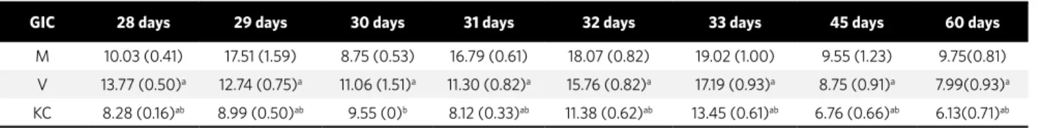
Table 2 shows fluoride release of materials after immersion in the sodium fluoride solution. On day 28, before application of fluoride, the amount of flu-oride released from Vidrion was higher than for the other materials, but there was a statistically signifi-cant difference among the groups (p < 0.05).
On day 29, the first day after application of fluo-ride, there was a statistically significant difference
Figure 1 - Silicone molds used to fabricate the test specimens.
Figure 2 - Amount of fluoride released for each one of the cements during the period assessed.
(F) Fluoride application Release of absorbed fluoride
F F
F F
32d 33d 45d 60d 70
60 50 40 30
1h 1d 3d 7d 14d 21d 30d 31d
Time
Fluoride r
elease (
µ
g
/c
m
2)
29d 28d 20
10 0
M V KC
Table 2 - Fluoride release of ionomer cements after application of fluoride.
N=10, Mean (standard deviation), a(p<0.05) compared to group M. b(p<0.05) compared to group V. Significant statistical difference observed between groups for
the same analyzed time.
GIC 28 days 29 days 30 days 31 days 32 days 33 days 45 days 60 days
M 10.03 (0.41) 17.51 (1.59) 8.75 (0.53) 16.79 (0.61) 18.07 (0.82) 19.02 (1.00) 9.55 (1.23) 9.75(0.81)
V 13.77 (0.50)a 12.74 (0.75)a 11.06 (1.51)a 11.30 (0.82)a 15.76 (0.82)a 17.19 (0.93)a 8.75 (0.91)a 7.99(0.93)a
KC 8.28 (0.16)ab 8.99 (0.50)ab 9.55 (0)b 8.12 (0.33)ab 11.38 (0.62)ab 13.45 (0.61)ab 6.76 (0.66)ab 6.13(0.71)ab
Table 1 - Fluoride release of ionomer cements during 28 days.
N=10, Mean (standard deviation), a(p<0.05) compared to group M. b(p<0.05) compared to group V. Significant statistical difference observed between groups for
the same analyzed time.
GIC 1hour 1 day 3 days 7 days 14 days 21 days 28 days
M 10.58 (1.20) 58.43 (2.89) 19.50 (2.17) 10.03 (0.41) 6.60 (0.56) 8.28 (0.41) 10.03 (0.41)
V 17.91 (2.81)a 31.68 (2.84)a 12.73 (1.76)a 7.80 (2.26)a 9.39 (0.56)a 6.21 (0.68)a 13.77 (0.50)a
Orthodontic cements: Immediate protection and fluoride release original article
among the groups (p < 0.05). On day 30, which cor-responds to 48 h after the first recharge of fluoride, there was no statistically significant difference be-tween the groups M and KC (p > 0.05).
There was a statistically significant difference between the amount of fluoride released after the applications of fluoride among the groups from the
31st day to the 60th day (p < 0.05). On days 32 and 33,
all the cements showed greater fluoride release, which shows the capacity of uptake and
accumula-tion of fluoride after recharge. On the 45th and 60th
day, significant decrease of fluoride release was ob-served for the three cements with values close to the
ones observed on the 7th and 28th day.
DISCUSSION
It has been widely reported that GIC or other mate-rials that contain fluoride present a cariogenic inhibito-ry effect when compared with composites without
fluo-ride, as observed by Kielbassa et al12 and this inhibition
is fundamental in orthodontic treatment. The fluoride solution used was at a concentration of 1000 ppm NaF, similar to the concentration of dentifrices used for
den-tal toothbrushing, according to Okuyama et al.16
Fluoride release was evaluated for 4 weeks with the purpose of observing the performance of the material during this period, since patients with fixed applianc-es normally visit the orthodontist once a month. In this study, a protocol of daily water change was used to assess fluoride release, for this protocol is better than
the accumulation of fluoride in a solution.20
Caves et al3 reported that the type of cement, the
geometric model and surface area may significantly influence fluoride release, but there is no standardized size for test specimens to assess fluoride release, which
are diverse in other studies.6 The present study used
disks measuring 4 mm in diameter and 4 mm high. Fluoride release found in glass ionomer ce-ments was higher 24 h after initial setting and
de-creased after 3 and 7 days. After the 7th day, there
was small variation and fluoride release was con-stant, which is similar to the findings of Komori
and Kojima13and Kuvvetli et al14(Fig 1).This
char-acteristic is clinically relevant for cementation materials and the 3 cements maintained a small
difference of fluoride release after the 7th day. The
cements that showed better performance were
Meron and Vidrion, which may mean a greater clinical effect for preventing enamel demineral-ization when compared with Ketac-Cem.
Meron cement showed good performance, which
corroborates the findings ofAkkaya et al,2a
perfor-mance close to the one found for Vidrion cement and better than Ketac-Cem, which showed a lower value
than the one found by Komori and Kojima.13
Fluo-ride release of Ketac-Cem was significantly lower than the one found for the other 2 ionomer cements, but release showed to be detectable during the
en-tire experiment which, according to Dijkman et al,7
is fundamental during orthodontic treatment. After one day of fluoride release, the 3 cements Meron, Ketac-Cem and Vidrion showed greater fluo-ride release when compared to the time interval of 1 h, which shows that these cements reach a maximum peak of fluoride release after 24 h of initial setting, with a statistically significant difference (p < 0.05).
On the 7th, 14th, 21st and 28th day, the cements showed
a similar fluoride release pattern, but with lower val-ues than at the time intervals of 1 h, 1 day and 3 days. This shows that despite the three cements presenting
lower values of fluoride releases after the 7th day, the
values were detectable during the entire experiment. The amount of fluoride recharge may depend on the capacity of intrinsic fluoride release of each ma-terial, since the sites occupied by intrinsic fluoride
are fixed and limited inside them.11 The cements that
showed greater initial fluoride release presented greater fluoride release during the entire experi-ment, which suggests a greater capacity of fluoride recharge, being in agreement with the findings of Xu
and Burgees.22 Fluoride released after the period of
exposure of recharge has a tendency to release the
same amount as the initial period,16 which may be
observed during the application period of fluoride from day 29 to day 33. Fluoride release observed af-ter 45 and 60 days showed values close to the ones
observed on the 7th and 28th day, which suggests that,
after these values, fluoride release tends to slowly di-minish, being detectable after longer periods.
Material porosity may influence the amount of fluoride released before and after recharge, in
accor-dance with Xu and Burgess.22 Obviously, greater
Resin-reinforced glass ionomer cements present less porosities than conventional GIC and therefore, lower fluoride release, which corroborates the
find-ings of Komori and Kojima13and Kuvvetli et al.14
In an in vivo study, Hallgren et al9 observed that
brackets and bands cemented with GIC signifi-cantly increased the concentration of fluoride in saliva. However, it is suggested that the orthodon-tic bands should be regularly checked because flu-oride release may not completely inhibit develop-ing caries lesion in the bands, which may be loose or in areas that are without GIC.
1. Aguiar DA, Silveira MR, Ritter DE, Locks A, Calvo MCM. Avaliação das propriedades mecânicas de quatro cimentos de ionômero de vidro convencionais utilizados na cimentação de bandas ortodônticas. Rev Dent Press Ortodon Ortop Facial. 2008 Maio-Jun;13(3):104-11.
2. Akkaya S, Uner O, Alaçam A, Değim T. Enamel fluoride levels after orthodontic band cementation with glass ionomer cement. Eur J Orthod. 1996 Feb;18(1):81-7. 3. Caves GR, Millett DT, Creanor SL, Foye RH, Gilmour WH. Fluoride release from
orthodontic band cements-a comparison of two in vitro models. J Dent. 2003 Jan;31(1):19-24.
4. Chuang SF, Jin YT, Tsai PF, Wong TY. Effect of various surface protections on the margin microleakage of resin-modified glass ionomer cements. J Prosthet Dent. 2001 Sep;86(3):309-14.
5. Cohen WJ, Wiltshire WA, Dawes C, Lavelle CL. Long-term in vitro fluoride release and rerelease from orthodontic bonding materials containing fluoride. Am J Orthod Dentofacial Orthop. 2003 Nov;124(5):571-6.
6. Creanor SL, Al-Harthy NS, Gilmour WH, Foye RH, Rogers I, Millett DT. Fluoride release from orthodontic cements-effect of specimen surface area and depth. J Dent. 2003 Jan;31(1):25-32.
7. Dijkman GE, de Vries J, Lodding A, Arends J. Long-term fluoride release of visible light-activated composites in vitro: a correlation with in situ demineralisation data. Caries Res. 1993;27(2):117-23.
8. Donly KJ, Nelson JJ. Fluoride release of restorative materials exposed to a fluoridated dentifrice. ASDC J Dent Child. 1997 Jul-Aug;64(4):249-50. 9. Hallgren A, Oliveby A, Twetman S. Salivary fluoride concentrations in children with
glass ionomer cemented orthodontic appliances. Caries Res. 1990;24(4):239-41. 10. Hattab FN, Amin WM. Fluoride release from glass ionomer restorative materials
and the effects of surface coating. Biomaterials. 2001 Jun;22(12):1449-58. 11. Itota T, Carrick TE, Yoshiyama M, McCabe JF. Fluoride release and recharge in
giomer, compomer and resin composite. Dent Mater. 2004 Nov;20(9):789-95. REFERENCES
CONCLUSION
It could be concluded that:
1) Immediate protection with fluoride varnish reduces the risk of syneresis and imbibition and do not inhibit fluoride release.
2) Meron and Vidrion cements presented greater capacity of fluoride uptake and release than Ketac-Cem cement.
3) The amount of fluoride release in the three cements increased after recharge. Thus, the use of fluoride mouthrinses is suggested as an alternative to increase the amount of fluoride released by GICs.
12. Kielbassa AM, Schulte-Monting J, Garcia-Godoy F, Meyer-Lueckel H. Initial in situ secondary caries formation: effect of various fluoride-containing restorative materials. Oper Dent. 2003 Nov-Dec;28(6):765-72.
13. Komori A, Kojima I. Evaluation of a new 2-paste glass ionomer cement. Am J Orthod Dentofacial Orthop. 2003 Jun;123(6):649-52.
14. Kuvvetli SS, Tuna EB, Cildir SK, Sandalli N, Gençay K. Evaluation of the fluoride release from orthodontic band cements. Am J Dent. 2006 Oct;19(5):275-8. 15. Mount GJ. Clinical performance of glass-ionomers. Biomaterials. 1998
Mar;19(6):573-9.
16. Okuyama K, Murata Y, Pereira PN, Miguez PA, Komatsu H, Sano H. Fluoride release and uptake by various dental materials after fluoride application. Am J Dent. 2006 Apr;19(2):123-7.
17. Suljak JP, Hatibovic-Kofman S. A fluoride release-adsorption-release system applied to fluoride-releasing restorative materials. Quintessence Int. 1996 Sep;27(9):635-8.
18. Thilander BL. Complications of orthodontic treatment. Curr Opin Dent. 1992 Dec;2:28-37.
19. Ian Matos Vieira IM, Louro RL, Atta MT, Navarro MFL, Francisconi PAS.O cimento de ionômero de vidro na odontologia. Rev Saúde com. 2006;2(1):75-84. 20. Wheeler AW, Foley TF, Mamandras A. Comparison of fluoride release protocols for
in-vitro testing of 3 orthodontic adhesives. Am J Orthod Dentofacial Orthop. 2002 Mar;121(3):301-9.
21. Wilson AD, Paddon JM, Crisp S.The hydration of dental cements. J Dent Res. 1979 Mar;58(3):1065-71.