an interview with
• Certiicate and Masters - Department of Orthodontics, Case Western Reserve University School of Dental Medicine. • Director of Orthodontics in the Case Western Reserve University School of Dental Medicine.
• Director, Craniofacial Imaging Center, Case Western Reserve University School of Dental Medicine. • Visiting Professor, Department of Orthodontics, University of Belgrade, Serbia.
• Active member of the American Association or Orthodontists (AAO) and the American Academy of Oral and Maxillofacial Radiology (AAOMR).
• Diplomate American Board of Orthodontics.
• Member of AAO Committee On Information Technology (COIT).
• Member of American Dental Association (ADA) Standards Committee on Dental Informatics [SCDI] Working Groups 12.1- Application of the DICOM Standard in Dentistry and 11.6 - Integration of Orthodontic Standards.
• Member of AAO/AAOMR Council on Creating Guidelines for the use of CBCT in Orthodontics. • Guest Editor - Seminar in Orthodontics – Cone Beam CT for the Orthodontist 2010.
• Editorial Review Board - Bone, American Journal of Orthodontics and Dentofacial Orthopedics, Angle Orthodontist, Euro-pean Journal of Orthodontics, Journal of Clinical Orthodontics, Journal of the American Dental Association, International Journal of Oral Science, Journal of Applied Oral Science, etc.
• American Association of Orthodontists Foundation (AAOF) Awards in 1999, 2001 and 2004. • Past President of the Omnicron Kappa Upsilon (OKU) National Honor Dental Society.
Professor J. Martin Palomo is a graduate of the Ponta Grossa State University and proudly hails from Ponta Grossa, Brazil. He was selected to specialize at the prestigious Case Western Reserve School of Dental Medicine (CWRU) in Cleveland Ohio and during his graduate training, began to feel that he could serve the profession more broadly through academics and speciically through imaging. Dr Palomo’s ambition has always been to serve his native Brazil; he has worked in Curitiba, the mayorship of Manoel Ribas, and the univer-sity of UNIPAR, in Umuarama. While working as an orthodontist in Brazil, he was invited to return to CWRU and appointed to Clinic Director and Research Fellow of the Bolton Brush Center. Dr Palomo earned several awards for his teaching from the American Associa-tion of Orthodontics and chaired their Council on EducaAssocia-tion Leadership Implementing Evidence Based Dentistry. Today Dr. Palomo is the Director of Orthodontics and Chair of the Graduate Studies Committee at CWRU. He has built the premier imaging center in the Midwest and is busy authoring the American Dental Association position paper on the subject. He has assembled an award winning team of top notch researchers, clinical orthodontists, physicians, and graduate students who contribute to the profession. His ambition is to serve his native Brazil through authorship, speaking, meetings, and collaborations. Dr. Palomo is married to a periodontist and they have a 7 year-old daughter and three dogs.
Lincoln I. Nojima
How to cite this section: Palomo JM. Interview. Dental Press J Orthod. 2012 July-Aug;17(4):4-11. Submitted: October 20, 2011 - Revised and accepted: March 01, 2012
» Patients displayed in this interview previously approved the use of their images.
Three-dimensional analysis require the incor-poration of didactic training during orthodontic residency, since orthodontists need training to analyze a CBCT image adequately. As a pioneer in 3D Imaging in academics, how is the 3D Diagnosis addressed at Case Western Reserve University?
(Lucia Cevidanes)
Our patients are three-dimensional entities, and a three-dimensional representation is not only more accurate, but also easier to understand. Our residents find it is easier to locate landmarks in 3D images than in cephalograms. We do still teach a whole cephalometric course, and perform cepha-lometric analyses on every patient, so at this point our residents get more education than when we did only 2D. We have courses that teaches them how to analyze a 3D image, going slice by slice and look-ing for pathology and abnormalities outside of nor-mal limits (Fig 1). I find it is great that while we are looking at these images, they are also taking cours-es on anatomy and pathology, and I noticed that they make more of a basic course to clinic connec-tion now, than when they would only see panoram-ic and cephalometrpanoram-ic radiographs. We also teach them how to use different software. We do find that we had to add more courses and class time, but this makes sense, since we are now working with more information, and doing more than before. In my opinion specialist need to do more, and provide a more comprehensive service. That to me is almost the definition of specialist.
What are you doing to interest young orthodon-tists in teaching and/or research after they inish their specialty programs? (Carla Evans)
I try to show our young residents and orthodon-tists, how rewarding it can be to be involved in aca-demics, mostly to oneself. To be in contact with col-leagues and have frequent discussions on current topics promotes mental stimulation, growth, coni-dence, and eficiency. It is like an ongoing continu-ing education course. It is an assurance that you will not do for the rest of your career the same thing you did as a fresh graduate. As G.V. Black said, “the pro-fessional has no right to be other than a continuous student”, and sometimes this is not easy unless you have contact with such environment.
What have you learned about facial growth and the response to orthodontic treatment from CBCT images? (Mark Hans)
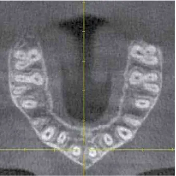
As far as facial growth, we think the additional information collected through density values may be useful in identifying areas of resorption and de-position in the mandible, and provide an insight on growth direction of growth. We have ongoing proj-ects on that area, and it looks promising. On the area of response to orthodontic treatment, there are a lot of tools available, that have shown how much change unknown to the orthodontist happens during treatment. Just the analysis of buccolingual inclination of molars show changes of 15 degrees that were not planned or desired. This may result in a less than stable result (Fig 2).
Have you implemented any innovative teaching methods in orthodontics? (Carla Evans)
Case Western Reserve University (CWRU) has a tradition of working with craniofacial imaging, with the irst Cephalometer being invented by Dr. Broad-bent, to the Bolton Standards, and pioneer work in three-dimensional imaging, which started with the combination of frontal and lateral cephalograms, and now CBCT. I am fortunate to be part of a team that has been implementing this pioneer work for a long time, and I think I have been able to contribute with cours-es such as “Advanced Craniofacial Imaging” given to orthodontic and pediatric dentistry residents where they work on their own computer on pre-selected im-ages, some with pathologies, others without, and then train them on how to handle different situations. I think the courses nowadays need to be hands-on and interactive, and to work with CBCT and 3D imaging software, the student can only learn by doing it.
In the “Seminars in Orthodontics” issue where you were the guest editor, Dr. Lysle Johnston made an analogy between 3D Images and the movie “The Matrix”, stating that we can only see what the programmers allow us to see. Can you comment on this statement and suggest care in the diagnostic interpretation to distinguish what can be artifacts of 3D rendering that shows or hides bone in the image? (Lucia Cevidanes)
One of the disadvantages of CBCT for both soft-ware and hardsoft-ware is the lack of standardization at the moment. The same image may look diagnostical-ly different in different software packages, and this is possible due to lack of regulation on what can be done by companies. The image in 3D may look pret-tier than the slices, but we teach at CWRU that if you want an answer you go to the slice mode and see it in the axial, sagittal, and coronal views. Analyses such as airway volume has also differences between soft-ware packages and unfortunately at this point cannot be used as cephalometric values, where we expect to see relative inter-software consistency. I am a big fan of Dr. Cevidanes, another Brazilian, who really leads the way in open source software packages, where to make an image pretty to sell more is not a priority.
Since this is one of your research lines in the Orthodontic Department at Case, in which mo-phologic and functional ways can the airways be analyzed through CBCT? (Matilde Nojima)
Even though the airway can be easily and auto-matically segmented by using most of the software packages available to orthodontists, it has not at this point shown diagnostic value. We have noted differ-ences in airway volume for different craniofacial pat-terns, and have noted the opening of the airway in
Figure 2 - The buccolingual inclination of posterior teeth can be accurately measured in the 3D images provided with CBCT. This can be valuable diag-nostic information, which can help in the decision between palatal expanders or archwire expansion, and provide more control on changes that may be occurring without the orthodontist’s knowledge. Such changes may play a role in the stability of the result.
102,8˚
102,4˚
cases of mandibular advancement, due to appliances, orthodontic treatment, or surgery, but there is still a lot of work that needs to be done to make airway as-sessment useful and diagnostic of sleep or breathing disorders. At this point, I do not think we would take a CBCT just for airway assessment, but if the CBCT was taken for other motives, we should investigate the airway as part of our protocol (Fig 3).
With the current knowledge base available would you recommend traditional orthodontic records to be replaced by a single CBCT? Is this possible in clinical orthodontics? (Dauro Oliveira)
I don’t think we can completely replace all orthodontic records with a CBCT image, but it can get very close (Fig 4). I think it could replace
impressions since electronic models can be created from CBCT images, but even though the patient’s face can be seen in a large field of view volume, it cannot replace the smile picture, the intra oral pic-tures, and lacks color (Fig 5).
Now, even though this is possible, I do not think that should be done in clinical orthodontics due to the ionizing radiation involved. The Board of Trust-ees of the American Association of Orthodontists (AAO) and American Academy of Oral and Maxillo-facial Radiology (AAOMR) have appointed a council committed to form guidelines on when to use CBCT. It includes 4 orthodontists: Dr. Carla Evans (Uni-versity of Illinois in Chicago), Dr. Kirt Simmons (Arkansas Children’s Hospital), Dr. Lucia Cevi-danes (University of Michigan), and myself, as well
Figure 3 - Airway assessment in 3D evaluate more than just the lateral view, and allow the creation of both volumes and areas of maximum constriction. This cases shows a patient with severe obstruction of the airways with and without a removable appliance that protrudes the mandible and slightly opens the vertical. The changes that can be seen in the axial slices would not be detected in a cephalometric radiograph.
Figure 5 - The CBCT created electronic model can also be used to replace models created from impressions, and can also be used in virtual treatment planning setups. This case shows the tipping back of cuspids after a bicuspid extraction, from both lateral and occlusal views.
as three radiologists: Dr. William Scarfe (University of Louisville), Dr. Mansur Ahmad (University of Min-nesota) and Dr. John Ludlow (University of North Carolina). Latest drafts already show the advantages of sometimes combining traditional 2D images with smaller ield of view 3D images. This is the direction where things are going in my opinion. We have to think of CBCT as a radiographic tool in our armamen-tarium. If we have a clinical question, we have to see which tool would better answer it. Sometimes a pan-oramic image is enough, some other times we would not be able to fully answer without a CBCT image. It is a clinical decision made by the healthcare provider.
Under what circumstances would you recom-mend a CBCT image be reviewed by an Oral and Maxillofacial Radiologist? (Mark Hans)
At CWRU we review every image in all three planes of space, which takes just a few minutes, following protocols where the image is irst oriented in space similar to what we do with cephalometrics. Anytime we see anything that does not look like it is within normal limits or have any doubt, we refer for a radio-logical review. We do not send every image, since as orthodontists we have the anatomical knowledge of how things should look and what can be a problem or not. We did not use to send our cephs and panos, but similarly would look for pathology and abnormalities and refer when we would ind something outside nor-mal limits. In addition to this protocol, we always offer a radiological reading to the patient or parent, in case they want it regardless.
What’s your position when related to risks of ion-izing radiation received by patients? Does any dose represent a cancer risk? (Lucia Cevidanes)
I don’t think this is a matter of opinion or belief, but of facts. One of the best descriptions I have seen lately is from a governmental commission called USNRC (United States Nuclear Regulatory Com-mission) whose ofice of public affairs puts out a fact sheet. This four page document does a great job showing the lack of knowledge we have on this topic at this time. It speculates that low levels of radiation would heal and not be cumulative, but since we do not have irm evidence at this point, we should act in a conservative way. There are groups that think ion-izing radiation has different effects depending on the dose, at low doses it can even be beneicial to the pa-tient, and at high doses detrimental. Since we do not have any reliable data on low dose effects, we should act in a worst case scenario event, and try to reduce as much as possible.
The use of CBCT as a diagnostic tool in clinical orthodontics has been growing, but not as rapid-ly. A possible obstacle seems to be commercially available software costs. What could be done to help with this problem? (Lincoln Nojima)
and employment of several people. If a company does a good job, puts a good quality product in the market, and stands behind it, I think the company deserves to be paid and supported for this.
Which beneits can 3D imaging ofer to the ortho-dontist to improve the stability of a inal result?
(Lincoln Nojima)
One of the challenges that the orthodontist faces for every patient is to start a cases that present with a malocclusion, but shows proper equilibrium and is presently stable, then change it, and reach again sta-bility and equilibrium in the inal result. The only way to do this is with proper knowledge and proper con-trol. 3D imaging can give more diagnostic information such as buccolingual inclination of teeth and position of root apices (Fig 6), that can be the additional infor-mation lacking in the proper knowledge section. The control still lies in the orthodontist’s hands.
Which are your recommendations for the standardization of 3D superimposition?
(Matilde Nojima)
3D superimposition in my opinion combines the old with the new. Our knowledge of stable areas and structures have to be used as places of superimposi-tion, but instead of manually selecting those points, and ending up with just a few landmarks, software packages are able to detect similarities not visible to the human eye, and superimpose on thousands of points. This can result in a more reliable superimposi-tion. The visualization at this point is still not optimal, and in my opinion is better assessed in the slice mode than in a 3D rendering. But software improvements are happening so fast that I would not be surprised if this is available at the time of this print.
Is there a reliable 3D cephalometric analysis? What has come from the expert’s discussions that happen every year in Cleveland in relation to these analysis? (Dauro Oliveira)
Several analyses have been suggested, and soft-ware packages already allow the users to pick right and left landmarks separately. To my knowledge, no technique has been widely accepted, in the way the Down’s analysis was for the lateral radiograph. But to put in perspective, it took 17 years since the
invention of the cephalometer for the Down’s analy-sis to be accepted, and it took 29 years for a super-imposition analysis. Since to see an image in 3D is more of a game changer, the speculation is that we cannot limit the information to what we used to do with images that showed less information. For 3D images we may need to think in volumes, areas, and density values, rather than lines and angles.
There has been a great deal of controversy over the use of CBCT for routine orthodontic cases. What is your opinion on the role CBCT will play in the future of orthodontics? (Mark Hans)
Routine means automatically for everybody. I don’t think routine should be applied for any radio-logical tool. There has to be a clinical examination prior to ordering any radiograph. That everybody received a ceph is as wrong as everybody receiving a CBCT, because of the ionizing radiation and our lack of knowledge on what low doses of radiation may do to our patient. I do think that if radiation was not an issue, we would probably only need CBCT, and would never take a pano or ceph again, but this is not the world we live in at this time.
How do you keep up with new techniques and technology related to CBCT? (Carla Evans)
I try to remain a constant student, and never as-sume I know anything. To me, the minute you think you know something, you stop learning. I still read journals, I go to meetings, I talk to other professionals, I talk to companies, etc... But I also make an effort to think outside of the box. Medicine has been working with 3D imaging and CT for a while now. I try to see
Carla A. Evans
» DDS, University of Michigan School of Dentistry.
» Specialty in Orthodontics in the Harvard
Univer-sity/Forsyth Dental Center.
» Doctor of Medical Sciences in Oral Biology degree
at Harvard University and University of Illinois at Chicago.
» Professor and Head in the Department of
Ortho-dontics, University of Illinois at Chicago.
» Professor of Bioengineering in the College of Engi-neering - University of Illinois at Chicago.
Dauro Oliveira
» DDS, Universidade Federal de Minas Gerais/RJ. » MSD, Marquette University.
» PhD in Orthodontics, Universidade Federal do Rio de Janeiro.
» Director of Orthodontics Pontifícia Universidade Católica-MG.
Lincoln Issamu Nojima
» DDS, Universidade de Passo Fundo/RS.
» MSc and PhD in Orthodontics, Universidade Federal do Rio de Janeiro/RJ.
» Associate Professor, Department of Orthodontics, Universidade Federal do Rio de Janeiro.
» Visiting Associate Professor, Department of Ortho-dontics, Case Western Reserve University/OH. » Post-doctoral stage at Case Western Reserve
Univer-sity/OH, Capes Scholarship 0906/11-6. » Diplomate, Brazilian Board of Orthodontics.
Lucia Cevidanes
» DDS, Universidade Federal de Goiás.
» MSc in Orthodontics, Methodist Institute for Higher
Education.
» PhD in Oral Biology, University of North Carolina at
Chapel Hill.
» Assistant Professor in the Department of
Ortho-dontics and Pediatric Dentistry at the University of Michigan.
» Diplomate, American Board of Orthodontics.
Mark G. Hans
» DDS, School of Dental Medicine, Case Western Re-serve University/OH.
» MSD, Department of Orthodontics, Case Western Reserve University/OH.
» Chairman Department of Orthodontics Case West-ern Reserve University/OH.
» Director, The Bolton-Brush Growth Study Center, Case Western Reserve University/OH.
» Diplomate, American Board of Orthodontics.
Matilde da Cunha Gonçalves Nojima
» DDS, Universidade Federal do Rio de Janeiro. » MSc and PhD in Orthodontics, Universidade Federal
do Rio de Janeiro/RJ.
» Associate Professor, Department of Orthodontics, Universidade Federal do Rio de Janeiro/RJ.
» Visiting Associate Professor, Department of Ortho-dontics, Case Western Reserve University/OH. » Post-doctoral at Case Western Reserve University/