Camila Leite Quaglio1, Karina Maria Salvatore de Freitas2, Marcos Roberto de Freitas3, Guilherme Janson4,
José Fernando Castanha Henriques5
Stability of maxillary anterior crowding treatment
original article
Objective: To evaluate the stability and the relapse of maxillary anterior crowding treatment on cases with premo-lar extraction and evaluate the tendency of the teeth to return to their pretreatment position.
Methods: The experimental sample consisted of 70 patients of both sex with an initial Class I and Class II malo-clusion and treated with first premolar extractions. The initial mean age was 13,08 years. Dental casts’ measure-ments were obtained at three stages (pretreatment, posttreatment and posttreatment of 9 years on average) and the variables assessed were Little Irregularity Index, maxillary arch length and intercanine. Pearson correlation coefficient was used to know if some studied variable would have influence on the crowding in the three stages (LII1, LII2, LII3) and in each linear displacement of the Little irregularity index (A, B, C, D, E) in the initial and post-retention phases.
Results: The maxillary crowding relapse ( LII3-2) is influenced by the initial ( LII1), and the teeth tend to return to their pretreatment position.
Conclusion: The results underline the attention that the orthodontist should be given to the maxillary anterior re-lapse, primarily on those teeth that are crowded before the treatment.
Keywords: Crowding. Angle Class I malocclusion. Angle Class II malocclusion. Corrective Orthodontics.
How to cite this article: Quaglio CL, Freitas KMS, Freitas MR, Janson G, Henriques JFC. Stability of maxillary anterior crowding treatment. Dental Press J Orthod. 2012 July-Aug;17(4):57-64.
Submitted: April 27, 2009 - Revised and accepted: August 19, 2009
» The authors report no commercial, proprietary or financial interest in the products or companies described in this article.
Contact address: Camila Leite Quaglio E-mail: [email protected]
1 Msc in Orthodontics, School of Dentistry, University of São Paulo, Bauru. (FOB-USP).
2 Head of Masters Course in Orthodontics, UNINGÁ, Professor of Orthodontics,
Centro Universitário do Norte Paulista (UNORP, São José do Rio Preto).
3 Head Professor of Orthodontics and Head of the PhD Course in Orthodontics, FOB-USP.
4 Head Professor of Orthodontics and Head of the Masters Course in Orthodontics,
FOB-USP.
5 Head Professor of Orthodontics, Head of the Undergraduate Course in Orthodontics,
Camila Leite Quaglio1, Karina Maria Salvatore de Freitas2, Marcos Roberto de Freitas3, Guilherme Janson4,
José Fernando Castanha Henriques5
Estabilidade do tratamento de apinhamento anterossuperior
Objetivo: avaliar a estabilidade e recidiva do tratamento de apinhamento dos dentes anterossuperiores em casos com extrações de pré-molares e avaliar a tendência dos dentes apinhados, no início do tratamento, a retornar à sua posição original.
Métodos: a amostra consistiu de 70 pacientes de ambos os sexos, com má oclusão inicial de Classe I e Classe II de Angle, tratados com extrações dos primeiros pré-molares. A idade média inicial foi de 13,08 anos. Foram
avalia-dos os modelos de estudo nas fases inicial (T1), final (T2) e, em média, 9 anos pós-tratamento (T3) de cada
pacien-te. As variáveis da arcada superior avaliadas e comparadas estatisticamente pela Análise de Variância (ANOVA) foram: índice de irregularidade de Little Superior (IRLS), comprimento da arcada (CAS) e distância intercaninos (DICS). O Teste de Correlação de Pearson foi utilizado para verificar se alguma variável estudada teria influência
sobre o apinhamento nas três fases (IRLS1, IRLS2, IRLS3) e em cada deslocamento de Little (A, B, C, D, E), na fase
inicial e pós-tratamento.
Resultados: a recidiva do apinhamento superior (IRLS3-2) é influenciada pelo apinhamento inicial (IRLS1), e os
den-tes tendem a voltar à posição original.
Conclusão: os resultados ressaltam a atenção que o ortodontista deve dar à recidiva anterossuperior, principalmente àqueles dentes que estavam apinhados antes do tratamento.
Palavras-chave: Recidiva. Má oclusão de Angle Classe I. Má oclusão de Angle Classe II. Ortodontia Corretiva.
Como citar este artigo: Quaglio CL, Freitas KMS, Freitas MR, Janson G, Henriques JFC. Stability of maxillary anterior crowding treatment. Dental Press J Orthod. 2012 July-Aug;17(4):57-64.
Enviado em: 27 de abril de 2009 - Revisado e aceito: 16 de agosto de 2009 » Os autores declaram não ter interesses associativos, comerciais, de propriedade ou financeiros, que representem conflito de interesse nos produtos e companhias des-critos nesse artigo.
Endereço para correspondência: Camila Leite Quaglio E-mail: [email protected]
1 Mestre em Ortodontia, FOB-USP.
2 Coordenadora do Mestrado Profissionalizante em Odontologia, área de concentração Ortodontia, UNINGÁ. Professora do curso de Especialização em Ortodontia, UNORP.
3 Professor Titular da disciplina de Ortodontia e Coordenador do curso de Pós-Graduação em Ortodontia em nível de Doutorado, FOB-USP.
4 Professor Titular da disciplina de Ortodontia e Coordenador do curso de Pós-Graduação em Ortodontia em nível de Mestrado, FOB-USP.
Stability of maxillary anterior crowding treatment
original article
INTRODUCTION
In the last decades many patients have been seeking orthodontic treatment for esthetics rea-sons. Orthodontic treatment can improve facial esthetics as well as the occlusion, but long-term stability of the aligned teeth is highly variable and unpredictable. A wide variability of long-term re-sults may be related to the amount of pretreatment crowding, treatment plan, patient’s age and coop-eration during and after treatment.22
For years, many papers on alignment stability researched mandibular anterior crowding, prob-ably because relapse of these teeth are greater than that of the maxillary anterior teeth.4 More
recent-ly, patient’s expectation have been considered,2,9,31
and for the patient, the alignment of the maxillary anterior teeth is especially important, since those teeth are the first to be shown on a smile.10,22 Since
there is greater concern on the esthetics, a small relapse could be a problem. This new scenario ob-ligates the orthodontists to seek out knowledge in order to inform their patients about this problem and to control the risk factors during and after orthodontic treatment.
Most studies have shown that crowding relapse appears to be multifactorial.10,15 The amount of
ini-tial crowding, the arch length, intercanine distance are the most studied factors. There is a consensus about the teeth’s tendency to return toward their original position,1,5,12,14,22 but these studies are, in
general, based on arches changes during the orth-odontic treatment.
Therefore, in order to seek out for more knowl-edge on long term stability of maxillary anterior teeth on the orthodontic treatment, this paper eval-uates the relapse of the maxillary anterior teeth in cases treated with premolar extractions and their tendency to return toward their original position.
MATERIAL AND METHODS
The sample was selected from the patient re-cords treated in the Department of Orthodontics at Bauru Dental School, University of São Paulo and in the ACOPEN (Assessoria e Consultoria em Ortodontia, Pesquisa e Ensino). To minimize the bias, the sample was selected by inclusion crite-ria based on the literature4,10,12,17,19,25,29,30 and on the
objective of this study. The selection criteria were patients with all permanent teeth erupted up to the first molars and under 15 years of age at pre-treatment (T1); no supernumerary teeth or tooth agenesis; no fiberotomy or interproximal strip-ping as part of the treatment plan; Class I or Class II Division 1 malocclusion of, at least, three-forths of a full step Class II molar relationship, no ante-rior open bite or crossbite and more than 3 mm of crowding on Little’s irregularity index9 in the
mandible (LIIMx). The pretreatment (T1), post-treatment (T2) and post-retention (T3) dental casts had to be in good condition to be evaluated. All patients were treated with fixed edgewise ap-pliances and they not underwent rapid maxillary expansion. The patients also had a maximum peer assessment rating (PAR) score of 5 and passive lip seal at posttreatment (T2). Retention included a maxillary Hawley plate, used it 24 hours per day, for 6 months minimal plus 3 months during sleep-ing, and a bonded lingual canine-to-canine retain-er in the mandibular arch. The post-retention den-tal casts (T3) had to be at least 5 years after treat-ment. The presence of third molars was not part of the inclusion criteria because there is no common sense that have shown these teeth really interfere in anterior crowding relapse.3,13,20,28
Therefore, the sample comprised 70 patients, 210 pairs of dental casts (pretreatment, posttreat-ment and post-retention).
Variable collection method
All dental cast measurements were made with a 0.01 mm precision digital caliper and capacity of 150 mm (Mitutoyo America, Aurora, Ill) by the same examiner. The examiner was blinded in relation to which group did the cast belong to in order to mini-mize the bias.
The assessed variables:
Little’s irregularity index (LII)
The index used to evaluate the mandibular an-terior crowding was proposed by Little18 and named
A B
C
D
F
H
G E
were adapted to be evaluated on the maxillary arch. This adaptation was used in previous studies.6,10,21
In order to measure the amount of crowding a digital caliper was positioned parallel to the occlu-sal plane. Each linear displacement between the 5 anatomic contact points (A, B, C, D, E) of the an-terior teeth were measure (Fig 1). Little’s irregu-larity index comprises the 5 linear displacements added up, which represents the amount of crowd-ing in the anterior teeth.
Arch length (AL)
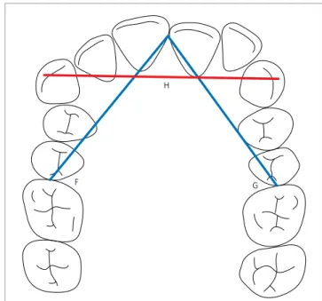
It is the total distance, in millimeters, from the contact point of the maxillary central incisors to the mesial of the first permanent molars in the right (F) and left side (G) (Fig 2).
Intercanine Width (ICW)
Distance from the cusp tip of the upper canines in millimeters. In cases of cusp wear, the tip was es-timated (Fig 2).
Statistical method
All statistical analyses were performed with Statis-tica software (version 6.0, Statsoft, Tulsa, Oklahoma). Normal distribution was verified by the Kolmogorov-Smirnov test and the results were considered significant
when p<0.05. The descriptive analysis found the mean, minimum, maximum, standard deviation, the groups and subgroups in all variables studied in the pretreat-ment (T1); posttreatment (T2), post-retention (T3) casts, as well as, the difference between the posttreatment and pretreatment phase (T2-T1) and the post-retention and posttreatment phase (T3-T2). The difference between T2 and T1 is considered to be treatment changes and the dif-ference between T3 and T2 is posttreatment changes.
Method error
The reliability of this study was evaluated by repeated measures of the variables of 15 patients (all phases) randomly chosen. The examiner had a month of interval between the first and the second measurement.
The systematic and casual error were evaluated for each variable. Systematic errors were evaluated with paired t tests at p<0.05, according to Houston.16
Casual errors were calculated according to Dahl-berg’s formula (Se2= Sd2/2n).7
Statistical analyses
Many studies in the literature are composed with Class I and Class II malocclusions in the same group. In this study, before the Class I and Class II maloc-clusion joined the same group the compatibility of
Figure 1 - Little’s Irregularity Index for the Upper Arch: Sum of the
dis-tances A+B+C+D+E.
Stability of maxillary anterior crowding treatment
original article
these two malocclusions was evaluated. The sample was divided into 3 groups: Group 1 (Class I maloc-clusion treated with 4 first premolar extraction), group 2 (Class II division 1 malocclusion treated with 4 first premolar extraction), group 3 (Class II division 1 malocclusion treated with 2 first maxil-lary premolar extraction). Intergroup comparisons of all variables were made by one-way analysis of variance (ANOVA). Intergroup sex distribution was evaluated with the chi-square test.
Pearson correlation coefficients were used to assess the relationships between the variables (LI-IMx, AL, ICW, posttreatment time and post-reten-tion time). Another evaluapost-reten-tion, was the correlapost-reten-tion between each linear displacement of Little’s irregu-larity index (A, B, C, D, E). In that way, it was pos-sible to verify the tendency of the teeth to return toward their original position (A1A3, B1B3, C1C3, D1D3, E1D3). The relapse also was evaluated in each linear displacement from Little’s irregularity index by using the scores in the pretreatment (T1) and post-retention (T3) phases. Any contact point that was correctly adjusted (0 mm), in any one of these phases (T1 or T3), was not considered. All other lin-ear displacements were used. The percentages of teeth that kept the same labiolingual direction at T1 and T3 were calculated.
The last comparison regarded the severity of the pretreatment crowding. The whole sample was divided in 2 groups (A and B). Group A comprised patients with LIIMx scores less than 7 mm, or mini-mal and moderate irregularity (19 patients), while group B had LIIMx scores equal to or greater than 7 mm, or severe and very severe irregularity (51 patients). The ratio between the post-retention changes (LIIMx3-2) and the correction amounts (LIIMx2-1) was called the relapse percentage. The absolute score of the correction amounts was used. The posttreatment changes that had negative scores (a greater alignment of the contact point) was con-sidered to be zero.
RESULTS Method error
Variables showed casual error smaller than 1 mm. Among all 30 variables, only width D at T2 showed a significant systematic error (96% precision).
Statistical analises
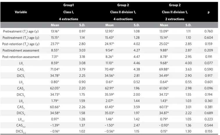
The table 1 and 2 show the compatibility be-tween the 3 groups regarding variables (Tab 1) and gender (Tab 2).
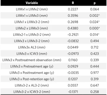
The table 3 shows the mean, minimum, maxi-mum, standard deviation and total sample. Pearson correlation coefficients were used to assess the re-lationships of LIIMx scores at different stages with the other variables (Tab 4). There was a positive correlation between LIIMx1 x LIIMx3, LIIMx1 x LI-IMx3-2 and LIIMx2 x LIIMx3. There was a negative correlation between LIIMx2-1 x LIIMx3-2. Table 5 shows the correlation between the linear distance in the post-retention (T3) and pretreatment (T1) phase in B, C and D.
The sample consisted of 70 patients, each pa-tient had 5 anatomic contact points described by Little18 (Fig 1); totalling of 350 contact points. In
the pretreatment phase, there were 309 linear dis-placements for labiolingual direction. The amount of linear displacement in post-retention phase was verified at the same contact points of the 309 ear displacements in pretreatment. The total of lin-ear displacement in post-retention phase was 184. These 184 linear displacements were used to evalu-ate the tendency that a tooth had to return toward its original position. A total of 142 anatomic contact points in the pretreatment phase had the same la-biolingual direction in the post-retention phase. These results have shown that 77% of the labiolin-gual linear displacement had the tendency to return toward their original position.
The total sample was divided in group A (LI-IMx1 < 7 mm) and group B (LI(LI-IMx1 > 7 mm) in order to evaluate the pretreatment severity crowding with the relapse percentage. The paired t test did not show a significant difference among these variables (Tab 6).
DISCUSSION
(LIIMx3-2) divided by the treatment changes (LI-IMx2-1) multiplied by 100. Others studies in the orthodontic literature had similar results10,11,15,21,22.
Our result showed a great stability of the maxillary anterior alignment, 88,12%.
Pearson correlation test was used to assess the re-lationships of LIIMx scores at different stages with the other variables. There was a significant positive correlation between Little’s irregularity index at pre-treatment (LIIMx1) and the postpre-treatment changes (LIIMx3-2). This result shows that pretreatment maxillary anterior crowding is correlated with the posttreatment maxillary anterior crowding (Tab 4), and this is supported by previous studies that have
shown that pretreatment maxillary anterior crowd-ing interfere in the crowdcrowd-ing relapse.1,8,24
The posttreatment Little’s irregularity index (LIIMx2) has a significant positive correlation with the post-retention Little’s irregularity index (LIIMx3). The posttreatment changes (LIIMx3-2) did not show a significant correlation with the amount of posttreatment crowding (LIIMx2) (Tab 4). Both results must be analyzed together because the crowding relapse would be influenced by qual-ity of treatment results (LIIMx2) only if there was a significant correlation between the amounts of posttreatment crowding (LIIMx2) and the post-treatment changes. Many previous studies, even
Variable
Group1
Class I,
4 extractions
Group 2
Class II division 1,
4 extractions
Group 2
Class II division 1,
2 extractions
p
Mean S.D. Mean S.D. Mean S.D.
Pretreatment (T1) age (y) 13.16
A 0.97 12.95A 1.08 13.09A 1.11 0.760
Posttreatment (T2) age (y) 15.15A 1.14 15.43A 1.28 15.14A 1.10 0.604
Post-retention (T3) age (y) 23.71A 2.80 24.97A 4.02 25.02A 2.85 0.159
Posttreatment assessment 8.55A 3.03 9.54A 4.27 9.88A 2.87 0.209
Post-retention assessment 7.31A 3.18 8.26A 4.47 8.78A 2.95 0.191
LII1 8.59
A 3.08 11.10A 4.46 9.68A 4.00 0.077
CAS1 71.04A 3.79 70.48A 4.38 69.88A 3.63 0.590
DICS1 34.78
A 2.25 34.56A 2.81 34.49A 2.90 0.917
LII2 0.80A 0.90 0.61A 0.52 0.64A 0.55 0.601
CAS2 62.05A 2.20 62.91A 1.96 61.06A 2.98 0.096
DICS2 34.73
A 1.75 35.59A 2.00 34.72A 1.55 0.194
LII3 1.79A 1.59 2.07A 1.44 1.43A 1.03 0.361
CAS3 60.66
A 2.26 61.40A 3.59 60.13A 3.01 0.381
DICS3 34.58A 1.58 35.03A 1.97 34.87A 2.22 0.689
LII3-2 0.97A 1.28 1.46A 1.42 0.79A 1.05 0.223
CAS3-2 - 1.39
A 1.37 - 1.50A 2.47 - 0.93A 1.36 0.544
DICS3-2 - 0.16A 1.02 - 0.56A 1.15 0.15A 1.30 0.155
Gender
Group 1 Class I,
4 extractions
Group 2 Class II division 1,
4 extractions
Group 2 Class II division 1,
2 extractions
Total
Male 12 11 11 34
Female 18 9 9 36
Total 30 20 20 70
chi-square=1.544 df = 2 P= 0.462
Table 1 - Intergroup comparisons (1-way ANOVA).
Stability of maxillary anterior crowding treatment
original article
though being about mandibular anterior crowd-ing, corroborated with this study.22,23,24 The
posi-tive significant correlation between posttreatment Little’s irregularity index (LIIMx2) and post-re-tention Little’s irregularity index (LIIMx3) is that posttreatment crowding is, at least, the same after a long-term evaluation.
Changes during treatment (LIIMx2-1) had a sig-nificant correlation with the post-retention changes (LIIM x 3-2) (Tab 4). Despite this negative correla-tion, it is considered to be a false negative. Since LI-IMx2-1 has a negative sign (posttreatment crowding minus pretreatment crowding) and the variable LI-IMx3-2 has a positive sign (post-retention crowd-ing minus pretreatment crowdcrowd-ing); when these two variables are correlated, the negative sign is main-tained as a positive correlation result.
The post-retention crowding (LIIMx3) was eval-uated with the posttreatment arch length and inter-canine width (AL3, ICW3), posttreatment observa-tion and age in all phases. These variables were cho-sen because the literature precho-sented some studies with significant positive correlation between the post-retention crowding (LIIMx3) and these vari-ables1,27,30 (Tab 4). None of these variables was
sig-nificantly correlated.
The Pearson correlation coefficients were used to assess the relationships between linear dis-placements of the anatomic contact points at T1 and T3. The percentages of teeth that kept the same labiolingual direction at T1 and T3 were calculated. There were significant positive correlations among linear displacements of anatomic contact point B (mesial surface of maxillary right lateral incisor with distal surface of maxillary right central inci-sor), contact point C (mesial surface of maxillary right central incisor with distal surface of maxil-lary left central incisor), and contact point D (me-sial surface of maxillary left lateral incisor with distal maxillary left central incisor) at T1 and T3 (Tab 5). These small proximal surfaces could cause a weak contact point which could increase suscep-tibility of misalignment over the years. The litera-ture has not evaluated the recurrence of crowding the way our study has, in each region of the Little Index (A, B, C, D and E), so our study should not be compared directly with any study published.
Variable Sample N =70
Mean Minimun Maximun S.D
LIIMx1 (mm) 9.62 3.21 20.38 3.87
LIIMx2 (mm) 0.70 0.00 3.44 0.71
LIIMx3 (mm) 1.77 0.10 6.87 1.41
LIIMx2-1 (mm) -8.92 -19.48 -2.94 3.77
LIIMx3-2 (mm) 1.07 -1.44 4.40 1.27
LIIMx3-1 (mm) -7.85 -18.57 -1.85 3.61
Pretreatment (T1) age (y) 13.08 10.63 15.02 1.03
Pretreatment (T1) age (y) 15.23 12.14 17.55 1.61
Pretreatment (T1) age (y) 24.44 18.84 33.11 3.22
Post-retention
observation (y) 9.21 5.00 17.23 3.39
Table 3 - Descriptive analysis of the sample.
Variable R p
LIIMx1 x LIIMx2 (mm) 0.2227 0.064
LIIMx1 x LIIMx3 (mm) 0.3596 0.002*
LIIMx1 x LIIMx3-2 (mm) 0.2698 0.024*
LIIMx2 x LIIMx3 (mm) 0.4380 0.000*
LIIMx2-1 x LIIMx3-2 (mm) -0.2921 0.014*
LIIMx3 x LIIMx3-2 (mm) -0.0832 0.494
LIIMx3x AL3 (mm) 0.0449 0.712
LIIMx3 x ICW3 (mm) -0.0973 0.423
LIIMx3 x Posttreatment observation (mm) 0.1160 0.339
LIIMx3 x Pretreatment age (y) 0.0929 0.444
LIIMx3 x Posttreatment age (y) -0.0035 0.977
LIIMx3 x Post-retention age (y) 0.1207 0.319
LIIMx3-2 x AL3-2 (mm) 0.0557 0.647
LIIMx3-2 x ICW3-2 (mm) -0.1371 0.258
Table 4 - Pearson correlation test.
*p< 0,05%.
Table 5 - Pearson correlation test between the variables A, B, C, D, E in
the post-retention (T3) and pretreatment (T1) phases.
*p< 0,05%.
Variable R p
A3 x A1 (mm) 0.1504 0214
B3 x B1 (mm) 0.4586 0.000*
C3 x C1 (mm) 0.3592 0.002*
D3 x D1 (mm) 0.4977 0.000*
E3 x E1 (mm) 0.0386 0.751
Table 6 - Paired t test between the pretreatment severity crowding with
the relapse percentage.
Variable
Group A
Severity
<7 mm S.D.
Group B
Severity
>7 mm
S.D. p
The literature has showned the teeth tendency to return to its original position by evaluating rotation or only by the assuming when there was a statistical correlation between total relapse and initial crowd-ing.1,8,22,24,30 If the Little’s irregularity index is
evalu-ated carefully, it does not show the teeth tendency to return to their original position. The Little’s ir-regularity index is the sum of 5 displacements (A, B, C, D, E) and it does not evaluate the direction of each anterior tooth in relation to its adjacent teeth (labial or lingual). To answer this question the mod-els were evaluated again taking into consideration the labiolingual direction in the pretreatment phase (T1) and in the post-retention phase (T3). From the 309 regions that showed labiolingual displacement in pretreatment phase, 184 regions showed labiolin-gual displacement in post-retention phase. There were 142 regions in post-retention phase that had the same pattern of displacement. This means that around 77% of the regions had the same pattern of displacement over the years, showing that the teeth have a tendency to return to the original position.
The significant correlation has shown that the pretreatment crowding pattern has some influence in posttreatment crowding, but this correlation does not show if the pretreatment crowding pattern has influence in posttreatment crowding percent-age, or in other words: “Does the teeth with greater pretreatment crowding tend to have more crowding relapse and vice-versa?” To answer this question the total sample was divided into two groups: Group A with moderate crowding (4-6 mm), and Group B with severe and very severe crowding (from 7 mm).18 The
comparison of the posttreatment crowding percent-age between these two groups showed no significant difference (Tab 6). This result does not show an in-fluence of the pretreatment crowding on the crowd-ing relapse percentage, even though the mean of the crowding relapse percentage of the Group A (sever-ity > 7 mm) was greater that crowding relapse per-centage of the Group B (severity < 7 mm). A similar study with this comparison showed significant difer-rences.12 That could be explained by the 8th Riedel’s
theorem26 which states that the further teeth have
been moved, they are less likely to relapse. Therefore, the relationship between the pretreatment crowd-ing and the posttreatment crowdcrowd-ing relapse might exist but this relationship should be carefully used when it comes to the pretreatment crowding sever-ity and posttreatment relapse. It means that 2 mm of posttreatment crowding in a case with 4 mm of pre-treatment crowding is 50% of relapse, but 4 mm of posttreatment crowding in a case with 12 mm of pre-treatment crowding is 33% of relapse. The amount of relapse shows that the case with the greater amount of pretreatment crowding had a greater relapse, but the percentage was lower.
Final considerations
Nowadays the patient is concerned about having an esthetic smile so many orthodontic treatments are aimed in correcting crowding. The increased demand for orthodontic treatment is a positive fact to the orthodontists but they cannot forget that the teeth appears to have a posttreatment ”agenda”. Or-thodontists have to be more careful in cases where the patients seek treatment only to align a tooth that bothers him. After treatment and over the years, the same tooth could be back to a similar po-sition. To avoid any failure, the orthodontist must be stringent in correcting this irregularity and on the retention plan. The patient has to be aware of the treatment and the posttreatment risks.
CONCLUSION
» The stability of maxillary anterior alignment in the whole sample was 88.12%, in an average of 9 years of posttreatment.
» There was a significant positive correlation between the amounts of pretreatment maxil-lary anterior crowding and the maxilmaxil-lary an-terior relapse. The greater was the amount of pretreatment crowding, the greater was the relapse.
Stability of maxillary anterior crowding treatment
original article
1. Artun J, Garol JD, Little RM. Long-term stability of mandibular incisors following successful treatment of Class II, Division 1, malocclusions. Angle Orthod. 1996;66(3):229-38.
2. Bedi R, Gulati N, McGrath C. A study of satisfaction with dental services among adults in the United Kingdom. Br Dent J. 2005 Apr 9;198(7):433-7.
3. Bishara SE. Third molars: a dilemma! Or is it? Am J Orthod Dentofacial Orthop. 1999 Jun;115(6):628-33.
4. Bondemark L, Holm AK, Hansen K, Axelsson S, Mohlin B, Brattstrom V, et al. Long-term stability of orthodontic treatment and patient satisfaction. A systematic review. Angle Orthod. 2007 Jan;77(1):181-91.
5. Burke SP, Silveira AM, Goldsmith LJ, Yancey JM, Van Stewart A, Scarfe WC. A meta-analysis of mandibular intercanine width in treatment and post-retention. Angle Orthod. 1998 Feb;68(1):53-60.
6. Canuto L. Avaliação da influência da realização da expansão rápida da maxila sobre a recidiva do apinhamento ântero-superior, em casos tratados ortodonticamente sem extrações [Dissertação]. Bauru (SP): Universidade de São Paulo, Faculdade de Odontologia de Bauru; 2006.
7. Dahlberg G. Statistical methods for medical and biological students. New York: Interscience; 1940.
8. de la Cruz A, Sampson P, Little RM, Artun J, Shapiro PA. Long-term changes in arch form after orthodontic treatment and retention. Am J Orthod Dentofacial Orthop. 1995 May;107(5):518-30.
9. de Oliveira CM, Sheiham A. Orthodontic treatment and its impact on oral health-related quality of life in Brazilian adolescents. J Orthod. 2004 Mar;31(1):20-7; discussion 15.
10. Destang DL, Kerr WJ. Maxillary retention: is longer better? Eur J Orthod. 2003 Feb;25(1):65-9.
11. Erdinc AE, Nanda RS, Işiksal E. Relapse of anterior crowding in patients treated with extraction and nonextraction of premolars. Am J Orthod Dentofacial Orthop. 2006 Jun;129(6):775-84.
12. Freitas KM, de Freitas MR, Henriques JF, Pinzan A, Janson G. Post-retention relapse of mandibular anterior crowding in patients treated without mandibular premolar extraction. Am J Orthod Dentofacial Orthop. 2004 Apr;125(4):480-7. 13. Harradine NW, Pearson MH, Toth B. The effect of extraction of third molars on
late lower incisor crowding: a randomized controlled trial. Br J Orthod. 1998 May;25(2):117-22.
14. Heiser W, Niederwanger A, Bancher B, Bittermann G, Neunteufel N, Kulmer S. Three-dimensional dental arch and palatal form changes after extraction and nonextraction treatment. Part 1. Arch length and area. Am J Orthod Dentofacial Orthop. 2004 Jul;126(1):71-81.
15. Heiser W, Richter M, Niederwanger A, Neunteufel N, Kulmer S. Association of the canine guidance angle with maxillary and mandibular intercanine widths and anterior alignment relapse: Extraction vs nonextraction treatment. Am J Orthod Dentofacial Orthop. 2008 May;133(5):669-80.
REFERENCES
16. Houston WJ. The analysis of errors in orthodontic measurements. Am J Orthod. 1983 May;83(5):382-90.
17. Huang L, Artun J. Is the post-retention relapse of maxillary and mandibular incisor alignment related? Am J Orthod Dentofacial Orthop. 2001 Jul;120(1):9-19. 18. Little RM. The irregularity index: a quantitative score of mandibular anterior
alignment. Am J Orthod. 1975 Nov;68(5):554-63.
19. Little RM, Riedel RA, Engst ED. Serial extraction of first premolars—post-retention evaluation of stability and relapse. Angle Orthod. 1990 Winter;60(4):255-62. 20. Little RM, Riedel RA, Stein A. Mandibular arch length increase during the
mixed dentition: post-retention evaluation of stability and relapse. Am J Orthod Dentofacial Orthop. 1990 May;97(5):393-404.
21. Martins P. Avaliação da influência da realização da expansão rápida da maxila sobre a recidiva do apinhamento ântero-superior, em casos tratados ortodonticamente com extrações de pré-molares [Dissertação]. Bauru (SP): Universidade de São Paulo, Faculdade de Odontologia; 2007.
22. Naraghi S, Andrén A, Kjellberg H, Mohlin BO. Relapse tendency after orthodontic correction of upper front teeth retained with a bonded retainer. Angle Orthod. 2006 Jul;76(4):570-6.
23. Nett BC, Huang GJ. Long-term posttreatment changes measured by the American Board of Orthodontics objective grading system. Am J Orthod Dentofacial Orthop. 2005 Apr;127(4):444-50; quiz 516.
24. Ormiston JP, Huang GJ, Little RM, Decker JD, Seuk GD. Retrospective analysis of long-term stable and unstable orthodontic treatment outcomes. Am J Orthod Dentofacial Orthop. 2005 Nov;128(5):568-74; quiz 669.
25. Pepicelli A, Woods M, Briggs C. The mandibular muscles and their importance in orthodontics: a contemporary review. Am J Orthod Dentofacial Orthop. 2005 Dec;128(6):774-80.
26. Riedel RA. A review of the retention problem. Angle Orthod. 1960 Oct;30:179-99. 27. Rothe LE, Bollen AM, Little RM, Herring SW, Chaison JB, Chen CS, Hollender LG.
Trabecular and cortical bone as risk factors for orthodontic relapse. Am J Orthod Dentofacial Orthop. 2006 Oct;130(4):476-84.
28. Southard TE, Southard KA, Weeda LW. Mesial force from unerupted third molars. Am J Orthod Dentofacial Orthop. 1991 Mar;99(3):220-5.
29. Taner TU, Haydar B, Kavuklu I, Korkmaz A. Short-term effects of fiberotomy on relapse of anterior crowding. Am J Orthod Dentofacial Orthop. 2000 Dec;118(6):617-23.
30. Vaden JL, Harris EF, Gardner RL Relapse revisited. Am J Orthod Dentofacial Orthop. 1997 May;111(5):543-53.