Comparison of two methods of visual magnification for
removal of adhesive flash during bracket placement using
two types of orthodontic bonding agents
Estefania Queiroga de Santana e Alencar1, Maria de Lourdes Martins Nobrega1, Fabio Roberto Dametto2,
Patrícia Bittencourt Dutra dos Santos3, Fabio Henrique de Sá Leitão Pinheiro4
1 Student, Master Degree Program in Dentistry, Universidade Potiguar (Laureate
International Universities), Natal, Rio Grande do Norte, Brazil
2 Assistant professor, Universidade Potiguar, (Laureate International Universities),
Master Degree Program in Dentistry and Faculty member of the Residence Program in Endodontics, Natal, Rio Grande do Norte, Brazil.
3 Assistant professor, Department of Orthodontics, Universidade do Estado do
Rio Grande do Norte (UERN), Caicó, Rio Grande do Norte, Brazil.
4 Assistant professor, Department of Preventive Dental Science (Orthodontic
Division), University of Manitoba, Winnipeg, Canada.
Submitted: September 19, 2015 - Revised and accepted: July 05, 2016
Objective: This study aimed to evaluate the effectiveness of two methods of visual magnification (operating microscope and light head magnifying glass) for removal of composite flash around orthodontic metal brackets. Material and Methods: Brackets were bonded in the center of the clinical crown of sixty well-preserved human premolars. Half of the sample was bonded with conventional Transbond XT (3M Unitek TM, USA), whereas the other half was bonded with Transbond TM Plus Color Change (3M Unitek TM, USA). For each type of composite, the choice of method to remove the flash was determined by randomly distributing the teeth into the following subgroups: A (removal by naked eye, n = 10), B (removal with the aid of light head magnifying glass, under 4x magnification, n = 10), and C (removal with the aid of an operating microscope, under 40x magnification, n = 10). Brackets were debonded and teeth taken to a scanning electron microscope (SS-x-550, Shimadzu, Japan) for visualization of their buccal surface. Quantification of composite flash was performed with Im-age Pro Plus software, and values were compared by Kruskal-Wallis test and Dunn’s post-hoc test at 5% significance level. Results: Removal of pigmented orthodontic adhesive with the aid of light head magnifying glass proved, in general, to be advantageous in comparison to all other methods. Conclusion: There was no advantage in using Transbond TM Plus Color Change alone. Further studies are necessary to draw a more definitive conclusion in regards to the benefits of using an operating microscope.
Keywords:Orthodontics. Composite resins. Lenses.
DOI: http://dx.doi.org/10.1590/2177-6709.21.6.043-050.oar
How to cite this article: Santana e Alencar EQ, Nobrega MLM, Dametto FR, Santos PBD, Pinheiro FHSL. Comparison of two methods of visual magnifica-tion for removal of adhesive flash during bracket placement using two types of orthodontic bonding agents. Dental Press J Orthod. 2016 Nov-Dec;21(6):43-50. DOI: http://dx.doi.org/10.1590/2177-6709.21.6.043-050.oar
» The authors report no commercial, proprietary or financial interest in the products or companies described in this article.
Contact address: Fabio Pinheiro, Department of Preventive Dental Science (Orth-odontic Division), D-341-780 Bannatyne Avenue, Winnipeg, MB R3E 0W2, Can-ada. E-mail: [email protected]
Objetivo: este estudo teve o objetivo de avaliar a eficácia de dois métodos de magnificação visual (microscópio cirúrgico e lupa de pala) para remoção da resina residual em torno de braquetes ortodônticos metálicos. Material e Métodos: os braquetes foram colados no centro da coroa clínica de 60 pré-molares humanos bem preservados. Metade da amostra foi colada com Transbond XT convencional (3M UnitekTM, EUA), enquanto a outra metade foi colada com TransbondTM Plus Color Change (3M-UnitekTM, EUA). Para cada tipo de resina, a escolha do método para remover o resíduo foi determinada por meio da distribuição aleatória dos dentes nos seguintes subgrupos: A (remoção a olho nu, n = 10), B (remoção com a ajuda de lupa de pala, sob uma ampliação de 4x, n = 10) e C (remoção com auxílio de um microscópio cirúrgico, sob uma ampliação de 40x, n = 10). Os braquetes foram descolados e os dentes, levados a um microscópio eletrônico de varredura (SS-X-550, Shimadzu, Japão) para visualização de sua superfície vestibular. A quantificação da resina residual foi realizada por meio do software Image Pro Plus, onde os valores foram comparados utilizando-se o teste de Kruskal-Wallis e o teste post-hoc de Dunn, ao nível de significância de 5%. Resultados: a re-moção da resina ortodôntica pigmentada com o auxílio da lente de aumento de pala provou, em geral, ser mais vantajosa, em comparação aos outros métodos testados. Conclusão: não houve vantagem em se usar a TransbondTM Plus Color Change sozinha. Estudos adicionais são neces-sários para se chegar a uma conclusão definitiva sobre os benefícios da utilização de microscópio cirúrgico.
INTRODUCTION
Although fixed appliances are effective in correct-ing malocclusions, plaque buildup (biofilm) tends to be an issue during prolonged orthodontic treatment. Some studies have demonstrated a clear association between biofilm and enamel demineralization.1,2,3 A linkage between biofilm and periodontal disease has also been established.4
Besides the regular use of oral hygiene aids by pa-tients,5,6,7 most clinical procedures in Dentistry aim at avoiding or eliminating the creation of retentive areas or undercuts. Unfortunately, ixed appliances have many retentive sites which can be considered a risk factor for the development of caries and periodontal disease.
Similarly, composite lash around orthodontic brackets can also work as a type of retentive factor that goes oten unnoticed. It has been demonstrated that unpolished and rough composite surfaces stimulate the accumulation of plaque and other debris.8,9,10
In addition, orthodontic bonding agents are known to be toxic to human gingival ibroblasts, thereby caus-ing inlammation and gcaus-ingival hyperplasia even in the presence of good oral hygiene.11 Therefore, careful re-moval of the bonding agent would reduce the risk of demineralization during orthodontic treatment.
To facilitate visualization of composite lash, the industry has developed a pigmented composite mate-rial of which pink color completely fades away during curing. The efectiveness of this type of material was assessed on typodonts in 2007, but the authors12 did not ind it to be clinically advantageous. No method of visual magniication was used in this study while removing composite lash.
Another way to remove the lash consists in using visual resources, such as magnifying glasses or operat-ing microscopes. The former was proposed in 2006 in a literature review13 on magniication devices that can be useful for Orthodontics. The operating microscope has been widely used in areas, such as Endodontics,14 Oral Surgery,15 and Periodontics.16 In Orthodontics; however, such methods have not been extensively put into practice. To date, studies in which clinical methods of mag-niication were used to help removing composite lash around orthodontic brackets are quite scarce. The fol-lowing search strategy was run in PubMed database in August, 2012: (orthod* AND bracket AND |ad-hesive OR composite| AND |lens OR microscope
OR magniication OR magnifying|). Only 75 arti-cles were retrieved; out of these, only one17 studied the inluence of magniication resources on the removal of orthodontic bonding agents, but its focus was on debonding. Alternative search strategies (“operating microscope AND orthodontic”; “loupe AND ortho-dontic”; “magnifying lens AND orthodontic”) also failed to retrieve relevant studies.
The present study assessed the eicacy of two mag-niication resources (the light head magnifying glass and the operating microscope) in removing composite lash around orthodontic brackets. Both conventional and pigmented resins were also tested. The following null hypothesis (H0) was postulated: “In comparison to the naked eye, there is no statistically signiicant diference in the amount of composite lash when using either method of visual magniication, regardless of the type of bonding agent which was applied to the bracket mesh base (conventional or pigmented).”
MATERIAL AND METHODS Sample selection
This study was reviewed and approved by the ethics committee (#008/2010) at Potiguar University (Lau-reate International Universities) in Natal, Brazil. Six-ty-two human premolars that had been extracted for orthodontic or periodontal reasons were stored in a solution of 0.1% thymol at room temperature. These teeth were selected according to the following inclusion criteria: 1) no enamel defect, 2) no color alteration.
Sample size for each group was calculated based on a previous study,12 since it was the only report on this matter (n = 10).
Sample preparation
Roots were removed cross-sectionally with a lex-ible diamond disk coupled to a low-speed hand piece. The cut was made at 3 mm from the cementoenamel junction. Ater sealing the root canals with composite (Flowable Restorative, 3M ESPETM, USA), each select-ed tooth was positionselect-ed inside 20 x 10-mm PVC cylin-ders, maintaining the buccal surface centered and paral-lel to the base of the cylinder. Polyester resin was poured into the cylinders for partial inclusion of teeth, leaving the buccal surface exposed.
for rehydration. There was no intention to simulate the oral environment, as specimens were expected to be as clean as possible to allow accurate measurement of the composite-containing area.
Slightly powdered vinyl gloves were used through-out the study. For complete removal of the powder, they were washed thoroughly with deionized water and later disinfected with 70% ethanol.
Teeth preparation
Dental prophylaxis with rubber cups and a mixture of deionized water and extra-thin pumice was per-formed for 10 seconds on the buccal surface of each tooth. The cups were replaced with new ones at every ten teeth. Specimens were washed for 30 seconds with an oil-free spray of deionized water and air. They were later dried out with oil-free air for 20 seconds.
The enamel was etched for 15 seconds with 37% phosphoric acid gel, then washed and dried, as de-scribed above. Etching was considered adequate based on the opaqueness of the surface. Shortly thereater, the primer of Transbond XT (3M UnitekTM, USA) was ap-plied with an extra ine brush.
Bracket bonding
Stainless steel 0.022 x 0.030-in Edgewise premolar brackets (Slim, Roth prescription, MorelliTM, Brazil) with a mesh base of 10.17 mm2 were used. The amount of resin to bond the brackets was standardized with a plunger-type dosimeter specially designed for this study.
Each bracket was centered on the buccal surface of each tooth using a self-locking bracket tweezer (Den-taurum, Germany). A pressure of 470 g was delivered against the bracket by a stainless steel needle adapted to the upper end of a mechanical press (Sot line, APEC, Brazil) in order to ensure maximum lash outlow.
Study groups
The combination of resin type (conventional or pig-mented), method of visual magniication (light head magnifying glass or operating microscope) and gold standard control (naked eye) generated six experimen-tal groups: Group A1 (conventional resin + naked eye), Group B1 (conventional resin + light head magnifying glass), Group C1 (conventional resin + operating micro-scope), Group A2 (pigmented resin + naked eye), Group B2 (pigmented resin + light head magnifying glass), and
Group C2 (pigmented resin + operating microscope). To comprise each group, ten premolars (ive maxillary and ive mandibular) were randomly selected.
Removal of composite flash
All brackets were held steady for 30 seconds by the opposite end of a bracket tweezer while a #5 dental ex-plorer was used to scrape of the composite lash around and over the edges of the base.
During flash removal with the naked eye, a con-ventional dental reflector was used to illuminate specimens. The reflector position was previously standardized with regards to distance and angulation. The same source of light was used when bonding the teeth with the help of the light head magnifying glass (TK600, under 4x magnification, Lohcus - Comér-cio e Tecnologia em Saúde Ltda., Brazil). For the operating microscope group (DF Vasconcellos SA, Brazil, under 40x magnification), the built-in lamp served as reflector.
All bonding procedures were performed by two calibrated orthodontists with neither previous expe-rience nor preference for a specific type of method, except for the gold standard (naked eye). Calibration sessions consisted in repeating each method until achieving consistency in terms of composite flash re-moval within the time span of 30 seconds. To avoid performance bias, the order with which each method was carried out was determined at random.
Following removal of the composite lash, samples were light-cured for 40 seconds (20 seconds mesial and 20 seconds distal) by a LED device (Radii Plus SDI, Brazil). The light unit tip was angulated 45 degrees and held as close as possible to the tooth surface. Debonding pliers were carefully used to successfully remove all the brackets in a way that the underlying composite was let intact, taking the shape (imprint) of the bracket mesh base. This helped the authors to develop a method to measure the composite lash area as it is explained below.
SEM preparation
The next step consisted of analyzing the images on a scanning electron microscope (SSX-550, Shimadzu, Ja-pan) of which settings were adjusted for back scattering function with acceleration voltage of 20 kv. This func-tion enables composifunc-tional contrast between the surface of the tooth (hydroxy apatite) and the resin (polymer).
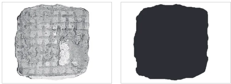
Magnification at 32x, although used in a previ-ous study,12 did not allow full visualization of brack-et surroundings. In order to enable analysis of the entire buccal surface on a single photograph, images of the four quadrants were individually captured and later assembled (Fig 1).
Quantification of composite flash
To serve as control and help distinguishing the dif-ference between composite and technical artifacts, the buccal surface of two teeth was demarcated and the
fol-lowing treatment modalities applied: upper let quad-rant = etching; upper right quadquad-rant = etching + bond-ing agent; lower let quadrant = etchetching + bond-ing + bondetching + bond-ing agent + conventional resin; and lower right quad-rant = etching + bonding agent + pigment resin (Fig 3).
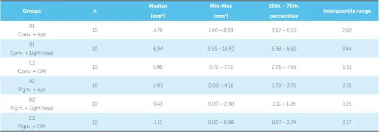
To isolate the area where only composite was pres-ent, any image suggestive of tooth structure (brighter areas) was removed (Fig 4). Contrast was adjusted by Image Pro Plus sotware (Fig 5) of which calibration was made possible by means of the scale available in each picture. The entire remaining dark area was measured in mm2. In order to quantify only the area of interest (resin around the bracket), it was necessary to mathe-matically subtract the value corresponding to the under-lying resin (bracket mesh base = 10.17 mm2).The oper-ator in charge of carrying out these measurements was not aware of the hypotheses being tested.
Figure 3 - Control sampl: In upper left quadrant, etching; in upper right quadrant, etching + bonding agent; in lower left quadrant, etching + bond-ing agent + conventional resin; in lower right quadrant, etchbond-ing + bondbond-ing agent + pigment resin.
Figure 1 - Image registration by quadrant (20x magnification each).
Statistical analysis
The values of the areas in each group were stored in a datasheet of SPSS (Statistical Package for the Social Sciences, SPSS Inc, Chicago, IL, version 9.0 for Mi-crosot Windows) for statistical analysis. Shapiro-Wilk test was used to analyze sample distribution. Given the absence of normal distribution in two groups (B2 and C2), intergroup comparison was performed with Kru-skal-Wallis test followed by Dunn’s post-hoc test for multiple comparisons. In all analyses, signiicance level was set at 5%.
RESULTS
Descriptive statistics containing sample size, medi-an, minimum and maximum values , 25th - 75th percen-tiles, and the interquartile range is available in Table 1. Kruskal-Wallis test at 5% with a KW statistic of 32.604
(corrected for ties) and 5 degrees of freedom detect-ed statistically signiicant diference between mdetect-edians (p < 0.0001). Dunn’s test for multiple comparisons iden-tiied ive pairs of groups with statistically signiicant diference (p ≤ 0.05) (Table 2).
According to data presented in Table 2, only the combination of pigmented composite and light head magnifying glass (B2) yielded a result that was superi-or to the combination of conventional composite and naked eye (A1). The combination of pigmented com-posite and naked eye (A2) was more beneicial than the combination of conventional composite and light head magnifying glass (B1). In comparison to the combina-tion of convencombina-tional composite and light head magnify-ing glass (B1), both the operatmagnify-ing microscope (C2) and the light head magnifying glass(B2) performed better when associated with the pigmented composite.
Groups n Median Min-Max 25th. - 75th. Interquartile range
(mm2) (mm2) percentiles
A1
Conv. + eye 10 4.76 1.40 – 8.68 3.62 – 6.23 2.62
B1
Conv. + Light head 10 6.94 3.53 – 14.50 5.38 – 8.82 3.44
C1
Conv. + OM 10 5.95 0.72 – 7.73 2.05 – 7.56 5.51
A2
Pigm. + eye 10 2.93 0.00 – 4.16 1.59 – 3.75 2.15
B2
Pigm. + Light head 10 0.43 0.00 – 2.30 0.11 – 1.26 1.15
C2
Pigm. + OM 10 1.11 0.00 – 6.98 0.57 – 2.74 2.17
Table 1 - Descriptive statistics
Table 2 - Intergroup comparison.
Conv = Conventional orthodontic adhesive; Pigm = Pigmented orthodontic adhesive; OM = Operating microscope.
Conv. = Conventional orthodontic adhesive; Pigm = Pigmented orthodontic adhesive; OM = Operating microscope; ns = non-significant (p > 0.05). *Statistically significant when p ≤ 0.05; **Statistically significant when p ≤ 0.01; *** Statistically significant when P ≤ 0.001.
Intergroup Comparison Median Diference Flash area Mean Rank
Diference P value
(1st. - 2nd.)
A1 (Conv. + eye) vs A2 (Pigm. + eye) 1.83 A1 > A2 13.867 ns
A1 (Conv. + eye) vs B1 (Conv. + Light head) -2.18 B1 > A1 -9.633 ns
A1 (Conv. + eye) vs B2 (Pigm. + Light head) 4.33 A1 > B2 27.567 **
A1 (Conv. + eye) vs C1 (Conv. + OM) -1.19 C1 > A1 0.3667 ns
A1 (Conv. + eye) vs C2 (Pigm. + OM) 3.65 A1 > C2 18.967 ns
A2 (Pigm. + eye) vs B1 (Conv. + Light head) -4.01 B1 > A2 -23.500 *
A2 (Pigm. + eye) vs B2 (Pigm. + Light head) 2.5 A2 > B2 13.700 ns
A2 (Pigm. + eye) vs C1 (Conv. + OM) -3.02 C1 > A2 -13.500 ns
A2 (Pigm. + eye) vs C2 (Pigm. + OM) 1.82 A2 > C2 5.100 ns
B1 (Conv. + Light head) vs B2 (Pigm. + Light head) 6.51 B1 > B2 37.200 ***
B1 (Conv. + Light head) vs C1 (Conv. + OM) 0.99 B1 > C1 10.000 ns
B1 (Conv. + Light head) vs C2 (Pigm. + OM) 5.83 B1 > C2 28.600 **
B2 (Pigm. + Light head) vs C1 (Conv. + OM) -5.52 C1 > B2 -27.200 **
B2 (Pigm. + Light head) vs C2 (Pigm. + OM) -0.68 C2 > B2 -8.600 ns
DISCUSSION
The pigmented composite Transbond TM Plus Color Change (3M UnitekTM, USA) is a good exam-ple of a material developed to facilitate visualization of composite lash during bracket placement. The irst study2 to test this material was published in June 2004. By the time this manuscript was written, only two more similar articles3,18 had been published, but none of them speciically addressed the advantages of pig-mented orthodontic adhesives to facilitate the removal of lash around orthodontic brackets.
Considering that the amount of composite left around orthodontic brackets tends to be quite sig-nificant, it is surprising that orthodontists are not so much concerned about it. In the present study, an area of up to 6.94 mm2 of remaining composite was observed. A large amount of composite flash was also described elsewhere.12
Busy clinical schedules and increased efort in ob-taining the most ideal bracket positioning may be two main reasons to explain why most orthodontists tend to overlook composite lash. Also, the color of con-ventional orthodontic adhesives matches quite well the color of enamel. This can certainly produce the false impression that lash was completely removed.
It was once thought that it could be advantageous to leave a certain amount of resin around brackets to seal any gaps between them and the enamel.19,20,21 However, the study by Farrow et al21 could not con-irm this hypothesis either when using luid resins or composites reinforced with inorganic particles.
The present study aimed to investigate the most effective method to remove composite adjacent to orthodontic brackets. Besides evaluating the influ-ence of the incorporation of pigments, this study also evaluated the advantages of two different methods of visual magnification: the operating microscope and the light head magnifying glass.
The method to measure the area of composite lash was based on the study by Armstrong et al.12 Unlike magniication used in their study, which was 32x, the present authors preferred to work with a magniication of 20x per quadrant, and then assemble the complete picture of the tooth by overlapping the four quadrants. This change proved necessary because the magniica-tion of 32x did not allow for full visualizamagniica-tion of the buccal surface containing the bracket.
Time spent on removal of flash composite was stringently controlled. In addition to avoiding per-formance bias, this was also helpful in assessing the clinical viability of each method. For orthodontists, turning a simple bracket placement procedure into a complex and time-consuming operation would not be economically feasible. In the present study, as far as flash removal with the naked eye is concerned, there was no advantage in using pigmented compos-ite. This finding is consistent with a previous report.12 However, removal of pigmented resin with the aid of the light head magnifying glass appeared to be the most advantageous method. Besides being more effective than removing conventional compos-ite with or without magnification, such low-cost and user-friendly method yielded a result very similar to removing pigmented composite with the aid of the operating microscope (Tables 1 and 2). In the era of excellence in Orthodontics, these data suggest that the combination of pigmented resin with light head magnifying glass should be encouraged when ortho-dontic assistants are removing composite flash. Or-thodontists could then check for bracket placement immediately thereafter with no need for visual aids.
In fact, a much better outcome was expected from the operating microscope. Both types of composites (conventional and pigmented), when removed with the aid of the operating microscope, performed very similarly to the removal of conventional composite with the naked eye (Table 2). However, it may be inaccurate to state that the operating microscope does not add precision to composite flash removal. In or-der to investigate this, methods without any time re-striction could be of great value, as this might have contributed to the poor performance of the operat-ing microscope. No matter how calibrated the op-erator may be, a longer bonding time will usually be necessary whenever using an operating microscope. This happens because the movements of the dental explorer require frequent focal adjustments. In ad-dition, considering that bracket positioning is quite an art which requires full visualization of the tooth, and that patients may move during the procedure, it is unlikely that the operating microscope will gain much popularity in the orthodontic community.
and light head magnifying glass, is in itself something that deserves consideration. Assessing the influence of the time spent on adjusting the operator’s position to the indirect vision transmitted by the operating microscope can finally decide whether it is advanta-geous to use this type of technology in Orthodontics.
CONCLUSIONS
1) The removal of a pigmented orthodontic ad-hesive with the aid of the light head magnifying glass proved, in general, to be advantageous compared to all other methods tested.
2) It was not possible to accurately assess the beneits from the combination of a pigmented composite and the operating microscope, thus eliciting the impor-tance of further studies designed to adapt its techni-cal requirements to the orthodontic clinitechni-cal setting.
Authors contribution
Conception or design of the study: FRD, FHSLP. Data acquisition, analysis or interpretation: EQSA. Writ-ing the article: EQSA, MLMN, PBDS, FHSLP. Critical revision of the article: FRD, PBDS. Final approval of the article: FHSLP. Overall responsibility: FHSLP.
1. Ogaard B, Rølla G, Arends J. Orthodontic appliances and enamel demineralization. Part 1. Lesion development. Am J Orthod Dentofacial Orthop. 1988 July;94(1):68-73.
2. Eliades T, Gioka C, Heim M, Eliades G, Makou M. Color stability of orthodontic adhesive resins. Angle Orthod. 2004 Jun;74(3):391-3. 3. Faltermeier A, Rosentritt M, Reicheneder C, Behr M. Discolouration of
orthodontic adhesives caused by food dyes and ultraviolet light. Eur J Orthod. 2008;30(1):89-93.
4. Mandel ID. Dental plaque: nature, formation and efects. J Periodontol. 1966 Sept-Oct;37(5):537-67.
5. Øgaard B, Alm AA, Larsson E, Adolfsson U.A prospective, randomized clinical study on the efects of an amine luoride/stannous luoride toothpaste/ mouthrinse on plaque, gingivitis and initial caries lesion development in orthodontic patients. Eur J Orthod. 2006 Feb;28(1):8-12. Epub 2005 Oct 17. 6. Kaklamanos E, Kalfas S. Meta-analysis on the efectiveness of powered
toothbrushes for orthodontic patients. Am J Orthod Dentofacial Orthop. 2008;133(2):187.e1-14.
7. Derks A, Frencken J, Bronkhorst E, Kuijpers-Jagtman AM, Katsaros C. Efect of chlorhexidine varnish application on mutans streptococci counts in orthodontic patients. Am J Orthod Dentofacial Orthop. 2008 Mar;133(3):435-9.
8. Kawai K, Urano M. Adherence of plaque components to diferent restorative
materials. Oper Dent. 2001 July-Aug;26(4):396-400.
9. Hannig M, Kriener L, Hoth-Hannig W, Becker-Willinger C, Schmidt H. Inluence of nanocomposite surface coating on bioilm formation in situ. J Nanosci Nanotechnol. 2007 Dec;7(12):4642-8.
10. Dezelic T, Guggenheim B, Schmidlin PR. Orthodontic adhesives induce human gingival ibroblast toxicity and inlammation. Oral Health Prev Dent. 2009;7(1):47-53.
11. Huang TH, Liao PH, Li HY, Ding SJ, Yen M, Kao CT. Orthodontic adhesives induce human gingival ibroblast toxicity and inlammation. Angle Orthod. 2008 May;78(3):510-6.
REFERENCES
12. Armstrong D, Shen G, Petocz P, Darendeliler MA. Excess adhesive lash upon bracket placement. A typodont study comparing APC PLUS and Transbond XT. Angle Orthod. 2007 Nov;77(6):1101-8.
13. Juggins KJ. The bigger the better: can magniication aid orthodontic clinical practice? J Orthod. 2006 Mar;33(1):62-6.
14. Carr GB, Murgel CA. The use of the operating microscope in endodontics. Dent Clin North Am. 2010 Apr;54(2):191-214.
15. García Calderín M, Torres Lagares D, Calles Vázquez C, Usón Gargallo J, Gutiérrez Pérez JL. The application of microscopic surgery in dentistry. Med Oral Patol Oral Cir Bucal. 2007 Aug 1;12(4):e311-6.
16. Hegde R, Sumanth S, Padhye A. Microscope-enhanced periodontal therapy: a review and report of four cases. J Contemp Dent Pract. 2009 Sept 1;10(5):E088-96.
17. Baumann DF, Brauchli L, van Waes H. The inluence of dental loupes on the quality of adhesive removal in orthodontic debonding. J Orofac Orthop. 2011 Mar;72(2):125-32.
18. Türkkahraman H, Adanir N, Gungor AY, Alkis H. In vitro evaluation of shear bond strengths of colour change adhesives. Eur J Orthod. 2010 Oct;32(5):571-4.
19. Joseph VP, Rossouw PE, Basson NJ. Some “sealants” seal–a scanning electron microscopy (SEM) investigation. Am J Orthod Dentofacial Orthop. 1994 Apr;105(4):362-8.
20. Palot C, Marzin I, Triconnet L. The peripheral joint: an unrecognized element in the bonding of orthodontic appliances. Orthod Fr. 1991;62 Pt 3:893-8. 21. Farrow ML, Newman SM, Oesterle LJ, Shellhart WC. Filled and unilled