Adult Hip and Orthopedic Oncology Groups, Department of Orthopedics, São Paulo University Medical School - São Paulo/SP, Brazil.
Email: [email protected] Received for publication on January 17, 2007. Accepted for publication on February 26, 2007.
CLINICAL SCIENCES
PROSPECTIVE STUDY OF THE TREATMENT OF
INFECTED HIP ARTHROPLASTIES WITH OR
WITHOUT THE USE OF AN ANTIBIOTIC-LOADED
CEMENT SPACER
Henrique Berwanger Cabrita, Alberto Tesconi Croci, Olavo Pires de Camargo, Ana Lúcia Lei Munhoz de Lima
Cabrita HB, Croci AT, Camargo OP de, Lima ALLM de. Prospective study of the treatment of infected hip arthroplasties with or without the use of an antibiotic-loaded cement spacer. Clinics. 2007:62(2):99-108.
PURPOSE: Our purpose was to compare 2 methods of treatment of chronic infection in hip arthroplasties—with or without an antibiotic-loaded cement spacer.
METHODS: In a prospective study, we treated 68 infected hip arthroplasties with discharging sinuses and bone loss, comparing 30 patients treated in 2 stages without the use of a spacer (control group) and 38 patients treated with a vancomycin-loaded spacer (study group). The average follow-up was 4 years (2-8.5 years). One patient died of unrelated causes 4 months after first-stage surgery and was excluded from the study.
RESULTS: The 2-stage surgery without spacer controlled the infection in 66.7% of patients, and the 2-stage surgery using the spacer controlled it in 89.1% (P < 0.05). At last follow-up, the average Harris Hip Score increased from 19.3 to 69.0 in the control group versus 19.7 to 75.2 in the study group (P > 0.05). The average leg length discrepancy was 2.6 cm in the control group and 1.5 cm in the study group (P < 0.05). The patients treated with a spacer had better clinical results (81.5% of patients with good results against 60.0% for the control group).
CONCLUSION: The use of an antibiotic-loaded spacer in the 2-stage treatment of infected hip arthroplasties provides better infection control with good functional results and is superior to treatment in 2 stages without a spacer. Level of Evidence: Therapeutic study, Level I-1.
KEYWORDS: Hip. Infection. Arthroplasty. Spacer. Bone cements.
INTRODUCTION
The prevalence of deep infection following hip arthro-plasty has been significantly reduced, although its levels still give cause for concern.1,2
The treatment of infection is a long process often re-quiring more than 1 surgery, causing suffering and giving rise to extremely high financial and socials costs. Never-theless, the prognosis for resolving infectious processes in
hip arthroplasties reaches about 80% to 90% with the cur-rent surgical techniques.3
The control of chronic infection in arthroplasties re-quires the removal of the prosthetic components and ex-tensive debridement. After this, there are 3 types of proce-dures that may be considered:4-6
1) Simple wound closure and maintenance of the patient without an implant, through the Girdlestone surgery;7
this is a safe method as regards infectious control, but functional outcome is poor.8,9
2) Immediate placement of a definitive prosthesis (1-stage revision), which was widely used by European surgeons during the 1980s and 1990s.7,10,11 Currently, this
dis-charging sinuses, poor condition of soft parts, and bone loss requiring allografts.8,12 Because a 1-stage revision
is indicated under conditions requiring the placement of cemented components, cementless arthroplasties should not be performed as a single-stage surgery.13
3) A 2-stage or a 3-stage surgery, which are safest and are used worldwide.14-18
However, the traditional 2-stage procedure has several disadvantages compared with the 1-stage surgery. During the intermediate period, patients stay in hospital for at least 3 weeks, which is the period of skeletal traction required to heal the soft tissues and start rehabilitation.19 Between
sur-geries, the patient has a shortened leg and is impaired re-garding rehabilitation and function. The patient’s loss of mobility increases the risk of pressure ulcers, osteoporosis, and thromboembolism.20 The second stage is more difficult
than an aseptic revision due to cicatricial retraction and mus-cular contractures that occur with leg shortening; bone land-marks are difficult to identify during surgery.19 Disuse
oste-oporosis impairs the mechanical conditions for the fixation of the permanent prosthesis and predisposes to fractures.21
At the beginning of the 1990s, 2 authors independently described the use of a cement block impregnated with an-tibiotics to fill large cavities in the acetabular and femoral regions of patients with an infected hip arthroplasty and severe bone loss.22,23 The spacer includes hip prosthesis
components coated with PMMA (polymethylmethacrylate) and may be articulated as a total prosthesis24,25 or unipolar
as a partial prosthesis.26,27
Regardless of the worldwide acceptance of the spacer as the main method of treatment of infected hip replace-ments, no objective data have been reported in the litera-ture to prove the real benefits of using a spacer in 2-stage surgeries.2,28
The purpose of this study was to prospectively compare 2 methods for treating chronic infection in hip arthroplast-ies: 2-stage surgery with and without using an antibiotic-loaded cement spacer.
MATERIALS AND METHODS
A prospective study was performed in 68 patients di-agnosed with chronic infected hip arthroplasties and treated in our Institution from April 1996 to January 2003.
As inclusion criteria, all of the following were required: 1. Previous surgery with total or partial hip prosthesis; 2. Diagnosis of infection, based on bacterial identification
in cultures of samples collected during the first surgery of the treatment;
3. Minimum time period between the arthroplasty and the infectious condition of 4 weeks;
4. Presence of a discharging sinus communicating with the prosthesis;
5. A written informed consent for the performance of the study procedures, where the patient states his/her aware-ness of the experimental character of the investigation and the possible complications secondary to the treat-ments.
Sixty-eight patients were randomly selected to receive treatment in 2 stages without a spacer (control group) or with the placement of an antibiotic-loaded cement spacer (study group).
The mean age of patients was 54.6 years (range 16-84 years), with no difference between the groups. The male sex prevailed, with 57.8% of the total number of patients. Etiologies requiring most of the primary arthroplasties had been trauma or trauma sequelae (32%), osteoarthrosis (27%), rheumatic disease (12%), and osteonecrosis (9%). This distribution with the high incidence of trauma patients may be explained by the fact that our facility is a major trauma referral center; most cases included in both groups (60.6%) were referred from other services. The preoperative functional evaluation of patients was performed using the
Harris Hip Score,29 and no differences were noticed
be-tween groups (average score of 16 in the control group and 18.5 in the study group, P = 0.02).
The evaluation of bone loss on the acetabular and femo-ral side after removal of the components was made accord-ing to the Gustilo and Pasternak classification30 (Tables 1
and 2). Most of the patients in both groups had severe femo-ral bone loss and mild to moderate acetabular bone loss. The bone loss was similar in both groups, with a trend of worse femoral losses for the study group.
The spacer was similar to a Thompson’s unipolar pros-thesis and was made in 2 phases: acetabular and femoral.28
The core of the spacer was a femoral component that was
Table 1 - Bone loss following removal of the acetabular component, per treatment group
Grade of bone loss I II III IV
Control group 10 (33.3%) 11 (36.7%) 5 (16.7%) 4 (13.3%) Study group 16 (42.1%) 15 (39.4%) 3 (8.0%) 4 (10.5%)
Table 2 - Bone loss following removal of the femoral component, per treatment group
Grade of bone loss I II III IV
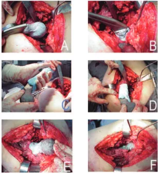
removed during a revision and sterilized (Figure 1); or, ide-ally, it was a Küntscher femoral nail bent according to the original angle between the neck and the diaphysis of the patient’s femur (Figure 2). We used 1.0 g of vancomycin hydrochloride powder (Vancocina CP®, Eli Lilly, Sao Paulo,
SP, Brazil ) per package of 40.0 g of acrylic cement
(Sim-plex P®, Striker Howmedica Osteonics, Rutherford, NJ,
USA ), as recommended by Penner et al31 and by Chohfi
et al.32 Typically, 2 packages of cement were used to make
the acetabular portion, and 2 to 4 packages were used to make the femoral portion.
The patients treated without a spacer remained for 3 weeks under skeletal traction to allow fibrosis formation at the Girdlestone resection.
Systemic antibiotics were given for 3 weeks, accord-ing to infectious disease protocols. The empiric therapy was 1.0 g of vancomycin and 2.0 g of ceftazidime daily until bacterial isolation. Oral antibiotics were indicated pre-scribed until 6 months after the first surgical stage, regard-less of whether the second stage was/was not completed.
The second stage was done only when erythrocyte sedi-mentation speed and C-reactive protein were at normal lev-els. In doubt, or if the increase of the values of the active phase tests persisted, samples taken by intra-articular punc-ture were collected for culpunc-ture. If any bacterial growth was noticed, the second surgical stage was not performed and
the treatment was considered as failed for this study18,33,34
For both stages, we report the bacteria that were iso-lated, the surgical length of time, blood reposition, hospi-tal stay, and complications.
Statistical Analysis
We used the chi-square test (χ2) and Fisher’s exact test to compare the class frequencies between the groups. To compare the magnitudes (quantitative data) between sam-ples of the groups, the Student t test was used for samsam-ples presenting parametric distributions, while the Mann-Whitney U-test was used for nonparametric distributions. To compare the magnitude of a same sample at 2 different times (ie, pre- and posttreatment), the Wilcoxon test was used.
RESULTS
None of the patients was lost to follow-up, which was 4 years (range, 2-8.5 years) in both groups. Three patients died of causes unrelated to the treatment after 48 months or more of follow-up; they were included in the study. The final results were as follows:
Thirty patients were treated in the control group, in 2-stages without a spacer. Of these, 1 died of septicemia ter first stage and 2 died of hemorrhagic complications af-ter the second stage. These patients account for a 10% mor-tality related to the treatment without a spacer.
Twenty-three patients underwent reimplantation. In one patient, this was not possible due to technical problems during surgery.
At last follow-up, of the 20 patients with successful im-plants, 2 reported recurrence of infection within less than 6 months after second stage and 2 had aseptic loosening of the acetabular cup requiring revision surgery. Eighteen
Figure 2 - A – Infected THR after interthrochanteric fracture in a 74 years old patient. Bone loss grade IV at femoral side and I at acetabular side; B – Spacer done with a Küntscher rnail bended to simulate femoral neck angle; C – After two years of second stage done with a wagner prosthesis and massive femoral bone allograft the patient has no signs of infection and a Harris hip Score of 87.
(60.0%) patients were infection-free and reported good functional results.
Thirty-eight patients were treated in the study group (with a spacer).
One patient died after pelvic migration of the spacer; this was classified as a method failure.
One patient died of nontreatment-related causes (acute cholecystitis) 4 months after the second stage and was ex-cluded from the study.
Thirty-three patients underwent a new total joint re-placement. Two patients had recurrent dislocations and were treated surgically. There were 2 aseptic loosenings: 1 cementless cup was loose requiring revision after 4 months and 1 cemented stem was loose without pain after 4 years.
At the last follow-up, 31 patients (81.5%) had a good functional prostheses with no infectious recurrences.
Bone allografts were used in 61.4% of all cases (35 of 57 second-stage surgeries), which confirms the bone loss stated at first stage surgery
Table 3 shows that the mean duration of the first surgi-cal stage was less in the control group (3 hours 12 mutes, P = 0.02), a finding that shows that the spacer in-creased the surgical time by 40.1 minutes. At the second stage, the mean duration of the surgery in the study group was virtually 1 hour less (3 hours 22 minutes, P = 0.001).
Table 4 displays hospital stay which was 34.6 days for the control group versus 24.7 days for the study group (P < 0.001), for the 1st stage operation. Once the second stage
was performed, a significant difference was noticed in the duration of patients’ hospitalization, with shorter hospital stays for the study group (11.7 days against 8.2 days, P = 0.004). The interval between surgeries was equivalent in both groups, with an average of 226.9 days (range, 70-610 days) for the control group and 162.8 days (range, 60-350 days) for the study group (P = 0.31).
Table 5 displays intensive care unit stay. No differences were noticed between the groups in terms of number of days during which the patients were cared for in the in-tensive care unit after the first surgical stage, although a trend exists towards a longer time in the hospital in the con-trol group. The use of the spacer allowed patients to have a shorter stay in the intensive care unit after the second sur-gical stage (1.4 days on the average, as compared to 4.1 days for the control group, P = 0.004).) No difference oc-curred between groups in the required amount of packed red cell transfusion (Table 6), but less fluid drained in both stages for the study group (Table 7).
Complications are listed in Table 8: Six spacer-related complications occurred: 3 dislocations, 2 pelvic migrations and 1 fracture. Of the dislocated spacers, 2 were from sur-geries performed in the beginning of the study. We observed that the necks of these spacers were too valgus, which fa-cilitated lateral migration. Although these 2 spacers re-mained dislocated, patients did not report any significant pain condition, and it was not difficult to perform the sec-ond surgical stage. Other complications were unrelated to the kind of treatment, such as femoral fractures at implant removal (9 cases), delirium (6 cases) and intestinal obsti-pation (6 cases).
There were 4 complications related to antibiotic therapy: 3 allergic reactions and 1 acute renal failure..
Table 3 - Duration of surgery, by treatment group (mean, minutes)
Control Study P
First stage 192.0 232.1 0.02
Second stage 265.3 202.9 0.001
Table 4 - Length of hospital stay, by treatment group (mean, days)
Control Study P
First stage 34.6 24.7 <0.001
Second stage 11.7 8.2 0.004
Table 5 - Length of stay in the intensive care unit, by treatment group (mean, days)
Control Study P
First stage 2.0 1.4 0.06
Second stage 4.1 1.4 0.004
Table 6 - Number of packs of concentrated red blood cells used after during and after surgery, by treatment group (mean)
Control Study P
First stage 4.1 3.1 0.12
Second stage 3.9 2.6 0.16
Table 7 - Drainage after surgery, by treatment group (mean, mL)
Control Study P
First stage 677.3 501.8 0.007
There was 1 case (5.0%) of recurrent dislocation in the control group and 2 cases (6.4%) in the study group.
The incidence of intra- and postoperative complications was not different between groups in both stages. During the first surgical stage, we had problems while removing the fixed femoral components, with femur fractures in 9 cases (23.7%).
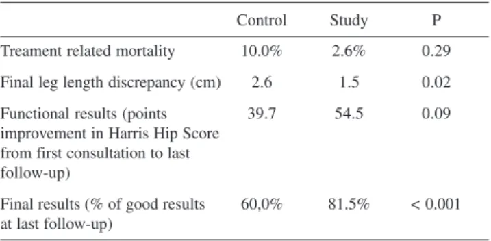
Outcome is displayed in Table 9, showing a significantly higher proportion of good results in the study group. The mortality related to the treatment was greater in the con-trol group (10% versus 2.6%), although without statistical significance (P = 0.29).
On the average, the leg length discrepancy was 2.6 cm in the control group and 1.5 cm in the study group (P = 0.02).
According to the Harris Hip Score, the final functional outcome was better in the study group, although with no statistical significance (P = 0.09). The average score ranged from 19.3 to 69.0 in the control group and from 19.7 to 75.2 in the study group.
The study group had a better final outcome, with 81.5% having good results versus 60.0% of the control group (P < 0.001).
Recurrence of infection (Table 10). was significantly higher for the control group, where it occurred in 7 patients (23.3%) 7 to 112 days (average, 36.0 days) after the first stage. These patients were treated with successive surgical debridements, muscular flaps, or antibiotic-loaded cement spacers. Three patients (8.1%) reported recurrence of fection after the second stage accounting to an overall in-fection failure of treatment in 33.3% of the patients.
In the study group recurrence of infection occurred in 2 patients after first stage (5.2%) and in 2 patients (6.1%) after second stage. The overall infection rate was 10.5% in the patients treated with a spacer.
Table 11 displays isolated bacterium species: Gram-positive bacteria were the most frequently isolated during the first surgical stage (68.5%), with prevalence of Staphy-lococcus aureus (31.5%), coagulase-negative staphilococci (13.7%), and Enterococcus faecalis (13.7%).
DISCUSSION
The use of a spacer reduced the mean duration of the second surgical stage in 1 hour. Reimplantation without a spacer is difficult because it is hard to find the surgical planes, identify the bone structures, and build the bed to accept the prosthesis. An extensive fibrosis results in ex-tended surgery time.
On average, after the first surgical stage, the control group patients stayed in hospital almost twice as long as patients of the study group The use of a spacer reduced the hospital stay, because the skeletal traction period is mandatory to allow healing of the soft parts in patients treated without a spacer.
Although we did not estimate the cost of treatment for
Table 8 - Incidence of complications, by treatment group and totals
Control Study Total
Spacer dislocation* 3 3
Intrapelvic migration of spacer* 2 2
Spacer fracture* 1 1
Femoral fracture at implant removal 4 5 9
Delirium 3 3 6
Intestinal obstipation 2 4 6
Deep vein thrombosis 2 2 4
Antibiotic allergy 2 1 3
Aseptic acetabular loosening 2 1 3
Recidivant disclocation 1 2 2
Thigh abcess 2 2
Nerve damage 2 2
Acute hemorrhage leading to death 2 2
Aseptic femoral loosening 1 1
Nausea and sickness 1 1
Retroperitoneal abscess 1 1
Genital edema 1 1
Acute renal failure due to antibiotics 1 1
Headache 1 1
Anaphylaxis due to morphine 1 1
Pneumonia 1 1
Femoral diaphysial pseudarthrosis 1 1
Herpes zoster 1 1
Acute colicystitis, sepsis, and death 1 1
Acute hemorrhage and hypovolemia 1 1
Inability to implant prosthesis at second stage 1 1 * Spacer related complication
Table 9 - Outcome of the treatment, by treatment group
Control Study P
Treament related mortality 10.0% 2.6% 0.29
Final leg length discrepancy (cm) 2.6 1.5 0.02
Functional results (points 39.7 54.5 0.09
improvement in Harris Hip Score from first consultation to last follow-up)
Final results (% of good results 60,0% 81.5% < 0.001 at last follow-up)
Table 10 - Recurrence of infection after first and second stages, by treatment group
Recurrence After After Overall
of infection first stage second stage
Control group 7 (23.3%) 3 (8.1%) 10 (33.3%)
Study group 2 (5,2%) 2 (6.1%) 4 (10.5%)
patients in this study, the shorter hospital stay, shorter op-erating room time, and shorter intensive care unit stay of the study group must definitely lead to a lower cost of the treatment with a spacer as compared to that without a spacer.
A technical impossibility of performing permanent ar-throplasty during the second surgical stage occurred in 1 patient of the control group, a fact also reported by Fitzgerald35 and Charlton et al.20 This difficulty is typical
of the second surgical stage in surgeries without a spacer, in which case it is difficult to dissect the muscular planes and identify bone landmarks.
In the initial trial with the PROSTALAC® Duncan and
Beauchamp27 reported 3 cases of spacer dislocation among
15 patients. After 9 years in the revision of 135 cases2,8 only
2 more of such complications exist, showing that the ex-perience acquired with the performance of the method pre-vents dislocation from occurring. Due to a pelvic migra-tion of a spacer and subsequent injury of the iliac vessels and death of 1 patient, we do not place the spacer as a uni-polar prosthesis in patients with acetabular bone weakness, particularly in obese and rheumatoid patients (Figure 3). In this case, we recommend the placement of a cement ball with antibiotics that fills the acetabular cavity and that ar-ticulates with the component implanted in the femoral re-gion.
It is important to note that severe bleeding occurred dur-ing the second surgical stage of the control group, with 2 deaths directly related to acute hemorrhage. The dead space
left after the removal of components is replaced by a hematoma when a spacer is not placed. This blood collec-tion causes continuous blood loss that is difficult to con-trol and forms the extensive fibrotic tissue found at reimplantation. The dissection of this fibrosis leads to ex-cessive bleeding and to hemorrhagic complications.
Tsukayama et al36 reported 2 infection-related deaths
among 98 patients. Fisman et al37 estimate that the
mor-tality associated with infected hip prostheses is 0.4% to 1.2% for 65-year-old patients and 2.0% to 7.0% for 80-year-old patients. During the 3 months that followed the prosthesis removal and the surgical cleaning, mortality dou-bled for both types of patients.
In both our groups of treatment the dislocation rate was similar to reported data, after the treatment of infected ar-throplasties, which is 7.3% to 12.7% of the cases14,16,21,29,30,37,39 regardless of the method employed.
The high incidence of severe femoral loss (up to 19 cases) in the study group demonstrates the long duration of the infectious activity in patients. Severe bone loss makes the treatment even more difficult, since more sophisticated reconstruction techniques are needed during the second sur-gical stage. It is hard to make a comparison with our se-ries due to the variety of classification systems used. Berry and Chandler38 and English et al18 showed variable bone
loss in all cases, and Alexeef et al40 found severe femoral
bone loss in 11 cases. Rudelli41 found severe bone loss in
65% of cases treated with 1-stage surgery and bone graft-ing.
Alexeeff et al40 used massive femoral grafts in 9 cases
without infectious recurrence after a 4-year follow-up. The Exeter technique was adopted by English et al18 with
in-fection resolution in 92.5% of 44 cases and good functional
Table 11 - Bacteria isolated on cultures during first stage (73 bacteria in 68 patients, 2 patients with 2 or more isolated microorganisms)
GRAM-POSITIVE BACTERIA 50 (68.5%)
Staphylococcus aureus 23 (31.5%)
Staphylococcus epidermidis, Staphylococcus cohnii and
coagulase negative Staphylococci 10 (13.7%)
Enterococcus faecalis 10 (13.7%)
Streptococcus viridans 3 (4.1%)
Streptococcus agalactiae 2 (2.7%)
Corinebacterium sp 1 (1.4%)
Streptococcus mitis 1 (1.4%)
GRAM-NEGATIVE BACTERIA 23 (31.5%)
Escherichia coli 4 (5.5%)
Enterobacter cloacae 4 (5.5%)
Proteus mirabilis 3 (4.1%)
Serratia marcenses 3 (4.1%)
Klebsiella sp 3 (4.1%)
Acinetobacter baumannii 1 (1.4%)
Aeromonas hydrophilia 1 (1.4%)
Citrobacter diversus 1 (1.4%)
Pseudomonas aeruginosa 1 (1.4%)
Providencia sp 1 (1.4%)
Stenotrophomonas maltophilia 1 (1.4%)
results. Hanssen and Osmon42 reported 4 infectious
recur-rences in 7 patients reconstructed with structured graft of the proximal femur. Rudelli used a homologous bone graft in 36 one-stage treated cases, 9 of them with discharging sinuses, and reached a 88.9% rate of infectious control with a minimum follow-up period of 2 years in the cases with discharging sinuses.41
In our patients, there were no mechanical complications with the massive allografts that were used in 14 patients in the study group and 8 patients in the control group. Main-taining and reconstructing bone stock is fundamental for young patients who will be subjected in the future to new revisions and require a mechanically resistant bone structure for the replacement of worn-out components (Figure 4).
The tendency towards better functional results in the study group might be due to the advantage that the reha-bilitation is performed immediately after the start of the intermediate period. In addition to early rehabilitation in the intermediate period, the use of the spacer led to less surgical aggression in the second surgical stage, with im-mediate, less painful postoperative handling and early func-tional recovery.
In both groups the mean leg length discrepancy was
ac-ceptable when compared to those of Alexeeff et al,40 who
found no discrepancy greater than 3.0 cm when they used a spacer and tissue bank in the second stage, and also to the data of Charlton et al20 who, after the treatment with a
spacer achieved full correction in only 50% of the patients. The identification of Gram-negative bacteria in 22 cases does not contradict the validity of the use of vancomycin added to acrylic cement. Gram-negative bacteria are not sensitive to a single type of antibiotics, a behavior that is adopted by Gram-positive bacteria against vancomycin. The sensitivity spectrum of Gram-negative bacilli varies be-tween aminoglycosides and first to fourth generation cephalosporins and quinolones; therefore, it is difficult to select 1 antibiotic to cover all Gram-negative bacteria. Koo et al43 tried to solve this problem by adding vancomycin,
gentamycin, and cefotaxime to the acrylic cement. They found infectious control in 95% of patients, but 4 cases (20%) reported side effects due to the antibiotic, with he-patic dysfunction and medullar depression. In our opinion, the addition of antibiotics to the cement to cover Gram-negative bacteria should be directed and implemented only in case of previous identification of the agent in an appro-priately collected culture. This concern is important from the perspective of hospital infection control, since it pre-vents the conditions favorable to proliferation of multire-sistant bacteria.
Infectious control, both after the first and second sur-gical stages, was better in the study group—this being the main subject of this article.
After the first surgical stage, the infectious control was poorer in the control group; this is probably due to the colo-nization of the extended hematoma that occupied the dead space left by the extraction of the metallic implants.
We believe that the follow-up time after second stage is still short and that more accurate analyses should be per-formed after 5 years of follow-up.
The overall success achieved in both groups was infe-rior to that reported by other authors who treated infections in 2 stages (72.0% to 97.7%) with a minimum 1-year fol-low-up period.2,3,9,15,18,37,42,44 Our results are similar to those
reported by Hunter and Dandy,45 with 33.3% of infectious
control and by Cierny and DiPasquale46 with 64.0%, who
treated patients with severe bone loss, as in our case. All our patients showed discharging sinuses on physi-cal examination, which characterizes the chronicity and
severity of infections.44 According to most
au-thors,2,8,15,16,22,28,39,40,47-50 the presence of discharging sinuses
contraindicates the performance of a 1-stage treatment. On the other hand, several studies,7,13,14,51 including the
com-munication by Rudelli et al,41 report performance of 1-stage
revisions even in the presence of discharging sinuses.
A
B
C
D
E
F
The indications for either 1-stage or 2-stage treatments are not very clear and vary a lot from one treatment center to the next. We agree with Elson,52 who states that a
dan-gerous situation occurs when a given author advocates a single treatment method. There should not be any competi-tive element in this issue, and it all will depend on the cor-rect comparison of personal results with those found by other investigators. The choice between 1-stage surgery and 2-stage surgery is not so important, since the experienced surgeon knows what is achieved using the selected method. A 2-stage approach should be used in chronically ill patients, especially those with bone loss and with
discharg-ing sinuses. Our overall infection control with both treat-ments was 73.1%, a value considered desirable in the lit-erature.1-4
CONCLUSIONS
We conclude that the 2-stage treatment of infected hip arthroplasties using antibiotic-loaded cement spacer is su-perior to the 2-stage surgery performed without the spacer, and that the use of the spacer provides better infectious con-trol, as well as better functional outcomes.
RESUMO
Cabrita HB, Croci AT, Camargo OP de, Lima ALLM de. Estudo prospectivo do tratamento das artroplastias infectadas do quadril sem e com o uso de espaçador de cimento com antibiótico. Clinics. 2007:62(2):99-108.
OBJETIVO: As revisões em dois tempos continuam sendo os métodos preferidos no tratamento das artroplastias infectadas do quadril. O procedimento em dois estágios apresenta várias desvantagens teóricas, ainda não comprovadas por estudos comparativos.
MATERIAIS E MÉTODOS: Em um estudo prospectivo, tratamos 68 pacientes com artroplastias infectadas de quadril com perdas ósseas e fístulas ativas, comparando 30 casos tratados em dois tempos sem espaçador (grupo controle) e 38 casos tratados em dois tempos com o uso de um espaçador de cimento adicionado a vancomicina (grupo de estudo). Um paciente faleceu após quatro meses da cirurgia e foi excluído do estudo. O seguimento médio foi de quatro anos (2-8,5 anos).
RESULTADOS: A cirurgia em dois tempos sem espaçador controlou a infecção em 66,7% dos casos comparada a 89,1% (p<0,05) nos casos tratados com espaçador. No último seguimento, o Escore de Harris para Quadril passou de 19,3 a 69,0 no grupo controle e de 19,7 para 75,2 no grupo do estudo (p>0,05). A média de discrepância de membros inferiores foi de 2,6cm no grupo controle e de 1,5cm nos grupo do estudo (p<0,05). O grupo tratado com espaçador teve melhores resultados clínicos ao final do estudo (81,5% de bons resultados comparados a 60,0% do grupo tratado sem espaçador).
CONCLUSÃO: O uso de espaçador adicionado a antibióticos no período intermediário do tratamento das artroplastias infectadas do quadril em dois tempos proporciona melhor controle de infecção, com bons resultados funcionais, sendo superior à cirurgia em dois tempos sem espaçador.
UNITERMOS: Quadril. Infecção. Artroplastia. Espaçador. Cimentos para ossos.
REFERENCES
1. Salvati EA, Della Valle AG, Masri BA, Duncan CP. The infected total hip arthroplasty. Instr Course Lect. 2003; 52:223-45.
2. Garvin KL, Hanssen AD. Infection after total hip arthroplasty. Past, present, and future. J Bone Joint Surg Am. 1995;11:1576-88. 3. Mitchell PA, Masri BA, Garbuz DS, Greidanus NV, Duncan CP.
Cementless revision for infection following total hip arthroplasty. Instr Course Lect. 2003;52:323-30.
4. Raut VV, Siney PD, Wroblewski BM. One-stage revision of infected total hip replacements with discharging sinuses. J Bone Joint Surg Br. 1994;76:721-4.
6. Schroder J, Saris D, Besselaar PP, Marti RK. Comparison of the results of the Girdlestone pseudarthrosis with reimplantation of a total hip replacement. Int Orthop. 1998;22:215-8.
7. Girdlestone GR. Acute pyogenic arthritis of the hip: an operation giving free access and effective drainage. Lancet. 1943:419-21.
8. Pagnano MW, Trousdale RT, Hanssen AD. Outcome after reinfection following reimplantation hip arthroplasty. Clin Orthop. 1997;338:192-204.
9. Castellanos J, Flores X, Llusa M, Chiriboga C, Navarro A. The Girdlestone pseudarthrosis in the treatment of infected hip replacements. Int Orthop. 1998;22(3):178-81.
10. Buchholz HW, Elson RA, Engelbrecht E, Lodenkamper H, Rottger J, Siegel F. Management of deep infection of total hip replacement. J Bone Joint Surg Br. 1981;63:342-53.
11. Wroblewski BM. One stage revision of infected cemented total hip arthroplasty. Clin Orthop. 1986;212:103-7.
12. Lecuire F, Collodel M, Basso M, Rubini J, Gontier D, Carrere J. Reprise des prothèses totales de hanche infectées par ablation réimplantation d´une prothèse sans ciment. Experiénce de 57 cas. Rev Chir Orthop Reparat Appar Mot. 1999;85:337-48.
13. Ure KJ, Amstutz HC, Nasser S, Schmalzried TP. Direct-exchange arthroplasty for the treatment of infection after total hip replacement. An average ten-year follow-up. J Bone Joint Surg Am. 1998;80:961-8. 14. Lentino JR. Prosthetic joint infections: bane of orthopedists, challenge
for infectious disease specialist. Clin Infect Dis. 2003;36:1157-61. 15. Colyer RA, Capello WN. Surgical treatment of the infected hip implant.
Two-stage reimplantation with a one-month interval. Clin Orthop. 1994; 298:75-9.
16. Mcdonald DJ, Fitzgerald RH Jr, Ilstrup DM. Two-stage reconstruction of a total hip arthroplasty because of infection. J Bone Joint Surg Am. 1989;71:828-34.
17. Morscher E. Solo espianto o reimplanto per la mobilizzazione settica? Chir Organi Mov. 1994;79:425-8.
18. English H, Timperley AJ, Dunlop D, Gie G. Impaction grafting of the femur in two-stage revision for infected total hip replacement. J Bone Joint Surg Br. 2002; 84:700-5.
19. Salvati EA, Chekofsky KM, Brause BD, Wilson PD Jr. Reimplantation in infection: a 12-year experience. Clin Orthop. 1982;170:62-75. 20. Charlton WP, Hozack WJ, Teloken MA, Rao R, Bissett GA.
Complications associated with reimplantation after girdlestone arthroplasty. Clin Orthop. 2003;407:119-26.
21. Clegg J. The results of the pseudarthrosis after removal of an infected total hip prosthesis. J Bone Joint Surg Br. 1977;59:298-301. 22. Zilkens KW, Casser HR, Ohnsorge J. Treatment of an old infection in a
total hip replacement with an interim spacer prosthesis. Arch Orthop Trauma Surg. 1990;109:94-6.
23. Abendschein W. Salvage of infected total hip replacement: use of antibiotic/PMMA spacer. Orthopedics. 1992;15:228-9.
24. Younger ASE, Masri BA, Duncan CP, Mcgraw RW. The outcome of two-stage arthroplasty using a custom-made interval spacer to treat the infected hip. J Arthroplasty. 1997;12:615-23.
25. Wentworth SJ, Masri BA, Duncan CP, Southworth CB. Hip prosthesis of antibiotic-loaded acrylic cement for the treatment of infections following total hip arthroplasty. J Bone Joint Surg Am. 2002;84(Suppl 2):123-8.
26. Kraay MJ, Goldberg VM, Figgie HE 3rd. Use of an antibiotic impregnated polymethyl methacrylate intramedullary spacer for complicated revision total hip arthroplasty. J Arthroplasty. 1992;7(Suppl):397-402.
27. Duncan CP, Beauchamp C. A temporary antibiotic-loaded joint replacement system for management of complex infections involving the hip. Orthop Clin North Am. 1993;24:751-9.
28. Migaud H, Chantelot C, Besson A, Gougeon F, Dubois HH, Duquennoy A. Temporary antibiotic-loaded cemented prosthesis for two- stage septic hip arthroplasty. Rev Chir Orthop Reparatrice Appar Mot. 1997;84:466-8.
29. Harris WH. Traumatic arthritis of the hip after dislocation and acetabular fractures: treatment by mold arthroplasty. An end-result study using a new method of result evaluation. J Bone Joint Surg Am. 1969;51:737-55.
30. Gustilo RB, Pasternak HS. Revision total hip arthroplasty with titanium ingrowth prosthesis and bone grafting for failed cemented femoral component loosening. Clin Orthop. 1988;235:111-9.
31. Penner MJ, Masri BA, Duncan CP. Elution characteristics of vancomycin and tobramycin combined in acrylic bone-cement. J Arthroplasty. 1996;11:939-44.
32. Chohfi M, Langlais F. O cimento ortopédico associado à vancomicina: comportamento mecânico e difusão do antibiótico . Rev Bras Ortop. 1994;29:363-70.
33. Wang JW, Chen CE. Reimplantation of infected hip arthroplasties using bone allografts. Clin Orthop. 1997;335:202-10.
34. McPherson EJ, Woodson C, Holtom P, Roidis N, Shufelt C, Patzakis M. Periprosthetic total hip infection: outcomes using a staging system. Clin Orthop. 2002;403:8-15.
35. Fitzgerald RH. Total hip arthroplasty sepsis. Prevention and diagnosis. Orthop Clin North Am. 1992;23:259-64.
36. Tsukayama DT, Estrada R, Gustilo RB. Infection after total hip arthroplasty. A study of the treatment of one hundred and six infections. J Bone Joint Surg Am. 1996;78:512-23.
37. Fisman DN, Reilly DT, Karchmer AW, Goldie SJ. Clinical effectiveness and cost-effectiveness of 2 management strategies for infected total hip arthroplasty in the elderly. Clin Infect Dis. 2001;32:419-30. 38. Berry DJ, Chandler HP, Reilly DT. The use of bone allografts in
two-stage reconstruction after failure of hip replacements due to infection. J Bone Joint Surg Am.1991;73:1460-8.
39. Miley GB, Scheller AD Jr, Turner RH. Medical and surgical treatment of the septic hip with one-stage revision arthroplasty. Clin Orthop. 1982;170:76-82.
40. Alexeeff M, Mahomed N, Morsi E, Garbuz D, Gross A. Structural allograft in two-stage revisions for failed septic hip arthroplasty. J Bone Joint Surg Br. 1996;78:213-6.
42. Hanssen AD, Osmon DR. Evaluation of a staging system for infected hip arthroplasty. Clin Orthop. 2002;403:16-22.
43. Koo KH, Yang JW, Cho SH, Song HR, Park HB, HA YC, et al. Impregnation of vancomycin, gentamicin, and cefotaxime in a cement spacer for two-stage cementless reconstruction in infected total hip arthroplasty. J Arthroplasty. 2001;16:882-92.
44. Carlsson AS, Josefsson G, Lindberg L. Revision with gentamicin-impregnated cement for deep infections in total hip arthroplasties. J Bone Joint Surg Am. 1978;8:1059-64.
45. Hunter G, Dandy D. The natural history of the patient with an infected total hip replacement. J Bone Joint Surg Br. 1977;59:293-7. 46. Cierny G 3rd, Dipasquale D. Periprosthetic total joint infections: staging,
treatment, and outcomes. Clin Orthop. 2002;403:23-8.
47. Hughes PW, Salvati EA, Wilson PD Jr, Blumenfeld EL. Treatment of subacute sepsis of the hip by antibiotics and joint replacement. Criteria for diagnosis with evaluation of twenty-six cases. Clin Orthop. 1979;141:143-57.
48. Garvin Kl, Salvati EA, Brause BD. Role of gentamicin-impregnated cement in total joint arthroplasty. Orthop Clin North Am. 1988;19:605-10.
49. Sanzén L, Carlsson AS, Josefsson G, Lindberg LT. Revision operations on infected total hip arthroplasties. Two- to nine-year follow-up study. Clin Orthop. 1988;229:165-72.
50. Elson RA. Exchange arthroplasty for infection. Perspectives from the United Kingdom. Orthop Clin North Am. 1993;24:761-7.
51. Loty B, Postel M, Evrard J, Matron P, Courpied JP, Kerboull M, et al. Remplacements en un temps des prothèses totales de hanches infectées et reconstructions osseuses par allogreffes. Etude de 90 reprises dont 46 avec allogreffes osseuses. Int Orthop. 1992;16:330-8.