Common recessive limb girdle muscular
dystrophies differential diagnosis: why and how?
Diagnóstico diferencial das distrofias musculares cintura-membros recessivas comuns:
como e por quê?
Ana Cotta1, Elmano Carvalho2, Antonio Lopes da-Cunha-Júnior3, Júlia Filardi Paim1, Monica M. Navarro4, Jaquelin Valicek2, Miriam Melo Menezes5, Simone Vilela Nunes5, Rafael Xavier Neto5, Reinaldo
Issao Takata6, Antonio Pedro Vargas5
ABSTRACT
Limb girdle muscular dystrophies are heterogeneous autosomal hereditary neuromuscular disorders. They produce dystrophic changes on muscle biopsy and they are associated with mutations in several genes involved in muscular structure and function. Detailed clinical, laboratorial, imaging, diagnostic flowchart, photographs, tables, and illustrated diagrams are presented for the differential diagnosis of common autosomal recessive limb girdle muscular dystrophy subtypes diagnosed nowadays at one reference center in Brazil. Preoperative image studies guide muscle biopsy site selection. Muscle involvement image pattern differs depending on the limb girdle muscular dystrophy subtype. Muscle involvement is conspicuous at the posterior thigh in calpainopathy and fukutin-related proteinopathy; anterior thigh in sarcoglycanopathy; whole thigh in dysferlinopathy, and telethoninopathy. The precise differential diagnosis of limb girdle muscular dystrophies is important for genetic counseling, prognostic orientation, cardiac and respiratory management. Besides that, it may probably, in the future, provide specific genetic therapies for each subtype.
Keywords:muscular dystrophies, ultrasonography, biopsy, magnetic resonance imaging, neuromuscular diseases. RESUMO
As distrofias musculares progressivas cintura-membros são desordens neuromusculares hereditárias autossômicas heterogêneas. Elas produzem alterações distróficas à biópsia muscular e estão associadas a mutações em diversos genes envolvidos na estrutura e função muscular. Fluxograma diagnóstico, fotos, tabelas e diagramas ilustrados dos aspectos clínicos, laboratoriais e de imagem são apresentados para o diagnóstico diferencial de distrofias musculares cintura-membros autossômicas recessivas comuns, diagnosticadas atualmente em um centro de referência no Brasil. Exames de imagem pré-operatórios direcionam o local da biópsia muscular. O padrão de envolvimento muscular difere de acordo com o subtipo de distrofia muscular cintura-membros. A substituição fibroadiposa do tecido muscular é mais acentuada no compartimento posterior da coxa na calpainopatia e proteinopatia relacionada à fukutina; anterior da coxa na sarcoglicanopatia; difusa na coxa na disferlinopatia e teletoninopatia. O diagnóstico diferencial preciso das distrofias musculares cintura-membros é importante para o aconselhamento genético, orientação prognóstica, tratamento cardíaco e respiratório. Além disso poderá, no futuro, provavelmente, propiciar terapias gênicas específicas para cada subtipo.
Palavras-chave:distrofias musculares, ultrassonografia, biópsia, imagem por ressonância magnética, doenças neuromusculares.
The limb girdle muscular dystrophies are a varied group of hereditary neuromuscular disorders. They receive this denomination due to their predominant pelvic and scapular muscle weakness, typically sparing distal and facial muscles1,2,3,4. They are usually characterized
by progressive course, symptoms beginning in child-hood or adult age, and dominant or recessive auto-somal inheritance.
The common muscle biopsy morphologic substrate to various types of muscular dystrophies is the “dystrophic
1Rede SARAH de Hospitais de Reabilitação, Departamento de Patologia, Belo Horizonte MG, Brazil;
2Rede SARAH de Hospitais de Reabilitação, Departamento de Neurofisiologia, Belo Horizonte MG, Brazil; 3Rede SARAH de Hospitais de Reabilitação, Departamento de Radiologia, Belo Horizonte MG, Brazil;
4Rede SARAH de Hospitais de Reabilitação, Departamento de Pediatria, Belo Horizonte MG, Brazil; 5Rede SARAH de Hospitais de Reabilitação, Departamento de Neurologia, Belo Horizonte MG, Brazil;
6Rede SARAH de Hospitais de Reabilitação, Departamento de Biologia Molecular, Brasília DF, Brazil.
Correspondence:Ana Cotta; Rede SARAH de Hospitais de Reabilitação; Av. Amazonas, 5953 Gameleira; 30510-000 Belo Horizonte MG, Brasil; E-mail: [email protected]
Conflict of interest:There is no conflict of interest to declare.
Received 12 March 2014; Received in final form 03 June 2014; Accepted 26 June 2014.
DOI:10.1590/0004-282X20140110
pattern”(Figure 1). Normal muscle biopsy is characterized by thin perimysial and almost imperceptible endomysial connective tissue, regular fiber size caliber, peripheral nuclei, and deep eosinophilic sarcoplasmic stain (Figure 1A). Dystrophic abnormalities are characterized by architectural disorder, pronounced variation in fiber caliber, atrophy, hypertrophy, necrosis, phagocytosis, regeneration, nuclear internalization, that progress, in late phases, to fat and fib-rous replacement of the muscular tissue4,5,6,7 (Figures 1B,
1C, 1D, 1E, 1F, 1G, 1H). Additional morphologic features and immunohistochemical evaluation of muscle frozen sections may provide either clues to limb girdle muscular
dystrophy diagnosis or present peculiar findings for each subtype (Figures 2 and 3).
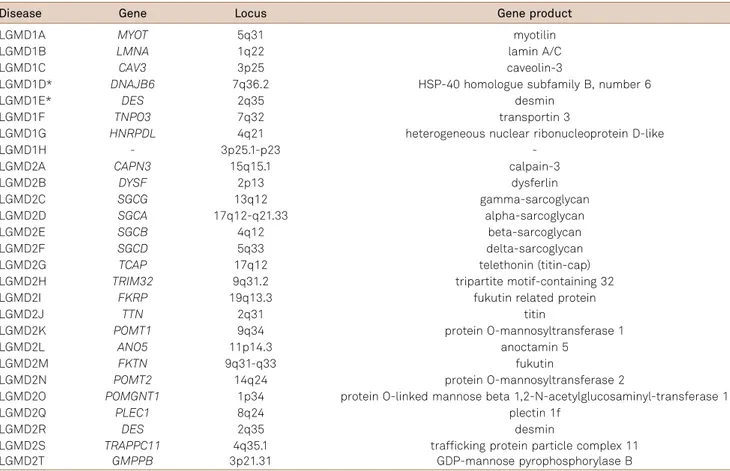
Limb girdle muscular dystrophies are known by the acronym “LGMD” (Limb Girdle Muscular Dystrophy). LGMD are classified by inheritance pattern as “LGMD1” for autosomal dominant and “LGMD2” for autosomal recessive disorders. They are subsequently classified with letters (LGMD1A, LGMD1B, etc.), in alphabetical order, in accordance to the chronological discovery of the cor-respondent mutated genes loci (Table 1)8,9. An updated
classification table is published every year8 ( freely available
at the URL: http://www.musclegenetable.fr). Until the Figure 1.Muscle biopsy morphologic patterns. Normal muscle biopsy: thin perimysial (arrow head) and endomysial (arrow) connective tissue (HE 200x) (A). Dystrophic muscle biopsies (B,C,D,E,F,G,H). Endomysial fibrosis (arrow), fiber splitting (arrow heads) of a LGMD2A patient (HE 100x) (B). Fiber hypertrophy (*), groups of atrophic fibers (arrow) of a LGMD2A patient (HE 100x) (C). Isolated atrophic fibers (arrow head), hypertrophy with fiber splitting (arrow) (D). Necrosis and phagocytosis (arrow) of a LGMD2A patient (D: HE 100x; E: HE 400x) (E). Fiber regeneration (arrow) of a LGMD2I patient (HE 200x) (F). Necrosis, phagocytosis, and regeneration foci (arrows) of a dysferlin-negative patient (reaction not shown) (HE 100x) (G). Hypertrophy with fiber splitting (arrows) and muscle tissue fat replacement (*) of a dysferlin-negative patient (HE 100x) (H).
publication of this article, 27 LGMD subtypes have been classified, and at least five additional entities are candidates for classification (Table 1). From these 27 LGMD subtypes,
19 are autosomal recessive (more than 90% of the patients) and eight are autosomal dominant (less than 10% of the patients) (Table 1).
Figure 3.Immunohistochemical findings (immunoperoxidase). A and B: Sarcolemmal integrity (arrow) (A), and dysferlin deficiency of a LGMD2B patient (arrow) (B) (insets are normal controls). Anti-spectrin RBC2/3D5 200x (A). Anti-dysferlin Ham1/7B6 200x (B). C, D, E, and F: gamma-sarcoglycan deficiency on muscle sarcolemma (arrow on E) of a LGMD2C patient. Anti-alpha-sarcoglycan (Adhalin Ad1/20A6) 200x (C). beta-sarcoglycan (BSarc/5B1) 200x (D). gamma-sarcoglycan (35DAG/21B5) 200x (E). Anti-delta-sarcoglycan (DSarc3/12C1) 200x) (insets are normal controls) (F). Spectrin (left arrow) and merosin (right arrow) serial frozen sections of the same fibers (*) with focal merosin deficiency of a LGMD2I patient (G). (G - left side: anti-spectrin (RBC2/3D5) 200x; G - right side: anti-merosin laminin alpha 2 chain (Mer3/22B2) 200x). Sarcomeric telethonin deficiency (arrow) of a LGMD2G patient (inset is normal control) (antitelethonin antibody G-11 sc-25327) (H).
Table 1.Classification of autosomal dominant (LGMD1) and autosomal recessive (LGMD2) limb girdle muscular dystrophies8,61.
Disease Gene Locus Gene product
LGMD1A MYOT 5q31 myotilin
LGMD1B LMNA 1q22 lamin A/C
LGMD1C CAV3 3p25 caveolin-3
LGMD1D* DNAJB6 7q36.2 HSP-40 homologue subfamily B, number 6
LGMD1E* DES 2q35 desmin
LGMD1F TNPO3 7q32 transportin 3
LGMD1G HNRPDL 4q21 heterogeneous nuclear ribonucleoprotein D-like
LGMD1H - 3p25.1-p23
-LGMD2A CAPN3 15q15.1 calpain-3
LGMD2B DYSF 2p13 dysferlin
LGMD2C SGCG 13q12 gamma-sarcoglycan
LGMD2D SGCA 17q12-q21.33 alpha-sarcoglycan
LGMD2E SGCB 4q12 beta-sarcoglycan
LGMD2F SGCD 5q33 delta-sarcoglycan
LGMD2G TCAP 17q12 telethonin (titin-cap)
LGMD2H TRIM32 9q31.2 tripartite motif-containing 32
LGMD2I FKRP 19q13.3 fukutin related protein
LGMD2J TTN 2q31 titin
LGMD2K POMT1 9q34 protein O-mannosyltransferase 1
LGMD2L ANO5 11p14.3 anoctamin 5
LGMD2M FKTN 9q31-q33 fukutin
LGMD2N POMT2 14q24 protein O-mannosyltransferase 2
LGMD2O POMGNT1 1p34 protein O-linked mannose beta 1,2-N-acetylglucosaminyl-transferase 1
LGMD2Q PLEC1 8q24 plectin 1f
LGMD2R DES 2q35 desmin
LGMD2S TRAPPC11 4q35.1 trafficking protein particle complex 11 LGMD2T GMPPB 3p21.31 GDP-mannose pyrophosphorylase B
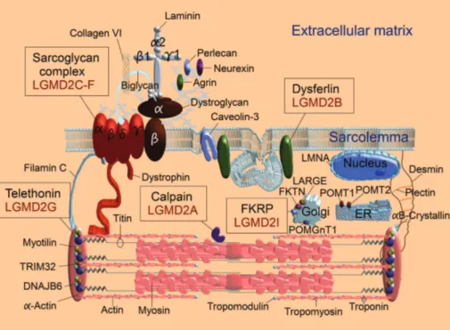
Genes or gene products from 21 autosomal dominant and recessive limb girdle muscular dystrophy subtypes are represented in Figure 4. Myotilin (LGMD1A), DNAJB6 (LGMD1D8), and TRIM32 (LGMD2H) are located on the
Z-disk of the sarcomere; myotilin and DNAJB6 are involved in protein aggregation4 and TRIM32 function is still
unknown4
. LMNA is the gene that codifies lamins A/C (LGMD1B), nuclear lamina associated proteins that provide structural support to the nuclear envelope. Caveolin-3 (LGMD1C) is a sarcolemma associated protein, component of the caveola4 (small invaginations of the plasma
mem-brane). Desmin (LGMD1E and LGMD2R) is an intermediate filament with protein aggregation function4. Plectin
(LGMD2Q) is a cytolinker, associated with desmin4. Titin
(LGMD2J) is a giant sarcomeric protein that spans from Z-disk to M-line; titin acts as an adjustable molecular spring during muscle contraction and it is essential for sar-comere assembly. POMT1 (LGMD2K), POMT2 (LGMD2N), POMGnT1 (LGMD2O), and FKTN (LGMD2M) are genes that codify putative glycosyltransferases4 located in the
endoplasmic reticulum (ER) (POMT1 and POMT2) and Golgi (POMGnT1 and FKTN) that are involved in the gly-cosylation of proteins of the extracellular matrix; these glycosyltransferases are important for the cytoskeleton-extracellular matrix link4.
The frequency of limb girdle muscular dystrophies varies worldwide10,11. The most common limb girdle muscular
dystrophy subtypes reported nowadays in Brazil are calpai-nopathy (LGMD2A) 32%, sarcoglycacalpai-nopathy (LGMD2C,
LGMD2D, LGMD2E, LGMD2F) 32%, dysferlinopathy (LGMD2B) 22%, fukutin related proteinopathy orFKRPathy (LGMD2I) 11%, and telethoninopathy (LGMD2G) 3%10,11.
This review will focus on the differential diagnosis of these eight limb girdle muscular dystrophy subtypes (Figure 4).
LGMD2A is caused by mutations in the calpain gene that codifies calpain, a proteolytic calcium activated enzyme, that in its inactive form, lies on titin and participates in sarco-mere repair and maintenance. LGMD2C-F (LGMD2C, LGMD2D, LGMD2E, LGMD2F) are caused by mutations in four genes that codify the structural proteins gamma, alpha, beta and delta-sarcoglycans, that are members of the dystro-phin associated glycoprotein complex and probably act as muscle membrane stabilizers during muscle contraction. LGMD2B is caused by mutations in the dysferlin gene that codifies dysferlin, a protein involved in vesicle-membrane fusion in order to repair membrane microlesions. LGMD2I is caused by mutations in theFKRPgene, that codifies fuku-tin related protein, a glycosyl transferase located in the Golgi complex and involved with glycosylation of diverse proteins such as alpha-dystroglycan and merosin (alpha 2 laminin) probably related to membrane stabilization. LGMD2G is caused by mutations in the TCAP (telethonin) gene, that codifies the protein telethonin that binds to titin, promoting sarcomere stabilization, during contraction, probably involved in sarcomere regulation and development (Figure 4).
The molecular diagnosis of a specific limb girdle mus-cular dystrophy subtype may be achieved in about 75% of the patients2.
HOW DO WE KNOW IF OUR PATIENTS HAVE A LIMB GIRDLE MUSCULAR DYSTROPHY?
It is important to be sure of the limb girdle muscular dys-trophy diagnosis in advance of subclassifying the disease. Therefore it is imperative to exclude both common and potentially treatable neuromuscular disorders (Figure 5).
The differential diagnosis of limb girdle muscular dystro-phies is performed through an integrated multiprofessional approach considering personal and familial history, physical examination with detailed manual muscle testing, laborato-rial, neurophysiological, and imaginological findings (Figure 5). There are some neuromuscular disorders that are relatively common (compared to limb girdle muscular dystrophies), that may be suspected from clinical findings. Among these com-mon disorders are dystrophinopathy, facioscapulohumeral muscular dystrophy, myotonic dystrophy (types 1 and 2), and spinal muscular atrophy12. Molecular studies usually
con-firm the diagnosis of these disorders12.
Even though dystrophinopathies are X-linked inherited disorders, they may be suspected in any patient with prox-imal weakness and increased serum creatine kinase levels. This is due to the high prevalence of dystrophinopathy in men and symptomatic women carriers compared to limb girdle muscular dystrophies12.
Facioscapulohumeral muscular dystrophy may present subtle facial weakness and clinical resemblance to limb gir-dle muscular dystrophy, demanding a high clinical suspicion level13. Asymmetric scapular weakness associated with distal
lower limb weakness, pronounced lower abdominal weak-ness, and lumbar hyperlordosis are suggestive of facioscapu-lohumeral muscular dystrophy in the differential diagnosis with limb girdle muscular dystrophy13. The diagnostic
con-firmation may be done, in the vast majority of the cases, with the detection of EcoRI and EcoRI/BlnI (with p13E-11 probe) restriction fragments between 10 and 35 Kb13,14. The deletion
of D4Z4 units in 4q35 impairs DNA methylation and alters the expression of theDUX4 gene13,14.
Patients with myotonic dystrophy type 1 may present subtle myotonia and it may be necessary to search for tenar muscle percussion myotonia15. Myotonic dystrophy type 2
presents clinical myotonia in less than half of the patients; myotonia may be absent even at neurophysiological invest-igation15. Few patients with myotonic dystrophy type 2
pre-sent cataracts15. Facial and jaw weakness, temporal atrophy,
and ptosis are common in myotonic dystrophy type 1 patients. These findings may be absent in myotonic dys-trophy type 2 patients, that may present predominant prox-imal weakness and clinical resemblance to limb girdle muscular dystrophy15. Distal weakness ( flexor digitorum
profundus), disabling myalgic pain, and prominent tremors, with normal or slightly elevated serum creatine kinase levels, may be the clues to myotonic dystrophy type 2, in the differ-ential diagnosis with limb girdle muscular dystrophy15.
Spinal muscular atrophy type 3 patients may achieve independent ambulation and present proximal weakness after childhood16. Spinal muscular atrophy type 4 symptoms
start after 18 years old with mild clinical course16. Muscle
weakness is usually symmetric, more proximal than distal, and worse in lower than in upper limbs16. Tongue
fascicula-tions and extremity tremor, as well as neurophysiological investigation, with neurogenic motor unit potentials, are very useful in the differential diagnosis with limb girdle mus-cular dystrophy. Molemus-cular confirmation is possible in most patients with the detection of exon 7 and 8SMN1gene dele-tion in subtypes 1, 2 and 316.
Potentially treatable neuromuscular disorders should be excluded from the differential diagnosis of limb girdle mus-cular dystrophy (Figure 5). These include myasthenia gravis; congenital myasthenic syndromes; glycogen-storage disease type 2 (Pompe disease); inflammatory (associated or not with rheumatologic diseases), endocrinological, toxic, meta-bolic, and mitochondrial myopathies, etc.
Myasthenia gravis and congenital myasthenic syndromes may be investigated in any patient with muscular weakness and ptosis that fluctuates along the day, affects active mus-cles and improves with rest17. Therefore, it is important to be
aware that there may be only slight weakness at the time of
Interdisciplinary neuromuscular patient evaluation Anamnesis including familial history
Complete neurological exam with Manual Muscle Testing Serum muscle enzymes; electromyogram
Suspicious of limb girdle muscular dystrophy
Rule out more common and potentially treatable diseases eg. dystrophinopathy, facioscapulohumeral muscular dystrophy, spinal muscular atrophy, myotonic dystrophy, myasthenic syndromes,
Pompe disease, inflammatory/ rheumatologic/ endocrine/ toxic/ metabolic/ mitochondrial myopathies, etc.
Muscle Image Studies (when available) or detailed Manual Muscle Testing
Muscle involvement pattern is used to direct molecular studies
Pathogenic mutations detected:
Definite diagnosis
Unavailable or normal molecular results
Liquid nitrogen frozen muscle biopsy: immunohistochemistry or immunofluorescence
Protein deficiency:
Phenotypic diagnosis
physical examination17. Some fatiguing maneuvers may
increase the chance to detect fatigue (such as sustained upgaze for 60 seconds, sustained abduction of the arms for 120 seconds, and sustained elevation of the legs in supine position for 90 seconds, among others17). Neurophysiological
examination with repetitive nerve stimulation is a valuable diagnostic tool in both myasthenia gravis and congenital myasthenic syndromes17. Serum anti-acetylcholine receptor
antibodies and anti-Musk antibodies may confirm myas-thenia gravis diagnosis.
Glycogen-storage disease type 2 (Pompe disease) may present prominent clinical resemblance to limb girdle mus-cular dystrophy, with predominant proximal weakness and autosomal recessive inheritance18. Some peculiar clinical
characteristics may suggest Pompe disease diagnosis such as respiratory insufficiency and increased tongue volume18.
Sometimes, respiratory insufficiency may manifest exclu-sively as increased susceptibility to respiratory infections, matinal headache, and daily somnolence, due to nocturnal hypoxia19. Diagnostic confirmation may be done through
alpha-glucosidase enzyme activity assays in both dried blood spots (DBS) and peripheral lymphocytes. Enzyme replacement therapy is available in Brazil and many parts of the world.
The differential diagnosis between limb girdle muscular dystrophy and inflammatory myopathies may be sometimes very difficult20. Both subacute rapid onset and negative
familial history suggest inflammatory myopathy20. Image
studies may be regarded with caution as hyperintensities observed on magnetic resonance STIR images may indicate both inflammation in myositis or they may precede fatty degeneration in muscular dystrophies20. Even in these cases,
image studies are a valuable tool to choose the most adequate muscle biopsy site (STIR hyperintensities on mag-netic resonance). Cryostat muscle sections may reveal either CD8 positive lymphocyte invasion of non-necrotic muscle fibers in polymyositis or perifascicular atrophy and mem-brane attack complex deposition in capillaries in dermato-myositis21. It is important to be aware that the absence of
classic signs of polymyositis or dermatomyositis does not exclude the diagnosis of inflammatory myopathy. Immune-mediated necrotizing myopathy may present necrotic mus-cle fibers with sparse inflammatory infiltrate, morphologi-cally mimicking muscular dystrophy20,21. Serologic
investigation should include the search for viral infections such as HTLV-1, HIV, HBV, and HCV, as well as autoantibo-dies for the differential diagnosis.
Toxic myopathies, associated with drugs, may be investi-gated in any patient without previous history of neuromus-cular disorder that develops myalgia, fatigue, weakness or myoglobinuria22. Toxic myopathies may be induced by statin
anticholesterol drugs; antirheumatic, anti-inflammatory, immu-nosuppressive drugs; nucleoside analogues; L-tryptophan
contaminated products, etc22. In Brazil, the most common
causes of toxic myopathies, in a neuromuscular reference center, were corticosteroids, propoxyphene, neuroleptics, zidovudine, and hypokalemiant diuretics23.
Serum creatine kinase and aldolase levels are often increased in limb girdle muscular dystrophies and are a valu-able diagnostic tool4
(Figure 5). Patients with congenital and mitochondrial myopathies usually present normal creatine kinase levels, and frequent ptosis. It is important to consider that limb girdle muscular dystrophy patients may present transaminase increase related to the muscular disease and not to any liver damage.
Neurophysiological exams reveal myopathic motor unit potentials in almost all limb girdle muscular dystrophy patients (Figure 5). In some dystrophinopathy, facioscapulo-humeral, and myotonic dystrophy patients, with typical clin-ical presentation, diagnostic molecular studies may be ordered by experienced clinicians at the first clinical exam-ination. In other patients, electroneuromyography may be very useful, demonstrating peculiar findings that may suggest specific disorders. Some examples are asymmetric muscular involvement in facioscapulohumeral muscular dystrophy, paraspinal involvement in Pompe disease, myo-tonic discharges in myomyo-tonic dystrophy / myomyo-tonic myopa-thies, distal involvement in hereditary distal myopamyopa-thies, and finger flexor weakness in inclusion body myositis, among others.
When personal and familial history, physical exam, neu-rophysiological studies and serum muscle enzymes point to the diagnosis of limb girdle muscular dystrophy, image studies may reveal particular muscular involvement pat-terns24 (Figures 6 and 7). Preoperative image studies
may guide muscle biopsy site selection and increase spe-cimen adequacy rate (Figure 8). When prominent muscle involvement ( fibrous and fat replacement) is observed on magnetic resonance image or computed tomography, cle ultrasound may locate the exact area of preserved mus-cle, suitable for histochemical and immunohistochemical studies (Figure 8).
represent fibrous and fat tissue replacement in advanced muscular disease and should be avoided for biopsy. A sur-gical pen was used for skin site demarcation, previous to muscle biopsy. Some years after muscle biopsy, molecular investigation became available, and revealed, in this patient, a c.390 G.A (p.Try130*) homozygous exon 3 mutation in the calpain (CAPN3) gene, confirming the diagnosis of cal-painopathy (LGMD2A).
When available, image studies should be performed prior to muscle biopsy. Careful manual muscle testing should be always performed. When image studies are unavailable, the muscle biopsy should be guided by detailed manual muscle
testing6. Grade 3 or (preferable) grade 4 Medical Research
Council (MRC) strength muscles should be selected6. This
practice may avoid“end-stage”muscle biopsies occurrence. Multiprofessional evaluation of clinical, laboratorial, neurophysiological, and image studies provide specific limb girdle muscular dystrophy subtype phenotypic diagnosis. Diagnostic confirmation is done according to available molecular or cryostat frozen immunohistochemical/immu-nofluorescence muscle biopsy studies (Figure 5).
WHY SHOULD WE PERFORM THE DIFFERENTIAL DIAGNOSIS OF SPECIFIC LIMB GIRDLE MUSCULAR DYSTROPHY SUBTYPES?
There are at least four main reasons to make specific limb girdle muscular dystrophy subtypes differential dia-gnosis: genetic counseling, cardiorespiratory risk evaluation, prognostic assumption, and future therapeutic possibilities. Adequate genetic counseling demands the correct identifica-tion of the specific inheritance pattern, either autosomal recessive or dominant (Table 1). Patients with sarcoglycano-pathy (LGMD2C, LGMD2D, LGMD2E, and LGMD2F), telethoninopathy (LGMD2G), and fukutin related proteino-pathy (LGMD2I) present increased risk of cardiac complica-tions1,25. Besides that, LGMD2I patients may present early
respiratory insufficiency, even while still ambulating. Patients with calpainopathy (LGMD2A) and dysferlinopathy (LGMD2B) characteristically present cardiac risk similar to the general population. Disease progression rate is usually slow in dysferlinopathy and telethoninopathy; moderate in calpainopathy and fukutin related proteinopathy, and rapid in sarcoglycanopathy4. Nowadays, there are many studies
considering specific therapeutic possibilities according to the particular limb girdle muscular dystrophy subtype. One example is the use of lymphocyte depletion treatments for dysferlinopathy26. There is great hope in future genetic
treat-ments directed to correct specific gene defects, as already tested in calpainopathy murine models27.
HOW CAN WE MAKE THE DIFFERENTIAL DIAGNOSIS OF COMMON LIMB GIRDLE MUSCULAR
DYSTROPHIES?
Calpainopathy (LGMD2A)
Calpainopathy is associated with pathogenic mutations in the calpain gene (CAPN3), located in 15q15.1, that codifies the enzyme calpain. Patients with calpainopathy usually pre-sent first symptoms around 13 years old, with an onset age range from 1 to 67 years28 (Table 2). First symptoms may
start either in lower or upper limbs. In a common presenta-tion, symptoms start almost simultaneously in lower and Figure 6.Schematic axial image diagram of part of the pelvis
upper limbs28. Muscular weakness usually starts in the lower
limbs and, in less than two years, it evolves to the upper limbs28. Disease progression is considered intermediate
among limb girdle muscular dystrophies and ambulation is usually lost around 35 years old or in the first 20 years of evolution3,28. No cardiac and respiratory complications
are common and life expectancy is similar to the general
population3. There is great phenotypic variability among
patients with calpainopathy, even among members of the same family with the same calpain mutation29. Physical exam
may reveal winging scapulae and there is usually no calf increase28. Serum creatine kinase is generally increased by 3
to 20 fold3,28. Image studies usually demonstrate prominent
and medial thigh (adductor magnus) (Figures 7 and 8) mus-cles involvement. In the legs, there is soleus and medial gas-trocnemius involvement24
. Rectus femoris is usually equally affected compared to other quadriceps femoris muscles24.
Muscle biopsy in calpainopathy usually presents variable grades of dystrophic abnormalities (Figures 1B, 1C, 1D, 1E). Severe endomysial fibrosis was the most striking abnormali-ty of a 16 years old female patient muscle biopsy, performed 6 years after her first symptoms (Figure 1B); later molecular studies demonstrated a c.2306G.A (p.Arg769Gln) exon 22 homozygous mutation in the calpain gene. Groups of atrophic fibers were the most prominent features of a 15 years old female patient submitted to muscle biopsy 6 years after her first symptoms (Figure 1C); later molecular studies
demonstrated a c.328C.T (p.Arg110) homozygous exon 2 mutation in the calpain gene. Slight variation in fiber caliber, with focal necrosis and phagocytosis (Figures 1D, 1E) were the principal changes of a 22 years old male patient submit-ted to muscle biopsy 8 years after his first symptoms; later molecular studies demonstrated a c.328C.T (p.Arg110) homozygous exon 2 mutation in the calpain gene. Even though the same mutation was found in the latter two patients, their muscle biopsies demonstrated distinctive morphologic abnormalities.
old female patient muscle biopsy (Figure 2E), 7 years after her first symptoms; later molecular studies revealed a c.390G.A (p.Try130*) homozygous exon 3 mutation in the calpain (CAPN3) gene. The same patient presented COX (cytochrome c oxidase) negative fibers on muscle biopsy (Figure 2H). COX negative fibers are not pathognomonic of primary respiratory chain disorders, and they may occasion-ally be found as a secondary phenomenon in other neuro-muscular disorders, as in this case.
Western blot studies may present abnormal calpain protein results29. Confirmatory molecular studies usually
demonstrate two pathogenic mutations in the calpain gene (CAPN3)28.
Calpain is a proteolytic calcium activated enzyme that, in its inactive form, lies on titin (giant protein with putative function of sarcomere stabilization during actin and myosin filaments contraction) (Figure 4). Therefore, it is believed that calpain plays an important role in sarcomere repair and maintenance30. The name
“calpain” derives from two words“calcium”and“papain”, describing its calcium activa-tion and its homology to the protease enzymes similar to “papain”(the papaya’s proteolytic enzyme)30.
Sarcoglycanopathy (LGMD2C, LGMD2D, LGMD2E, LGMD2F)
Patients with sarcoglycanopathies present pathogenic mutations in any of the four subtypes of sarcoglycan genes, that have gene products expressed in the sarcolemma:SGCG (LGMD2C), SGCA(LGMD2D),SGCB (LGMD2E), andSGCD (LGMD2F), respectively located in 13q12, 17q12-q21.33, 4q12, and 5q33, that codify gamma, alpha, beta, and delta-sarcoglycan proteins. Symptoms onset usually occurs
around 6 years old, with an age range from 1 to 30 years in all forms, except for LGMD2D, with first symptoms circa 13 years old31,32(Table 2).
Clinical presentation is generally similar to dystrophino-pathy with early predominant proximal weakness, frequent falls, Gowers maneuver, and rapid evolution to gait loss and cardiac complications25. Muscle enzymes are usually
5 to more than 40 times elevated1,3 (Table 2). Image
studies usually demonstrate severe involvement of adductor magnus and biceps femoris, and moderate involvement of vastus lateralis, vastus intermedius, vastus medialis, adduc-tor longus, semimembranosus and semitendinosus mus-cles24(Figure 7).
On the contrary to Duchenne muscular dystrophy patients, sarcoglycan patients present equal male and female frequency, winging scapula, preserved cognitive functions and early true calf hypertrophy (initial increased calf muscle volume contrary to the early calf fat replacement observed in Duchenne’s pseudohypertrophy). Diagnostic confirmation is done either by multiplex PCR (polymerase chain reaction) directed to the most common mutations according to the patient geographic area33or SGCG/ SGCA/SGCB/ SGCD
genes sequencing32.
Muscle biopsy in sarcoglycanopathy patients usually shows a dystrophic pattern. On the contrary to calpainopa-thy and telethoninopacalpainopa-thy, lobulated fibers are not a usual feature in sarcoglycanopathy. Even though, a 29 years old female patient, submitted to muscle biopsy 25 years after her first symptoms, presented lobulated fibers on her muscle biopsy (Figure 2G). Later molecular investigation demon-strated a c.525delT homozygous exon 6 mutation in the gamma-sarcoglycan (SGCG) gene. In this case, lobulated Table 2.Differential diagnosis of the most common autosomal recessive limb girdle muscular dystrophy subtypes reported in Brazil10,11.
Clinical and laboratorial findings LGMD
2A 2C-F* 2B 2I 2G
Mean onset age (years)28,32,35,36,45,49 13 6 19 19 12
Typical onset age range (years)28,6,35,45,11 8-15 6-8 17-25 2-40 9-15
Wide onset age range (years)28,32,36,37,46,51 1-67 1-30 1-58 2-50 1-20
Disease progression rate3 ++ +++ + ++ +
Subacute start mimicking polymyositis3,38,45,46,49 -/+ - ++ -
-Ambulation loss (age in years)28,11,6,38,45,49 21-40 12-16 18-58 .12 .30
Cardiomyopathy3,28,25,1,38,45,46,11 - +++ - + +
Early respiratory abnormality28,32,1,38,45,46,11 - - - +
-Upper and lower limbs interval (years)28,6,38,45,11
,2** ** .6** ** **
Increased calf1,3,28,11,32,35,45,46 -/+ ++ -/+ ++ ++
Winged scapula3,28,11,32,38,45,46 ++ ++ - -/+ +
“Dystrophinopathy-like”phenotype3,11,32,38,45,46 -/+ ++ - ++
-Serum creatine kinase (CPK) times increase1,51,62 3-20x 5-40x 10-70x 10-20x 1-30x
Distal myopathy phenotype1,35,38,11 - - -/+ - -/+
Contractures1,45,11 + + - -
-Onset age: age at first symptoms. 2A: LGMD2A: calpainopathy. 2C-F: LGMD2C, LGMD2D, LGMD2E, LGMD2F: sarcoglycanopathy. 2B: LGMD2B: dysferlinopathy. 2I: LGMD2I: fukutin related proteinopathy. 2G: LGMD2G: telethoninopathy. Grading:“+”slow/slight,“++”intermediate/moderate,“+++”
fibers could be, perhaps, related to the long duration of her symptoms.
When molecular tests are unavailable, a phenotypic dia-gnosis may be rendered through immunohistochemical studies on muscle biopsy frozen sections, directed to the four gene products, using commercially available antibodies to gamma, alpha, beta and delta-sarcoglycan proteins. A 29 years old male patient, submitted to muscle biopsy 17 years after his first symptoms, presented complete gamma-glycan immunohistochemical deficiency on muscle sarco-lemma (Figure 3E) with preserved expression of the other sarcoglycan proteins (Figures 3C, 3D, 3F). Later molecular studies demonstrated a c.525delT homozygous exon 6 muta-tion in the gamma-sarcoglycan (SGCG) gene.
There is no universal correlation between the sarcoglycan subtype immunohistochemical deficiency and the sarcogly-can mutated gene. A mutation in one gene may generate a secondary deficiency of the other proteins of the com-plex32. Therefore, it is not possible to precisely infer the
specific sarcoglycanopathy subtype, based on the immuno-histochemical finding of deficiency of one specific protein32.
The four sarcoglycans, associated with LGMD2C-F sarco-glycanopathies, are “sarco-lemmal” “glyco-proteins”, that are components of the dystrophin associated glycoprotein complex. They probably act as muscle membrane stabilizers during muscle contraction34(Figure 4).
Dysferlinopathy (LGMD2B)
Dysferlinopathy is caused by pathogenic mutations in the dysferlin gene (DYSF), located in 2p13, that codifies the protein dysferlin. First symptoms usually begin in a narrow age range around 19 years old, with exceptional cases starting from birth to 58 years old35,36,37(Table 2). Different
from other limb girdle muscular dystrophies, subacute pre-sentation may occur in about 25% of the patients. It may simulate both clinically and histologically inflammatory myopathies such as polymyositis38, as observed on a muscle
biopsy of a 16 years old dysferlin-negative female patient (Figure 2C).
Some dysferlinopathy patients present predominant dis-tal weakness, others proximal and disdis-tal weakness38. Rare
patients may present predominant anterior compartment distal weakness39. Besides that, there are oligo symptomatic
patients with creatine kinase increase38. Usually, there is
lower limb weakness that, after a period of about 6 years, is followed by upper limb weakness, but this interval may vary from 1 to 16 years35. Even though decreased calf volume
is the most common clinical presentation, calf volume increase may be observed in about 28% of the patients35.
A frequent clinical finding on physical examination is the relative deltoid muscle volume preservation, compared to biceps brachialis lower third35. Clinical and pathological
exam revealed prominent distal biceps brachialis atrophy
of a 18 years old dysferlin-negative male patient. The worse distal biceps brachialis involvement may be noticed on mus-cle biopsy (Figures 2A and 2B). At this time of the investiga-tion it is necessary to remind that deltoid volume preservation may be observed in facioscapulohumeral mus-cular dystrophy, that has already been excluded from the dif-ferential diagnosis (Figure 5).
Muscle enzymes are usually excessively elevated (more than 10 to 70 fold reference values)1,3(Table 2). Image studies
may demonstrate diffuse involvement of both anterior and posterior thigh compartments, with moderate involvement of the vastus lateralis, vastus medialis, adductor magnus, adductor longus, biceps femoris, semitendinosus, semimem-branosus, soleus, medial and lateral gastrocnemius muscles24
(Figure 7). When magnetic resonance imaging is performed, fat suppressed T2 and STIR weighted sequences may dem-onstrate hyperintensities, difficult to differentiate from inflammatory myopathies40.
Muscle biopsy in dysferlinopathy patients usually shows a dystrophic muscle pattern and the variability in morpholo-gic findings may be related to the duration of symptoms. Necrosis, phagocytosis, and regeneration foci were the most prominent muscle biopsy features of a 20 years old female dysferlin-negative patient, submitted to muscle biopsy 4 years after her first symptoms (Figure 1G). On the other hand, fiber caliber variation, atrophy, hypertrophy with fiber splitting were severe on a muscle biopsy of a 33 years old dysferlin-negative female patient, submitted to muscle biopsy 15 years after disease onset (Figure 1H).
Phenotypic diagnosis is usually suggested through com-plete or partial38 dysferlin deficiency with commercially
available antibodies on muscle biopsy, as observed on a 33 years old female patient muscle biopsy (Figures 3A and 3B). Dysferlin deficiency may also be detected through peri-pheral monocytes Western blot38,41. Genotypic diagnosis is
confirmed through dysferlin gene sequencing38,41.
Dysferlin is a protein that anchors on the sarcoplasmic membrane and it is necessary to repair membrane microle-sions42 (Figure 4). This occurs through vesicle formation
and fusion with the sarcolemma42(Figure 4). Transmission
electron microscopy in patients with dysferlinopathy demonstrates plasma membrane microlesions and subsarco-lemmal vesicle accumulation43. The name
“dysferlin” is derived from“dys-”from“dystrophy”and“fer-lin”from its homology to the“fer-1”( fertility factor 1), involved in mem-brane fusion during spermatogenesis44.
FKRPathyor Fukutin related proteinopathy
(LGMD2I)
years, with a mean onset age around 19 years45,46(Table 2).
Most patients present clinical symptoms and signs that mimic dystrophinopathy (both “Duchenne-like” and “Becker-like” cases), with predominant proximal muscle weakness and calf volume increase in about 76% of the patients45,46. Other muscles may present increased volume
such as the brachioradialis45,46
. Unexpected to limb girdle muscular dystrophies, about 20% of the patients with fukutin related proteinopathy may present facial weakness45.
About 30% (15% to 46%) of the patients present cardiac complications45,46. Respiratory abnormalities are common
and occur in about 65% of the cases, even in ambulant patients, on the contrary to most muscular dystrophies45,46
(Table 2). It is important to remind that Pompe disease (gly-cogen storage disease type 2) may present respiratory insuf-ficiency and has already been excluded from the differential diagnosis (Figure 5).
Muscle biopsy may demonstrate dystrophic pattern and secondary merosin deficiency45 (Figures 1F and 3G).
Muscle biopsy may present, in some patients, inflammatory infiltrate63, as observed on a 11 years old female patient
submitted to muscle biopsy 4 years after her first symp-toms (Figure 2D); later molecular studies demonstrated two pathogenic mutations, c.826 C.A (p.Leu276Ile) and c.1384 C.T (p.Pro462Ser), in the fukutin related protein (FKRP) gene.
Serum creatine kinase is usually elevated. Image studies may demonstrate severe involvement of the posterior thigh muscles, mainly biceps femoris, and adductor muscles24
(Figure 7). There is usually slight involvement of the quad-riceps femoris with relative preservation of the rectus femoris24. Moderate involvement of the posterior leg
mus-cles, with abnormalities of both medial and lateral gas-trocnemius may be observed24. At this time of the
investigation, molecular studies may be performed in accordance to muscle involvement pattern (Figures 5 and 7). Fukutin related protein is located in the Golgi complex and it is involved with glycosylation of diverse proteins such as alpha-dystroglycan and merosin (alpha 2 laminin)47
(Figure 4). Alpha-dystroglycan connects extracellular mem-brane proteins, such as merosin, with beta-dystroglycan that resides in the sarcolemma and is part of the dystrophin associated glycoprotein complex (Figure 4). Therefore, the putative function of the fukutin related protein is to promote the correct glycosylation of extracellular matrix proteins, essential to membrane stabilization during muscle contrac-tion. The name “fukutin related protein” derives from its proximity to the “fukutin protein” in the Golgi complex. The name “fukutin” is an acknowledgment to Yukio Fukuyama, that described the first cases of Fukuyama con-genital muscular dystrophy, associated with mutations in the fukutin (FKTN) gene, later related to fukutinopathy (LGMD2M)2,3,4,48.
Telethoninopathy (LGMD2G)
Telethoninopathy is caused by mutations in the teletho-nin (TCAP)gene, located in 17q12, that codifies the protein telethonin. Symptoms usually start between 9 and 15 years old; exceptionally there may be congenital and around 20 years old onset49,50,51 (Table 2). Ambulation loss usually
occurs around the fourth decade of life51
. Patients usually present proximal and distal muscular weakness. Early foot drop, related to tibialis anterior muscle weakness, may be the first disease presentation51. Cardiac abnormalities are
common11. Image studies may demonstrate diffuse muscle
involvement of the thigh (Figure 7). Severe adductor mag-nus, biceps femoris, semitendinosus, semimembranosus, and tibialis anterior muscles involvement may be observed, as well as moderate involvement of the vastus lateralis, vas-tus intermedius, recvas-tus femoris, vasvas-tus medialis, adductor longus and gracilis muscles (Figure 7)51,52,53. Muscle biopsy
may demonstrate dystrophic pattern with rimmed vacuoles51. Lobulated fibers have been commonly described
on telethoninopathy patients and they were observed on the muscle biopsy of a 54 years old female patient muscle biopsy, 46 years after her first symptoms (Figure 2F); molecular investigation, on a research basis, revealed a c.157C.T (Q53X) homozygous mutation in the telethonin (TCAP) gene (patient previously described)51.
A phenotypic diagnosis of telethoninopathy may be per-formed through immunofluorescence, Western blot or immunohistochemistry (Figure 3H), with commercially available antibodies. Diagnostic confirmation may be per-formed through direct sequencing of the telethonin gene.
Telethonin binds to titin and received the name of“ titin-cap”(Figure 4). Titin is a giant elastic protein that extends from the“Z”disk to the“M”line in the sarcomere, promot-ing sarcomere stabilization durpromot-ing actin and myosin slidpromot-ing. The putative function of telethonin is associated with sarco-mere regulation and development mechanisms54. Telethonin
received its name after its identification, in a cooperative brazilian-italian research, that received donations from the Italian“Telethon”(“tele”from“television”and“thon”from “marathon”)49,51.
SUMMARY OF MUSCLE INVOLVEMENT PATTERNS OF COMMON RECESSIVE LIMB GIRDLE MUSCULAR DYSTROPHIES
Image studies and schematic diagrams are very useful for the differential diagnosis of common autosomal recessive limb girdle muscular dystrophies (Figures 6 and 7)24,40.
posterior thigh, soleus and medial gastrocnemius muscles (LGMD2A) (Figure 7A). Magnetic resonance image of a 16 years old, male patient, 4 years after his first symptoms, with homozygous c.525delT (p.F175fs) exon 6 mutation in the gamma-sarcoglycan (SGCG) gene, showed involvement of the glutei, adductor magnus, biceps femoris, and quadriceps femoris muscles (LGMD2C)24,40
(Figure 7B). Computed tomography image of a 23 years old female patient, 4 years after her disease onset, with complete immunohistochemical dysferlin deficiency in muscle biopsy frozen sections, pre-sented moderate diffuse involvement of vastus lateralis, vastus medialis, adductors, posterior thigh and posterior leg muscles (LGMD2B) (Figure 7C). Magnetic resonance image of a 11 years old female patient, 4 years after her first symptoms, with two pathogenic c.826C.A (p.Leu276Ile) and c.1384C.T (p. Pro462Ser) mutations in the fukutin related protein (FKRP) gene, showed severe adductor magnus and biceps femoris muscles involvement, with rectus femoris signal preservation (LGMD2I)24,40 (Figure 7D). Computed tomography image of
a 54 years old female patient, 46 years since her first symp-toms, with complete immunohistochemical and immuno-fluorescence telethonin deficiency and c.157C.T mutation in the telethonin (TCAP) gene (LGMD2G), disclosed severe diffuse involvement of pelvis, thigh and legs (Figure 7E). The LGMD2G schematic diagram was based on previous LGMD2G publications, with diffuse thigh and early tibialis anterior involvement51,52,53.
CONCLUSIONS
In conclusion later studies describing the molecular mechanisms (Figure 4) involved in limb girdle muscular dys-trophies will be necessary to elucidate the physiopathogenic mechanisms of these diseases4,9,29,34,42,57,58,59. The precise
differ-ential diagnosis of limb girdle muscular dystrophies may be achieved through an integrated clinical, laboratorial, neurophys-iological and image studies approach. Immunohistochemical muscle biopsy frozen section analysis contributes to the phenotypic diagnosis of sarcoglycanopathy, dysferlinopathy, and telethoninopathy; it may reveal secondary merosin defi-ciency in fukutin related proteinopathy. Muscle Western blot may reveal calpain decrease in calpainopathy.
Muscle image studies are very useful to select muscle biopsy site in order to provide specimen adequacy. Besides that, careful manual muscle testing and image studies may direct confirmatory molecular studies. It is necessary to exclude most common or potentially treatable neuromuscu-lar conditions prior to the diagnosis of limb girdle muscuneuromuscu-lar dystrophy. The differential diagnosis of a specific limb girdle muscular dystrophy subtype is important for adequate gen-etic counseling, intervention in treatable cardiac and respir-atory complications, and prognostic considerations. There is a hope that, in the future, the diagnosis of a specific limb gir-dle muscular dystrophy subtype may improve”quality of life, with the advent of specific new therapies.
References
1. Norwood FL, de Visser M, Eymard B, Lochmüller H, Bushby K and Members of EFNS Guideline Task Force. EFNS guideline on diagnosis and management of limb girdle muscular dystrophies. Eur J Neurol 2007;14:1305-1312.
2. Bushby K. Diagnosis and management of the limb girdle muscular dystrophies. Pract Neurol 2009;9:314-323.
3. Nigro V, Aurino S, Piluso G. Limb girdle muscular dystrophies: update on genetic diagnosis and therapeutic approaches. Curr Opin Neurol 2011;24:429-436.
4. Mitsuhashi S, Kang PB. Update on the genetics of limb girdle muscular dystrophy. Semin Pediatr Neurol 2012;19:211-218.
5. Dubowitz V, Sewry C. Muscle biopsy. A practical approach. Third edition. Printed in China: Saunders Elsevier, 2007:1-600.
6. Engel AG, Franzini-Armstrong C. Myology 3rd ed. New York: McGraw-Hill, 2004:1-1960.
7. Sewry CA, Molnar MJ. Chapter 5. Histopathology and immunoana-lysis of muscle. In: Karpati G, Hilton-Jones D, Bushby K, Griggs RC. (EDS) Disorders of Voluntary Muscle 8th edition. Cambridge: Cambridge University Press, 2010:93-127.
8. Kaplan JC, Hamroun D. The 2014 version of the gene table of monogenic neuromuscular disorders (nuclear genome). Neuromuscul Disord 2013;23:1081-1111.
9. Torella A, Fanin M, Mutarelli M, et al. Next-generation sequencing identifies transportin 3 as the causative gene for LGMD1F. PLoS One 2013;8:e63536:1-7.
10. Zatz M, de Paula F, Starling A, Vainzof M. The 10 autosomal recessive limb-girdle muscular dystrophies. Neuromuscul Disord 2003;13:532-544.
11. Vainzof M, Bushby K. Chapter 11. Muscular dystrophies presenting with proximal muscle weakness. In: Karpati G, Hilton-Jones D, Bushby K, Griggs RC.(EDS) Disorders of Voluntary Muscle 8th edition. Cambridge: Cambridge University Press, 2010:230-256.
12. Norwood FL, Harling C, Chinney PF, Eagle M, Bushby K, Straub V. Prevalence of genetic muscle disease in Northern England: in-depth analysis of a muscle clinic population. Brain 2009;132:3175-3186.
13. Tawil R, Van Der Maarel SM. Facioscapulohumeral muscular dystrophy. Muscle Nerve 2006;34:1-15.
14. Sacconi S, Camaño P, de Greef JC, et al. Patients with a phenotype consistent with facioscapulohumeral muscular dystrophy display genetic and epigenetic heterogeneity. J Med Genet 2012;49:41-46.
15. Udd B, Krahe R. The myotonic dystrophies: molecular, clinical, and therapeutic challenges. Lancet Neurol 2012;11:891-905.
16. D’Amico A, Mercuri E, Tiziano FD, Bertini E. Spinal muscular atrophy. Orphanet J Rare Dis 2011;6:71.
17. Rowin J. Approach to the patient with suspected myasthenia gravis or ALS: a clinician’s guide. Continuum Lifelong Learning Neurol 2009;15:13-34.
18. van der Ploeg AT, Reuser AJ. Pompe’s disease. Lancet 2008;372 (9646):1342-1353.
19. Bembi B, Cerini E, Danesino C, et al. Diagnosis of glycogenosis type II. Neurology 2008;71:(Suppl)S4-S11.
20. Benveniste O, Romero NB. Myositis or dystrophy? Traps and pitfalls. Presse Med 2011;40:249-255.
with the exception of inclusion body myositis, 10-12 October 2003, Naarden , The Netherlands. Neuromuscul Disord 2004;14:337-345.
22. Dalakas MC. Toxic and drug-induced myopathies. J Neurol Neurosurg Psychiatry 2009;80:832-838.
23. Scola RH, Pereira ER, Lorenzoni PJ, Werneck LC. Toxic myopathies: muscle biopsy features. Arq Neuropsiquiatr 2007;65:82-86.
24. Straub V, Carlier PG, Mercuri E. TREAT-NMD workshop: pattern recog-nition in genetic muscle diseases using muscle MRI: 25-26 February 2011, Rome, Italy. Neuromuscul Disord 2012;22:(Suppl)S42-S53.
25. Fanin M, Melacini P, Boito C, Pegoraro E, Angelini C. LGMD2E patients risk developing dilated cardiomyopathy. Neuromuscul Disord 2003;13:303-309.
26. Lerario A, Cogiamanian F, Marchesi C, et al. Effects of rituximab in two patients with dysferlin-deficient muscular dystrophy. BMC Musculoskelet Disord 2010;11:157.
27. Bartoli M, Roudaut C, Martin S, et al. Safety and efficacy of AAV-mediated calpain 3 gene transfer in a mouse model of limb-girdle muscular dystrophy type 2A. Mol Ther 2006;13:250-259.
28. Sáenz A, Leturcq F, Cobo AM, et al. LGMD2A: genotype-phenotype correlations based on a large mutational survey on the calpain 3 gene. Brain 2005;128:732-742.
29. Zatz M, Starling A. Calpains and disease. N Engl J Med 2005;352:2413-2423.
30. Beckmann JS, Spencer M. Calpain 3, the“gatekeeper”of proper sarcomere assembly, turnover and maintenance. Neuromuscul Disord 2008;18:913-921.
31. Eymard B, Romero NB, Leturcq F, et al. Primary adhalinopathy (alpha-sarcoglycanopathy): clinical, pathologic, and genetic correla-tion in 20 patients with autosomal recessive muscular dystrophy. Neurology 1997;48:1227-1234.
32. Klinge L, Dekomien G, Aboumousa A, et al. Sarcoglycanopathies: can muscle immunoanalysis predict the genotype? Neuromuscul Disord 2008;18:934-941.
33. Gouveia TL, Paim JF, Pavanello RC, Zatz M, Vainzof M. Sarcoglycanopathies: a multiplex molecular analysis for the most common mutations. Diagn Mol Pathol 2006;15:95-100.
34. Ozawa E, Mizuno Y, Hagiwara Y, Sasaoka T, Yoshida M. Molecular and cell biology of the sarcoglycan complex. Muscle Nerve 2005;32:563-576.
35. Rosales XQ, Gastier-Foster JM, Lewis S, et al. Novel diagnostic features of dysferlinopathies. Muscle Nerve 2010;42:14-21.
36. Paradas C, González-Quereda L, De Luna N, et al. A new phenotype of dysferlinopathy with congenital onset. Neuromuscul Disord 2009;19:21-25.
37. Takahashi T, Aoki M, Suzuki N, et al. Clinical features and a mutation with late onset of limb girdle muscular dystrophy 2B. J Neurol Neurosurg Psychiatry 2013;84:433-440.
38. Nguyen K, Bassez G, Krahn M, et al. Phenotypic study in 40 patients with dysferlin gene mutations: high frequency of atypical pheno-types. Arch Neurol 2007;64:1176-1182.
39. Illa I, Serrano-Munuera C, Gallardo E, et al. Distal anterior compartment myopathy: a dysferlin mutation causing a new muscular dystrophy phenotype. Ann Neurol 2001;49:130-134.
40. Degardin A, Morillon D, Lacour A, Cotten A, Vermersch P, Sojkovic T. Morphologic imaging in muscular dystrophies and inflammatory myopathies. Skeletal Radiol 2010;39:1219-1227.
41. Gallardo E, de Luna N, Diaz-Manera J, et al. Comparison of dysferlin expression in human skeletal muscle with that in monocytes for the diagnosis of dysferlin myopathy. PLoS One 2011;6:e29061:1-9.
42. Han R. Muscle membrane repair and inflammatory attack in dysferlinopathy. Skelet Muscle 2011;1:10:1-8.
43. Selcen D, Stilling G, Engel AG. The earliest pathologic alterations in dysferlinopathy. Neurology 2001;56:1472-1481.
44. Bashir R, Britton S, Strachan T, et al. A gene related to Caenorhabditis elegans spermatogenesis factor fer-1 is mutated in limb-girdle muscular dystrophy type 2B. Nat Genet 1998;20:37-42.
45. Poppe M, Cree L, Bourke J, et al. The phenotype of limb-girdle muscular dystrophy type 2I. Neurology 2003;60:1246-1251.
46. Boito CA, Melancini P, Vianello A, et al. Clinical and molecular characterization of patients with limb-girdle muscular dystrophy type 2I. Arch Neurol 2005;62:1894-1899.
47. Esapa CT, Benson MA, Schröder JE, et al. Functional requirements for fukutin-related protein in the Golgi apparatus. Hum Mol Genet 2002;11:3319-3331.
48. Voit T, Tomé FMS. Chapter 44. The congenital muscular dystrophies. In:Engel AG, Franzini-Armstrong C. Myology 3rd ed. New York: McGraw-Hill, 2004:1203-1238.
49. Moreira ES, Wiltshire TJ, Faulkner G, et al. Limb-girdle muscular dystrophy type 2G is caused by mutations in the gene encoding the sarcomeric protein telethonin. Nat Genet 2000;24:163-166.
50. Ferreiro A, Mezmezian M, Olivé M, et al. Telethonin-deficiency initially presenting as a congenital muscular dystrophy. Neuromuscul Disord 2011;21:433-438.
51. Paim JF, Cotta A, Vargas AP, et al. Muscle phenotypic variability in limb girdle muscular dystrophy 2G. J Mol Neurosci 2013;50:339-344.
52. Olivé M, Shatunov A, Gonzalez L, et al. Transcription-termination mutation in telethonin causing autosomal recessive muscular dystrophy type 2G in a European patient. Neuromuscul Disord 2008;18:929-933.
53. Negrão L, Matos A, Geraldo A, Rebelo O. Limb-girdle muscular dystrophy in a Portuguese patient caused by a mutation in the telethonin gene. Acta Myol 2010;29:21-24.
54. Gregorio CC, Trombitás K, Centner T, et al. The NH2 terminus of titin spans the Z-disc: its interaction with a novel 19-kD ligand (T-cap) is required for sarcomeric integrity. J Cell Biol 1998;143:1013-1027.
55. Valle G, Faulkner G, De Antoni A, et al. Telethonin, a novel sarcomeric protein of heart and skeletal muscle. FEBS Lett 1997;415:163-168.
56. Sandell SM, Mahjneh I, Palmio J, Tasca G, Ricci E, Udd BA. ‘Pathognomonic’ muscle imaging findings in DNAJB6 mutated LGMD1D. Eur J Neurol 2013;20:1553-1559.
57. Mercuri E, Muntoni F. Muscular dystrophies. Lancet 2013;381:845-860.
58. Dalakas MC, Park KY, Semino-Mora C, Lee HS, Sivakumar K, Goldfarb LG. Desmin myopathy, a skeletal myopathy with cardiomyopathy caused by mutations in the desmin gene. N Engl J Med 2000;342:770-780.
59. Reed UC. Congenital muscular dystrophy. Part I: a review of phenotypical and diagnostic aspects. Arq Neuropsiquiatr 2009;67:144-168.
60. Worman HJ. Nuclear lamins and laminopathies. J Pathol 2012;226:316-325.
61. Vieira NM, Naslavsky MS, Licinio L, et al. A defect in the RNA-processing protein HNRPDL causes limb-girdle muscular dystrophy 1G (LGMD1G). Hum Mol Genet 2014 [Epub ahead of print].
62. Nigro V, Savarese M. Genetic basis of limb-girdle muscular dystrophies: the 2014 update. Acta Myol 2014;33:1-12.