799
CLINICS 2007;62(6):799-802
LETTER TO THE EDITOR
Serviço de Endoscopia Digestiva do Hospital das Clínicas da Faculdade de Medicina da Universidade de São Paulo, São Paulo, SP-Brazil
Email: [email protected]
ECHOGUIDED HEPATICO-GASTROSTOMY: A CASE
REPORT
Everson L.A. Artifon, Dalton M. Chaves, Shinichi Ishioka, Thiago F. Souza, Sérgio E. Matuguma, Paulo Sakai
INTRODUCTION
Endoscopic retrograde cholangiopancreatography (ERCP) with biliary drainage has become the gold stand-ard procedure for decompression of obstructed biliary ducts.1,2 Skilled endoscopists are expected to achieve
suc-cessful drainage in 90% to 95% of cases.3 However,
anatomic variation, such asperiampullary diverticulum, tumor invasion and surgical diversions are all situations that may result in failure.4,5 In cases where such failure occurs,
available options include repeat ERCP at a tertiary care center,6 percutaneous transhepatic drainage (PTD),7,8 and
surgery.9 PTC has complication rate of up to 32%, with
pos-sible fistula formation, cholangitis, peritonitis, empyema, hematoma, and liver abscesses.10,11 Surgery, although
de-finitive, is associated with increased morbidity and mor-tality.12 The echoguided hepatico-gastrostomy technique
was first described in 2003 by Giovannini et al.13 and may
be seen as a variation of the intrahepatic approach, but without selective drainage through the ampulla.
In terms of a minimally invasive concept and low com-plication rate, this is the first presentation of hepatico-gas-trostomy drainage using both endoscopic ultrasound and fluoroscopy guidance performed at the Gastrointestinal En-doscopy Unit in the Hospital das Clínicas – University of São Paulo School of Medicine.
PATIENT AND METHOD
Preparation
The patient was placed in a supine decubitus position, and 10% lidocaine solution was sprayed into the pharynx for local anesthesia. An adequate level of sedation was managed by an anesthesiologist, who administered intra-venous midazolan and/or propofol.
Case report
An 82-year-old man presented with a history of discom-fort in the right upper quadrant of the abdomen, weight loss and jaundice in the last 2 months. Laboratory data showed abnormal liver functions with serum bilirubin of 31mg/dl. US and CT scan showed a large hilar solid mass, with di-lated intrahepatic bile ducts and portal vein invasion. ERCP showed an irregular stop at common hepatic duct and the guide wire attempts to pass through the hilar stenosis did not have success. The patient was judged to have an inop-erable locally advanced hilar cancer and indicated for a non-surgical palliative drainage by means of echoguided hepatico-gastrostomy assisted with fluoroscopy.
Technique
The procedure was performed using a linear array echoendoscope with a working channel with 3.8mm (GF-UCT-160, Olympus – Mellvile – NY, USA). This instru-ment is coupled with an ultrasound processor machine. A 19 – gauge needle (EUSN-19T, Wilson-Cook, Winston Salem, NC) was used. Although correct orientation of the 19-gauge needle is more challenging than the 22-gauge, it permits easy passage of a 0.035 – inch guidewire.
The fistula between the gastrointestinal tract and the intrahepatic biliary tree can be enlarged using either a 4 or 6mm wire-guided balloon catheter (Max Force, microvasive); 6 or 7 French bougie (SBDC-6 or 7, Wilson Cook), or passage of the covered biliary metallic stent (Wallstent 60 / 100mm, Boston Scientific) directly through the gastric fistula as done in the present case.
800
CLINICS 2007;62(6):799-802 Echoguided hepatico-gastrostomy: a case report
Artifon ELA et al.
was obtained (figure 3). An important technique detail is to adequate the tip of the needle into the dilated intrahe-patic duct punctured synchronized with diaphragm breath movements. The needle is exchanged for a guidewire and the image was controlled by both EUS and fluoroscopy guidance. A flexible metallic stent was passed over the guide through gastric fistula to the left intra-hepatic duct without any dilation procedure. The deployment of metal-lic stent occurred successfully and bile flow to stomach demonstrated the method’s effectiveness. However as the patient has an esophageal-gastric hernia, the gastric open-ing of metallic stent occurred in the cardia, very close to the esophageal-gastric junction (figure 4). Up to the 8th
fol-low up day, the patient did not present abdominal pain, his liver enzymes and bilirubin decreased significantly and his conventional abdominal US was normal with no bile leak-age. At the 30th follow-up day, the patient did not present
jaundice and a normal conventional US was obtained. The patient presented an uneventful recovery.
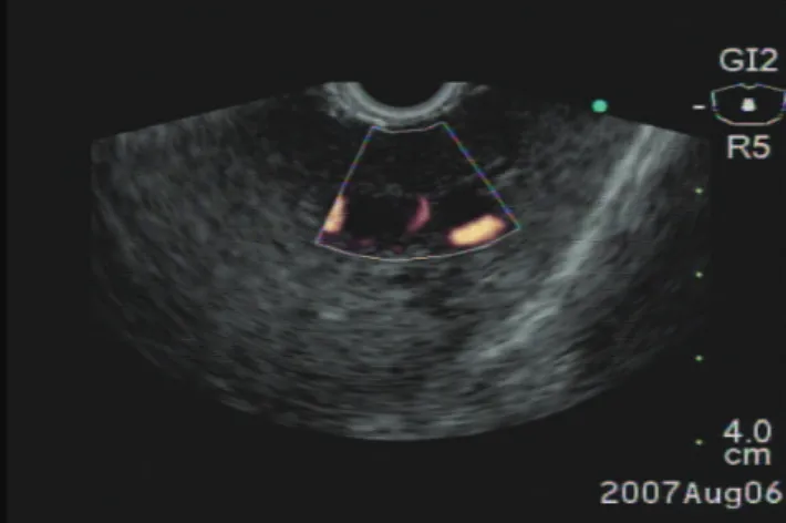
Figure 1 – EUS color Flow Doppler before performing an intrahepatic approach.
Figure 2 – EUS image demonstrating a 19 – gauge needle puncturing the left hepatic biliary branch undcer endosonography, intrahepatic approach.
Figure 3 – Fluoroscopic image demonstrating intrahepatic cholangiogram after contrast media injection under EUS guidance
Figure 4b – Endoscopic view of the metallic stent opened in the cardia.
Figure 4a – Fluoroscopy image demonstrating the metallic stent opened.
DISCUSSION
Burmester et al.14 attempted ecoguided
place-801
CLINICS 2007;62(6):799-802 Echoguided hepatico-gastrostomy: a case report
Artifon ELA et al.
ment of 8.5 French stents in three and with one bile leak as a complication. In two cases, Mallery et al.15 performed
EH by cannulation adjacent to a EUS-placed wire, with a minor complication of wire passage outside the bile duct lumen. Puspok et al.16 reported successful (lower than
100%) EH with no immediate complications. Another bile leak was reported by Bories and Will et al.17,18 in a case of
malignant biliary obstruction treated with EUS-guided hepaticogastrostomy followed by the deployment of a cov-ered Wallstent, similar to the one used in the present case. One could argue about alkaline gastritis due to bile in the gastric mucosa but these patients have poor survival and the quality of life comes to be the most important factor among many clinical features raised in the palliative method used in patients with advanced biliary cancer.
The overall reported success rate of EH was 89% with an overall complication rate of 18% that included three major complications (bile leak in 8%).16
Technical problems encountered by the endoscopist are similar to those encountered via the percutaneous route. These include difficulties associated with advancement of the guide wire through tortuous ducts and high-grade
ob-structions requiring judicious use of bougies and other di-lator catheters.2,16
Advantages of EH over percutaneous transhepatic drain-age include puncture of the biliary tree with real-time ul-trasound guidance using color Doppler information, thus avoiding the possibility of vascular injury. In addition, as-cites is not seen in the interventional field when present in the peritoneum. Finally, with EH there is no need for an external drain and simultaneous staging of the tumor is pos-sible. The limitation of EH is that access to the right he-patic ductal system generally does not permit advancement of the guidewire into the distal common bile duct.3,14,15
In conclusion, EH has been shown to have high effi-cacy with an acceptable complication rate. Consequently, EH has become a credible alternative to the palliative bil-iary internal drainage at tertbil-iary care centers. Improvement in the design of available large-channel echoendoscopes, as well as devices that permit the performance or one-step procedures, will probably improve the safety of the proce-dure, and lead to wider use. However, multicenter studies comparing EH with PTC are needed to further define the utility and indications of this technique.
REFERENCES
1. Fogel EL, Sherman S, Devereaux BM, Lehman GA. Therapeutic biliary endoscopy. Endoscopy. 2001;33:31-38.
2. Carr-Locke DL. Overview of the role of ERCP in the management of diseases of the biliary tract and the pancreas. Gastrointest Endosc. 2002;56:S157-S160(suppl).
3. Huibregtse K, Kimmey MB. Endoscopic retrograde cholangiopancreatography, endoscopic sphincterotomy and endoscopic biliary and pancreatic drainage, in Yamada T (ed): Textbook of Gastroenterology. Philadelphia, PA, J.B. Lippincott, 1995, pp 2590-2617.
4. Lobo DN, Balfour TW, Lftkhar SY. Periampullary diverticula: consequences of failed ERCP. Ann R Coll Surg Engl. 1998;80:326-331.
5. Wright BE, Cass OW, Freeman ML. ERCP in patients with long-limb Roux-en-Y gastrojejunostomy and intact papilla. Gastrointest Endosc. 2002;56:225-232.
6. Choudari CP, Sherman S, Fogel EL, Phillips S, Kochell A, Flueckiger J, et al. Success of ERCP at a referral center after a previously unsuccessful attempt. Gastrointest Endosc. 2000;52:478-483.
7. Ferrucci JT Jr, Mueller PR, Harbin WP. Percutaneous transhepatic biliary drainage: technique, results, and applications. Radiology. 1980;135:1-13.
8. Harbin WP, Mueller PR, Ferryucci JT Jr. Transhepatic cholangiography: complications and use patterns of the fine-needle technique – a multiinstitutional survey. Radiology. 1980;135:15-20.
9. Smith AC, Dowsett JF, Russel RC, Hatfield AR, Cotton PB. Randomised trial of endoscopic stenting versus surgical bypass in malignant low bileduct obstruction. Lancet. 1994;344:1655-1660.
10. Lameris JS, Stoker J, Nijs HG, Zonderland HM, Terpstra OT, van Blankenstein M, et al. Malignant biliary obstruction: percutaneous use of self-expandable stents. Radiology. 1991;179:703-707.
11. Beissert M, Wittenberg G, Sandstede J, Beer M, Tschammler A, Burghardt W, et al.. Metalic stents and plastic endoprostheses in percutaneous treatment of biliatry obstruction. Z Gastroenterol. 2002;40:503-510.
802
CLINICS 2007;62(6):799-802 Echoguided hepatico-gastrostomy: a case report
Artifon ELA et al.
13. Giovannini M, Dotti M, Bories E, Moutardier V, Pesenti C, Danisi C, et al. Hepaticogastrostomy by echoendoscopy as a palliative treatment in a patient with metastatic biliary obstruction. Endoscopy. 2003;35:1076-1078.
14. Burmester E, Niehaus J, Leineweber T, Huetteroth T. EUS-cholangio-drainage of the bile duct: report of 4 cases. Gastrointest Endosc. 2003;57:246-250.
15. Mallery S, Matlock J, Freeman ML: EUS-guided rendezvous drainage of obstructed biliary and pancreatic ducts: report of 6 cases. Gastrointest Endosc. 2004;59:100-107.
16. Puspok A. Biliary therapy: are we ready for EUS-guidance? Minerva medica. 2007;98:379.
17. Bories E, Pesenti C, Caillol F, Lopes C, Giovannini M. Transgastric endoscopic ultrasonography-guided biliary drainage: results of a pilot study. Endoscopy. 2007;39:287-91.