The effects of ghrelin on colonic anastomosis healing
in rats
Canan Ceran,IRauf Tug˘rul Aksoy,IO¨ zlem Gu¨lbahar,IIFigen O¨ ztu¨rkIII
IIno¨nu¨ University Medical School, Department of Pediatric Surgery, Malatya, Turkey.IIGazi University Medical School, Department of Biochemistry, Ankara, Turkey.IIIErciyes University Medical School, Department of Pathology, Kayseri, Turkey.
OBJECTIVES: In addition to its roles in the stimulation of growth hormone secretion and the regulation of appetite and metabolism, ghrelin exerts immunomodulatory, anti-inflammatory and antioxidant actions in several organ systems. In this study, we investigated the effects of ghrelin on the healing of experimental colonic anastomoses.
METHODS:Wistar rats were randomly divided into two groups (n = 10 in each). A segment of colon was excised, and an end-to-end anastomosis was performed in the distal colon. The Ghrelin Group received 10 ng/kg/day IP ghrelin for seven days postoperatively, whereas the Control Group received an identical volume of saline. On the seventh postoperative day, the anastomotic bursting pressures and hydroxyproline levels were measured, and adhesion formation around the anastomoses was examined. Histopathological analyses were performed to evaluate inflammatory cell infiltration, fibroblast infiltration, collagen density and neovascularization. RESULTS:In the Ghrelin Group, the bursting pressure and hydroxyproline levels were significantly higher than in the Control Group. The adhesion formation scores were lower in the Ghrelin Group than in the Control Group. Although the inflammatory cell infiltration was diminished in the Ghrelin Group, the degrees of fibroblast infiltration, collagen density and neovascularization were not significantly different between the groups.
CONCLUSION:Our results indicate that ghrelin improves the healing of colonic anastomoses in rats.
KEYWORDS: Ghrelin; Colonic Anastomosis; Anastomosis Healing.
Ceran C, Aksoy RT, Gu¨lbahar O¨ , O¨ztu¨rk F. The effects of ghrelin on colonic anastomosis healing in rats. Clinics. 2013;68(2):239-244.
Received for publication onJuly 27, 2012;First review completedSeptember 1, 2012;Accepted for publication onOctober 1, 2012 E-mail: [email protected]
Tel.:+90 422 341 0660-3407
& INTRODUCTION
Intestinal resection and the subsequent creation of an anastomosis are common gastrointestinal operations. Anastomotic healing has been extensively investigated in many clinical and experimental studies over the past two centuries (1-3). The patient’s age and condition, the blood supply and microbial flora of the colon, the presence of inflammation at the anastomotic site and the choice of surgical technique are the primary factors involved in the healing of intestinal anastomoses (3,4). The available techni-ques have improved considerably, and the potential adverse effects of multiple local and systemic factors (e.g. radiation, antineoplastic drugs) have been documented (1). Despite the amount of progress that has been made, anastomotic dehiscence remains a major complication of this procedure.
The process of colon anastomosis healing involves complex interactions between peptide growth factors and the collagen turnover process during the phases of inflammation, fibrosis and maturation (5,6). Ghrelin, an orexigenic hormone, was first identified in the rat stomach in 1999 as an endogenous ligand of the growth hormone secretagogue receptor type 1a (GHSR-1a, a.k.a. ghrelin receptor) (7). In addition to its roles in growth hormone (GH) secretion stimulation and appetite regulation, ghrelin exerts immunomodulatory, anti-inflam-matory and antioxidant actions in several organ systems (8-10). In addition, ghrelin has protective effects against ischemia/reperfusion injury (11).
Because of the immunomodulatory and anti-inflamma-tory properties of ghrelin and its role in GH secretion, we tested the effects of ghrelin on the healing of colonic anastomoses in rats. We assessed bursting pressure (BP), hydroxyproline (HP) content, adhesion formation and histopathological changes to evaluate the effects of ghrelin on colonic anastomosis healing.
& MATERIALS AND METHODS
Male Wistar rats weighing 200-250 g were used in this study. All of the animals were maintained under conditions
Copyrightß2013CLINICS– This is an Open Access article distributed under the terms of the Creative Commons Attribution Non-Commercial License (http:// creativecommons.org/licenses/by-nc/3.0/) which permits unrestricted non-commercial use, distribution, and reproduction in any medium, provided the original work is properly cited.
No potential conflict of interest was reported.
of controlled temperature (23.2
˚
C) and humidity (55.5%) and a 12-hour light/dark cycle. The rats were fed regular rat chow and had ad libitum access to tap water. The rats were randomly divided into two groups.In the Control Group, a segment of colon was excised, and an anastomosis was created. The animals received 1 ml of saline solution via intraperitoneal (IP) injection for seven days postoperatively.
In the Ghrelin Group, a segment of colon was excised, and an anastomosis was created. The animals received 10 ng/ kg/day of rat ghrelin (Sigma-Aldrich Corp, St. Louis, MO, USA) dissolved in 1 ml of saline solution via IP injection for seven days postoperatively.
In both groups, a 1-cm-long colonic segment was excised prior to the creation of the anastomosis to measure the basal HP.
Surgical procedures
All of the animals underwent an overnight fast before the surgery. The animals were anesthetized with 40 mg/kg of ketamine (Ketalar, Eczacıbas¸ı, I˙stanbul) and 8 mg/kg of xylazine (Rompun, Bayer AG, Leverkusen, Germany). A 3-cm midline incision was created in the abdomen. The sigmoid colon was divided 3 cm from the peritoneal reflection while the vascular arcades were preserved. Approximately 1 cm of colon was excised for basal HP measurements. A single-layer, end-to-end anastomosis was created using 6-8 sutures (6-0 polypropylene; ProleneH; Ethicon, Edinburgh, Scotland) placed 1 mm apart. The fascia and skin were closed separately with running 4-0 silk sutures (MersilkH; Ethicon). All of the procedures were performed by the same surgeon using sterile surgical techniques.
Adhesion formation scores
All of the rats were euthanized on postoperative day seven. Postmortem, the anastomoses were examined macro-scopically, and the integrity of the anastomosis, the presence of perianastomotic abscesses or peritonitis and the forma-tion of any adhesions were recorded.
The results were evaluated in a blind fashion according to the scoring scale of van der Hamm et al. (12): 0 = no adhesions; 1 = minimal adhesions occurring mainly between the anastomosis and the omentum; 2 = moderate adhesions occurring between the omentum and the anastomotic site and between the anastomosis and a loop of small bowel; and 3 = severe and extensive adhesions, including abscess formation.
Bursting Pressure Measurements
An anastomotic segment approximately 10 cm in length, with a centrally situated suture line, was maintained in vivo without adhesiolysis. Two feeding catheters (6 Fr) were placed into the lumen through each end of the colon. Both ends of the colon were occluded with 2-0 silk sutures. A saline solution infusion was begun at a rate of 2 ml/min using an IVAC 770 infusion pump (IVAC Corp., San Diego, California, USA), while the pressure within the lumen was continuously monitored via the transducer of a pressure monitoring system connected to the other catheter (Harvard Rodent Ventilator, Model 683) (13). Data were recorded on a computer using AcqKnowledge version 3.7.2. The pressure that caused anastomotic leakage was recorded as the BP.
Determination of HP content
After the bursting pressure was measured, a 5-mm-wide ring of tissue that included the anastomosis was removed. One-half of the removed tissue was wrapped in aluminum foil and was stored at220
˚
C for the subsequent measure-ment of HP content. The remaining half of the anastomotic site was stored in 10% formaldehyde for later histopatho-logical evaluations. After the samples had been warmed to room temperature, their dry weights were recorded, and their HP content was determined using the method reported by Jamall et al. (14).Histopathological assessment
After hematoxylin and eosin staining, the colonic tissues and anastomoses were examined under a light microscope and were scored in a blind fashion using the modified 0-to-4 Ehrlich and Hunt numerical scale (15) (Table 1). The parameters evaluated were inflammatory cell infiltration, fibroblast ingrowth, neovascularization and collagen deposi-tion. Each parameter was individually assessed (Table 1).
Statistical analysis
The results are expressed as the mean¡standard devia-tion of the mean for BP, HP, and histologic scores. The results of adhesion formation scores were expressed as the mean¡standard error of the mean. The Shapiro-Wilk normality test indicated that the data had a normal distribution (p,0.05). The bursting pressures of the two groups were compared with an unpaired t-test. The HP contents of the groups were compared using ANOVA. The adhesion scores and histologic scores were compared using a one-way ANOVA, and multiple comparisons between the groups were performed using the least significant difference (LSD) post-hoc test. Differences were considered statistically significant if p,0.05. The data were analyzed with com-mercial statistical software (SPSS for Windows 17; SPSS, Chicago, IL, USA).
Ethics
The study protocol was approved by the Animal Ethics Committee of Inonu University Medical School.
& RESULTS
Adhesion formation
No anastomotic dehiscence was detected in either group. The mean adhesion formation scores were 1.5¡0.56 (standard error) for the Control Group and 0.2¡0.13 for
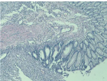
the Ghrelin Group. Loose adhesions were noted in the EO Ghrelin Group; however, four of the Control Group animals exhibited thicker and more numerous adhesions around the anastomoses (Figure 1). The adhesion formation scores were significantly higher in the Control Group than in the Ghrelin Group (p= 0.037).
Table 1 -Histological Grading Scale.
Score Characteristic
0 No evidence
1+ Occasional evidence
2+ Light scattering
3+ Abundant evidence
Bursting pressure
The mean BPs were significantly higher in the Ghrelin Group (168.3¡20.8 mmHg) compared with the Control Group (117.8¡46.5 mmHg) (Figure 2) (p,0.05).
HP content
The mean HP contents were 0.96¡0.06mg/mg wet tissue
in the Control Group and 1.04¡0.09mg/mg in the Ghrelin
Group. The HP content was significantly decreased in the Control Group compared with the mean control HP value of 1.8¡0.26mg/mg that was obtained in the control colonic
tissues prior to anastomosis creation (p= 0.012). In the
Ghrelin Group, the HP content was similar to the basal HP value (Figure 3).
Histopathological assessments
The mean inflammatory cell infiltration, fibroblast infil-tration, collagen density and neovascularization scores are presented in Table 2.
A comparison of the mean histological scores of the groups revealed a significant difference between the inflammatory cell infiltration scores (p,0.05). In the Ghrelin Group inflammatory cell infiltration scores were found to be lower than the Control Group. (Figure 4). There were no significant differences in fibroblast infiltration, collagen intensity or capillary formation between the groups.
& DISCUSSION
Ghrelin has been described as a natural ligand of GHS receptor type 1a. GHS receptors are expressed in several regions of the central nervous system and its periphery; this widespread distribution indicates that ghrelin has various effects in addition to stimulating the secretion of GH (16-19). The results of the current study, which included mechan-ical, biochemmechan-ical, macroscopic and microscopic evaluations, demonstrated a beneficial effect of ghrelin on the healing of colonic anastomoses in an experimental model.
Ghrelin is mainly produced by X/A cells in the fundic glands of the stomach. Small amounts of ghrelin are also generated in the small intestine, pancreas, kidney and hypothalamus (7,16,17). Takachi et al. (20) found that following total gastrectomy, gastric cancer patients displayed
Figure 1 - Thicker and more numerous adhesions around an anastomosis from a Control Group animal.
significant weight loss and an immediate and remarkable decline in serum ghrelin, which did not recover even after a long postoperative period. In addition, gastrectomy patients are prone to anastomotic rupture and fistulas. Yu et al. (21) demonstrated in a rat model that in the early recovery phase after partial gastrectomy, exogenous ghrelin treatment increased food intake and body weight and promoted anastomotic healing. Ghrelin may have clinical implications for recovery from gastrectomy.
Various growth factors have been shown to enhance wound healing in several experimental models. Wheeless et al. (5) evaluated the effect of GH on the BP of radiation-injured terminal ileal anastomoses in a rat model. The BP in the GH-treated rats was significantly greater than that of the control group. Karahasanog˘lu et al. (22) have suggested that GH strengthens left colonic anastomoses in rats. It has been shown that ghrelin stimulates GH release in humans and rats (23,24), which implies that the observed effect of ghrelin on anastomotic healing may be due to its GH secretagogue properties. The spectrum of biological activities attributed to
growth factors includes mitogenic and chemoattractant properties, the ability to release cytokines, the promotion of angiogenesis and the stimulation of extracellular matrix production (25-27). Because ghrelin is a natural ligand of GHS receptor type 1a and stimulates GH release in humans and rats, we predicted that ghrelin would improve anastomotic healing. Consistent with this hypothesis, we found that anastomotic healing was improved following ghrelin administration. GH accelerates cell proliferation and enhances amino acid efflux into cells for protein synthesis or
Figure 3 -The mean HP contents (mg/mg wet tissue) of the Ghrelin and Control Groups compared to the control HP levels. In the Control
Group, the tissue HP contents were significantly decreased during anastomosis healing compared to the measurements performed prior to anastomosis creation (p,0.05). In the Ghrelin Group, the tissue HP contents during anastomosis healing were not different from those measured prior to anastomosis creation.
Table 2 -Mean histological scores (*p,0.05).
Control Group Ghrelin Group
Mean
Std.
Deviation Mean
Std. Deviation
Inflammatory cell infiltration
2.1* 0.57 1.2* 0.42
Fibroblast infiltration 1.6 0.97 1.9 0.87
Collagen density 1.5 0.85 1.7 0.67
Neovascularization 1.5 0.53 1.3 0.95
gluconeogenesis (28). In contrast, ghrelin, a natural ligand of GHSR, did not alter fibroblast infiltration or neovasculariza-tion at the anastomotic site of the colon in the present study. Collagen synthesis, which may be evaluated by measur-ing the tissue HP content, is an essential feature of anastomotic healing. Therefore, we measured the tissue HP content. The HP levels were higher in the Ghrelin Group compared with the control group, suggesting increased collagen synthesis and therefore improved anastomotic wound healing following ghrelin administration. BP mea-surements indicate the mechanical strength of an anasto-mosis. It has been reported that the BP increases progressively immediately after the formation of a colonic anastomosis (29). The present study showed that the anastomotic BPs were increased in the Ghrelin Group compared with the Control Group. In a similar experimental study of post-gastrectomy rats, ghrelin effectively increased the BPs and HP contents of anastomoses on post-operative day seven (21).
In addition to measuring HP and BP, we evaluated the anastomotic area both macroscopically and microscopically. Macroscopic adhesion formation around the anastomosis was scored quantitatively, and the adhesion scores were found to be decreased following ghrelin administration. Anastomotic healing is affected by the strength of the primary inflammatory response; the rate of mucosal re-epithelialization; the amount, strength and maturation rate of the new collagen; and the presence of collagenolysis in the initial three days of the postanastomotic period (30). Inflammation is an integral element of wound healing. However, inflammation is aggravated, wound healing is delayed due to increased collagenolysis. It has been shown that ghrelin has anti-inflammatory properties (7-11), and this may be the cause of the reduced adhesion formation. In support of this hypothesized connection, the inflammatory cell infiltration scores were lower in the Ghrelin Group than in the Control Group.
The principal source of collagen during wound healing is fibroblasts. Fibroblast influx to the wound increases markedly on postoperative day two (29), after which it decreases to the normal level. In the present study, the extent of fibroblast infiltration did not differ between the two groups at the examined time point (Day 7). We believe that this result might be different at other time points, which merits further investigation.
The HP levels were higher in the Ghrelin Group compared with the control group, which suggests an increase in collagen synthesis. The anastomotic BP was also elevated in the Ghrelin Group. The observed effects of ghrelin on anastomotic healing may be partially due to the GH secretagogue properties of this substance. However, ghrelin, a natural ligand of GHSR, did not alter the fibroblast infiltration, collagen density or neovascularization at the anastomotic site of the colon on the seventh day after anastomosis formation. Interestingly, ghrelin administration reduced the adhesion formation around the anastomosis, which may be a result of the anti-inflammatory effect of ghrelin. In the Ghrelin Group, the inflammatory cell infiltration scores were found to be lower than in the Control Group. The results of this study indicate that ghrelin improves anastomotic healing. We propose that the underlying cause of this effect is, at least in part, a mechanism other than the GHS receptor agonist activity of ghrelin.
& AUTHOR CONTRIBUTIONS
Ceran C planned the study, performed the animal experiments and wrote the manuscript. Aksoy RT performed the animal experiments and wrote the manuscript. Gu¨lbahar O performed the biochemical evaluations. O¨ ztu¨rk F performed the histopathological assessments.
& REFERENCES
1. Herndon DN, Hayward PG, Rutan RL, Barrow RE. Growth hormones and factors in surgical patients. In: Cameron JL, editors. Advances in Surgery. St Louis, Missouri: Mosby Year Book. 1992;65-93.
2. Schrock TR, Deveney CW, Dunphy JE. Factor contributing to leakage of colonic anastomoses. Ann Surg. 1973;177(5):513-8, http://dx.doi.org/10. 1097/00000658-197305000-00002.
3. Karatepe O, Kurtulus I, Yalcin O, Battal M, Kamali G, Aydin T. Adrenomedulline improves ischemic left colonic anastomotic healing in an experimental rodent model. Clinics. 2011;66(10):1805-10, http://dx. doi.org/10.1590/S1807-59322011001000021.
4. Inan A, Sen M, Su¨rgit O, Ergin M, Bozer M. Effects of the histamine H2 receptor antagonist famotidine on the healing of colonic anastomosis in rats. Clinics. 2009;64(6):567-70.
5. Wheeless CR Jr, Zanagnolo V, Bowers D, Brenner MJ, Lilley R. The effect of growth hormone on the bursting strength of ileal anastomotic segments in radiation-injured rat bowel. Gynecol Oncol. 1998;70(1):121-2, http://dx.doi.org/10.1006/gyno.1998.5045.
6. Ekiz F, Kirca V, Seker A, Ko¨ksoy FN, Igdem AA. The effect of epidermal growth factor on anastomosis, fascia, and skin wound healing. Ulus Travma Acil Cerrahi Dergisi. 2005;11(2):96-101.
7. Kojima M, Hosoda H, Date Y, Nakazato M, Matsuo H, Kangawa K. Ghrelin is a growth-hormone-releasing acylated peptide from stomach. Nature. 1999;402(6762):656-60, http://dx.doi.org/10.1038/45230. 8. Sehirli O, Sener E, Sener G, Cetinel S, Erzik C, Yeg˘en BC. Ghrelin
improves burn-induced multiple organ injury by depressing neutrophil infiltration and the release of pro-inflammatory cytokines. Peptides. 2008;29(7):1231-40, http://dx.doi.org/10.1016/j.peptides.2008.02.012. 9. Chang L, Du JB, Gao LR, Pang YZ, Tang CS. Effect of Ghrelin on septic
shock in rats. Acta Pharmacol Sin. 2003;24(1):45-9.
10. Wu R, Dong W, Zhou M, Cui X, Hank Simms H, Wang P. Ghrelin improves tissue perfusion in severe sepsis via downregulation of endothelin-1. Cardiovasc Res. 2005;68(8):318-26, http://dx.doi.org/10. 1016/j.cardiores.2005.06.011.
11. Wu R, Dong W, Ji Y, Zhou M, Marini CP, Ravikumar TS, Ping W. Orexigenic Hormone Ghrelin Attenuates Local and Remote Organ Injury after Intestinal Ischemia-Reperfusion. PLoS One. 2008;3:e2026. http:// www.plosone.org/article/info%3Adoi%2F10.1371%2Fjournal.pone.0002026, http://dx.doi.org/10.1371/journal.pone.0002026.
12. van der Ham AC, Kort WJ, Weijma IM, van den Ingh HF, Jeekel J. Effect of fibrin sealant on the healing of colonic anastomosis in the rat. Br J Surg. 1991;78(1):49-53.
13. Demirog˘ullari B, So¨nmez K, Tu¨rkyilmaz Z, Ekingen G, Dursun A, Bor V, et al. Comparison of consequent small bowel anastomoses after transient ischemia: an experimental study in rats. J Pediatr Surg. 1998;33(1):91-3, http://dx.doi.org/10.1016/S0022-3468(98)90369-4.
14. Jamall IS, Finelli VN, Que Hee SS. A simple method to determine nanogram levels of 4-hydroxyproline in biological tissues. Anal Biochem. 1981;112(1):70-5, http://dx.doi.org/10.1016/0003-2697(81)90261-X. 15. Ehrlich HP, Tarver H, Hunt TK. Effects of vitamin A and glucocorticoids
upon inflammation and collagen synthesis. Ann Surg. 1973;177(2):222-7, http://dx.doi.org/10.1097/00000658-197302000-00017.
16. Gnanapavan S, Kola B, Bustin SA, Morris DG, McGee P, Fairclough P, et al. The tissue distribution of the mRNA of Ghrelin and subtypes of its receptor, GHS-R, in humans. J Clin Endocrinol Metab. 2002;87(6):2988, http://dx.doi.org/10.1210/jc.87.6.2988.
17. Brzozowski T, Konturek PC, Drozdowicz D, Konturek SJ, Pawlik M, Sliwowski Z, et al. Role of central and peripheral Ghrelin in the mechanism of gastric mucosal defence. Inflammopharmacology. 2005;3(1-3):45-62, http://dx.doi.org/10.1163/156856005774423971. 18. Chang L, Zhao J, Yang J, Zhang Z, Du J, Tang C. Therapeutic effects of
Ghrelin on endotoxic shock in rats. Eur J Pharmacol. 2003;473(2-3):171-6. 19. Leite-Moreira AF, Soares JB. Physiological, pathological and potential therapeutic roles of Ghrelin. Drug Discov Today. 2007;12(7-8):276-88, http://dx.doi.org/10.1016/j.drudis.2007.02.009.
20. Takachi K, Doki Y, Ishikawa O, et al. Postoperative Ghrelin levels and delayed recovery from body weight loss after distal or total gastrectomy. J Surg Res. 2006;130(1): 1-7, http://dx.doi.org/10.1016/j.jss.2005.08.003. 21. Yu S, Wang ZG, Yang CG, Yan J, Qiu WC, Zheng Q. Ghrelin promote
partial the early recovery of rats after subtotal gastrectomy. Af J Pharm Pharmacol. 2011;5(5):658-63.
23. Date Y, Murakami N, Kojima M, Kuroiwa T, Matsukura S, Kangawa K, et al. Central effects of a novel acylated peptide, Ghrelin, on growth hormone release in rats. Biochem Biophys Res. Commun. 2000;275(2):477-80, http://dx.doi.org/10.1006/bbrc.2000.3342. 24. Takaya K, Ariyasu H, Kanamoto N, Iwakura H, Yoshimoto A, Harada M,
et al. Ghrelin strongly stimulates growth hormone release in humans. J Clin Endocrinol Metab. 2000;85(12):4908-11, http://dx.doi.org/10. 1210/jc.85.12.4908.
25. Itoh T, Suzuki M, Mitsui Y. Keratinocyte growth factor as a mitogen for primary culture of rat hepatocytes. Biochem Biophys Res Commun. 1993;192(3):1011-15, http://dx.doi.org/10.1006/bbrc.1993.1517. 26. Raines EW, Dower SK, Ross R. Interleukin-1 mitigenic activity of
fibroblasts and smooth muscle cells is due to PDGF-AA. Science. 1989;243(4889):393-6, http://dx.doi.org/10.1126/science.2783498. 27. Schreiber AB, Winkler ME, Derynck R. Transforming growth
factor-alpha: A more potent angiogenic mediator than epidermal growth factor.
Science. 1986;232(4755):1250-3, http://dx.doi.org/10.1126/science.2422 759.
28. Hammarqvist F, Stromberg C, von der Decken A, Vinnars E, Wernerman J. Biosynthetic human growth hormone preserves both muscle protein synthesis and the decrease in muscle-free glutamine, and improves whole-body nitrogen economy after operation. Ann Surg. 1992;216(2):184-91, http://dx.doi.org/10.1097/00000658-199208000-00009.
29. Christensen H, Chemnitz J, Christensen BC, Oxlund H. Collagen structural organization of healing colonic anastomoses and the effect of growth hormone treatment. Dis Colon Rectum. 1995;38(11):1200-5, http://dx.doi.org/10.1007/BF02048337.