https://doi.org/10.1590/0004-282X20170176
REVIEW
Autoimmune encephalitis: a review of
diagnosis and treatment
Encefalites autoimunes: uma revisão sobre diagnóstico e tratamento
Lívia Almeida Dutra1,2, Fabiano Abrantes1, Fabio Fieni Toso1, José Luiz Pedroso1, Orlando Graziani Povoas
Barsottini1, Romana Hoftberger3
Autoimmune encephalitis (AIE) is considered one of the most common causes of noninfectious acute encephalitis. It is estimated that 20% of all encephalitis cases in northern Europe are immune-mediated1. he California Encephalitis Project
found that anti-N-methyl-D-aspartate receptor (anti-NMDAR) encephalitis occurred in 47% of patients under 30 years of age2.
Autoimmune encephalitis is typically an acute or subacute onset and that may become chronic later3. Suggested
mech-anisms that may trigger AIE include tumors (paraneoplastic), infections (parainfectious), or it may be cryptogenic4.
Autoimmune encephalitis has a wide variety of clinical manifestations including behavioral and psychiatric symp-toms, autonomic disturbances, movement disorders and sei-zures3,5. We reviewed common causes of AIE and discuss their
pathophysiology, diagnostic approach and management.
Pathophysiology of autoimmune encephalitis: an overview
Autoimmune encephalitis presents an immune response against neuronal autoantigens with production of antibodies6.
Anti-neuronal antibodies are classiied into antibodies against cell surface antigens (CSAab), antibodies against syn -aptic antigens (SyAab) and antibodies against intraneuronal antigens (INAab), also known as onconeural antibodies6,7.
he CSAab (i.e., anti-NMDAR antibodies) target mole -cules involved in neurotransmission leading to neuronal dys-function7. hey may have agonistic or antagonistic efects on
the receptors, block ion channel pores or disrupt the
interac-tion with neighboring molecules. hey could also alter recep -tor localization at the membrane or cause recep-tor internal-ization, thus reducing cell surface expression of receptors7.
Moreover, they could lead to complement deposition and activation of natural killer cells resulting in cell death7.
he SyAab are believed to contribute to alteration of neu -rotransmitter release. In contrast, INAab (i.e., anti-Hu, anti-Yo, anti-Ma) are most likely not directly pathogenic and probably an epiphenomenon of T-cell-mediated immune response7.
he term AIE is used to describe a group of neurological dis -orders with symptoms of limbic and extra-limbic dysfunction in
association with CSAab or SyAab8. Patients may have antibodies
1Universidade Federal de São Paulo, Departamento de Neurologia e Neurocirurgia, Divisão de Neurologia Geral, São Paulo SP, Brasil; 2Hospital Israelita Albert Einstein, São Paulo SP, Brasil;
3Medical University of Vienna, Institute of Neurology, Vienna, Austria.
Correspondence: Lívia Almeida Dutra; Rua Pedro de Toledo, 650; 04039-002 São Paulo SP, Brasil; E-mail: [email protected]
Conflict of interest: There is no conflict of interest to declare.
Received 18 March 2017; Received in final form 31 August 2017; Accepted 18 September 2017.
ABSTRACT
Autoimmune encephalitis (AIE) is one of the most common causes of noninfectious encephalitis. It can be triggered by tumors, infections, or it may be cryptogenic. The neurological manifestations can be either acute or subacute and usually develop within six weeks. There are a variety of clinical manifestations including behavioral and psychiatric symptoms, autonomic disturbances, movement disorders, and seizures. We reviewed common forms of AIE and discuss their diagnostic approach and treatment.
Keywords: encephalitis; antibodies, neoplasm; status epilepticus; anti-N-Methyl-D-Aspartate receptor encephalitis; immunoglobulin; rituximab.
RESUMO
As encefalites autoimunes (EAI) são a principal causa de encefalite não-infecciosa. As manifestações neurológicas são variadas, incluindo alterações comportamentais ou psiquiátricas, disautonomia, transtornos do movimento e epilepsia. Habitualmente a instalação dos sintomas ocorre em até 6 semanas, de forma aguda ou subaguda. As EAI podem ser desencadeadas por tumores, quadros infecciosos virais ou ainda apresentar etiologia criptogênica. Este artigo revisa as principais EAI, estratégias de diagnóstico e tratamento.
of more than one type, and CSAab and SyAab may be present
together with anti-INAab, especially in paraneoplastic AIE.
Patients with AIE associated with CSAab have a more
favorable prognosis. In contrast, those with AIE associ-ated with INAab often show limited response to immuno-therapy and their symptoms are mostly irreversible due to T-cell-mediated neuronal damage6,7.
Clinical manifestations
Patients with AIE may present with a variety of move -ment disorders such as ataxia, dystonia, myoclonus, and orofacial dyskinesia. Seizures are the most common
symp-tom and diferent types of seizures may be seen, including
refractory status epilepticus9. Autonomic disturbances are
also frequently reported such as sudoresis, hypertension, tachycardia and hypoventilation. Some patients may show involvement of the myenteric plexus and develop gastroin-testinal manifestations (diarrhea, gastroparesis, and consti-pation). Sleep disturbances such as agrypnia excitata, insom-nia, abnormal sleep movements and behaviors, sleep apnea, and hypersomnia are also found10. Some of these indings are
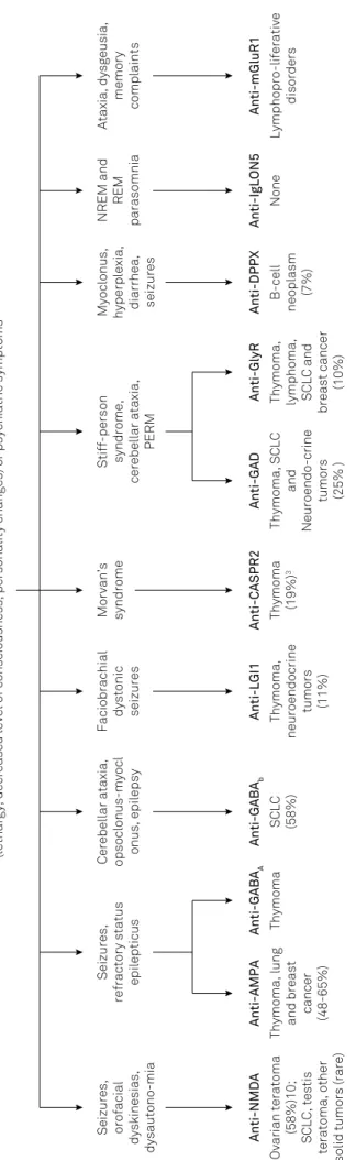
suggestive of a certain type of encephalitis and may indicate
a speciic underlying antibody and tumor (Figure 1).
Table 1 summarizes the diagnostic criteria for limbic
encephalitis. hese include working memory deicits, mood
changes, and often seizures within three months from onset. Many patients with AIE present with a broader spectrum of
neurological symptoms. For improving the recognition of
immune-mediated cases, diagnostic criteria for suspected AIE were recently established including criteria for
seronega-tive AIE (Table 2). Deinite soroposiseronega-tive AIE requires typical
clinical picture and positive anti-neuronal antibodies
Anti-NMDAR encephalitis
Anti-N-methyl-D-aspartate receptor encephalitis is
one of the most common causes of AIE and was originally described in 2007 in a cohort of 12 patients, 11 of them with ovarian teratomas11. his condition predominantly afects
children and young female patients3. Underlying
malignan-cies are found mainly in patients between the age of 12–45 years; most of them are ovarian teratomas (94%), followed by extraovarian teratomas (2%), and other tumors (4%). Herpes simplex virus-1 encephalitis appears to be a trigger for anti-NMDAR encephalitis; most postherpetic AIE cases are now believed to be anti-NMDAR encephalitis.
Approximately 70% of patients present with prodromal symptoms such as fever, headache, nausea, vomiting,
diar-rhea, and lu-like symptoms, two weeks before the onset of
neurological manifestations. Behavioral complaints, psy-chosis, delusions, hallucinations and paranoia,
accompa-nied with memory deicits and language disturbance, are
frequently found at an early stage3,12. he most common
movement disorders are orofacial dyskinesias,
choreoatheth-osis, and dystonia12. Patients may progress to catatonia or Fig
ur
e 1
.
Clinical findings in a
ut
oimmune encephalitis with associa
tion with tumor
s
.
Rapid progression of working memory deficits, altered mental status
(lethargy, decreased level of consciousness, personality changes) or psychiatric symptoms
Seizures,
refractory status
epilepticus
Stiff-person syndrome,
cerebellar ataxia,
PERM
Seizures, orofacial
dyskinesias,
dysautono-mia Anti-NMDA Ovarian teratoma
(58%)10;
SCLC, testis
teratoma, other
solid tumors (rare)
Anti-AMPA
Thymoma, lung and breast
cancer
(48-65%)
Anti-GABA
A
Thymoma
Cerebellar ataxia, opsoclonus-myocl onus, epilepsy
Anti-GABA
b
SCLC (58%)
Faciobrachial
dystonic seizures Anti-LGI1 Thymoma,
neuroendocrine
tumors (11%) Morvan’s syndrome
Anti-CASPR2 Thymoma
(19%)
3
Anti-GAD
Thymoma, SCLC
and
Neuroendo-crine
tumors (25% ) Anti-GlyR Thymoma, lymphoma, SCLC and
breast cancer
(10%)
Myoclonus, hyperplexia, diarrhea, seizures Anti-DPPX
B-cell
neoplasm
(7%)
NREM and
REM
parasomnia Anti-IgLON5
None
Ataxia, dysgeusia,
memory
complaints Anti-mGluR1
Lymphopro-liferative
mutism, followed by an altered level of consciousness and
autonomic instability. An important diferential diagnosis
of anti-NMDAR encephalitis is neuroleptic malignant syn-drome, because many patients are initially treated with neu-roleptics for behavioral symptoms3,13. Table 3 summarizes the
diagnostic criteria for anti-NMDAR encephalitis.
Seizures are seen in 16% of female and 34% of male patients; they can be focal seizures or patients may present
with status epilepticus. Extreme delta brush is a speciic elec -troencephalogram (EEG) pattern found in 30% of patients with anti-NMDAR encephalitis14. Children more frequently present
with behavioral symptoms and movement disorders, whereas adults present with psychiatric symptoms and seizures.
Brain magnetic resonance imaging (MRI) is abnormal in 35% of patients at disease onset, but it may show late
abnor-malities in 50%, mainly nonspeciic hyperintense lesions in
the grey and white matter. Rare cases show lesions suggestive of demyelination and overlap with demyelinating syndromes such as neuromyelitis optica spectrum disorders (NMOSD) associated with anti-aquaporin-4 antibodies or demyelinat-ing diseases associated with myelin oligodendrocyte glyco-protein antibodies(MOG-ab)13.
Anti-AMPAR encephalitis
Patients with anti-α
-amino-3-hydroxy-5-methyl-4-isoxazolepropionic acid receptor (anti-AMPAR) encephali -tis characteristically present with seizures, memory impair-ment and psychosis. Some may develop sleep disturbances
and movement disorders. Anti-AMPAR encephalitis is para -neoplastic in etiology in 64% of cases, mostly associated with thymoma, ovarian teratoma and lung and breast cancer.
Brain MRI shows T2 and FLAIR hyperintensities, particularly
in the medial temporal lobe. Subcortical and cortical lesions, which are sometimes suggestive of demyelination, may also be found. Cerebrospinal luid (CSF) examination may show
pleocytosis and oligoclonal bands15.
Anti-GABA-AR encephalitis
Anti-gamma-aminobutyric acid A receptor (anti-GABA-AR)
encephalitis was irst reported in 2014 in six patients (two
male children, one female teenager and three male adults)16.
hey developed a rapidly progressive encephalopathy with
early behavioral or cognitive changes that evolved with refrac-tory seizures and multifocal lesions as seen on brain MRI16.
In most of these patients, CSF analysis showed lymphocytic
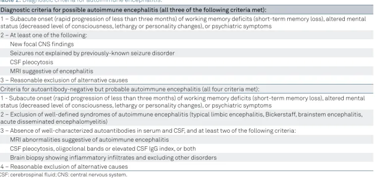
Table 2. Diagnostic criteria for autoimmune encephalitis.
Diagnostic criteria for possible autoimmune encephalitis (all three of the following criteria met):
1 – Subacute onset (rapid progression of less than three months) of working memory deficits (short-term memory loss), altered mental status (decreased level of consciousness, lethargy or personality changes), or psychiatric symptoms
2 – At least one of the following: New focal CNS findings
Seizures not explained by previously-known seizure disorder CSF pleocytosis
MRI suggestive of encephalitis
3 – Reasonable exclusion of alternative causes
Criteria for autoantibody-negative but probable autoimmune encephalitis (all four criteria met):
1 - Subacute onset (rapid progression of less than three months) of working memory deficits (short-term memory loss), altered mental status (decreased level of consciousness, lethargy or personality changes), or psychiatric symptoms
2 – Exclusion of well-defined syndromes of autoimmune encephalitis (typical limbic encephalitis, Bickerstaff, brainstem encephalitis, acute disseminated encephalomyelitis)
3 – Absence of well-characterized autoantibodies in serum and CSF, and at least two of the following criteria: MRI abnormalities suggestive of autoimmune encephalitis
CSF pleocytosis, oligoclonal bands or elevated CSF IgG index, or both Brain biopsy showing inflammatory infiltrates and excluding other disorders 4 – Reasonable exclusion of alternative causes
CSF: cerebrospinal fluid; CNS: central nervous system.
Table 1. Diagnostic criteria for definite autoimmune limbic encephalitis.
Diagnosis can be made when all four of the following criteria have been met:
1 – Subacute onset (rapid progression of less than three months) of working memory deficits, seizures, or psychiatric symptoms suggesting involvement of the limbic system
2 – Bilateral brain abnormalities on MRI T2-weighted FLAIR sequence restricted to the medial temporal lobes 3 – At least one of the following:
EEG with epileptic or slow-wave activity involving the temporal lobes CSF pleocytosis
4 – Reasonable exclusion of alternative causes
pleocytosis with or without oligoclonal bands. A recent study
identiied an underlying neoplasia in 27% of these patients,
mostly thymomas17. Similar to that seen in patients with
gamma-aminobutyric acid B receptor (GABA-BR) and
anti-AMPAR antibodies, they may also present with coexisting
autoimmune disorders such as thyroiditis or myasthenia18.
Anti-GABA-BR encephalitis
Anti-GABA-BR encephalitis is characterized by cognitive symptoms with severe seizures or status epilepticus19. Other
presentations include ataxia and opsoclonus-myoclonus. In a small series of 20 patients with anti-GABA-BR, about 50% were found to have small-cell lung cancer20. Males and
females appear to be equally afected. he long-term progno -sis in anti-GABA-BR encephalitis is determined by the pres-ence of an underlying malignancy21.
Anti-LGI1 and anti-CASPR2 encephalitis (formerly known as anti-VGKC-associated encephalitis)
he irst reports of anti-voltage-gated potassium chan
-nel-complex antibodies (anti-VGKC) date back to 2001 and
described patients with neuromyotonia, Morvan’s syndrome and limbic encephalitis22. Other rare phenotypes included
epilepsy and painful polyneuropathy23. Anti-VGKC antibod
-ies, in fact, later turned out to be directed against proteins
that form a complex with VGKC called leucine-rich glioma-inactivated 1 (LGI1 and contactin-associated protein-like 2 (CASPR-2)24,25. Each of these antibodies presents with
spe-ciic clinical symptoms.
The LGI1 is a secreted synaptic protein that interacts
with transmembrane proteins ADAM22 and ADAM23 to form a trans-synaptic complex involving potassium
channels and AMPAR. Genetic disruption of LGI1 pro -tein in humans causes autosomal dominant lateral tem-poral lobe epilepsy26. The clinical spectrum of anti-LGI1
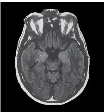
encephalitis usually comprises limbic encephalitis, hyponatremia and seizures. Half of the patients develop faciobrachial dystonic seizures, which are characterized by brief unilateral contractions of the arm (often evolv-ing into the ipsilateral face or leg) that are shorter than three seconds and occur several times a day. Two-thirds of patients present with brain MRI hyperintensities in
the medial temporal lobe (Figure 2). Paraneoplastic anti-LGI1 encephalitis is uncommon; however, patients
should be screened for lung cancer27. Relapses may occur
in up to 20% of patients28. Table 3. Diagnostic criteria for anti-NMDA receptor encephalitis.
Probable anti-NMDA receptor encephalitis
Diagnosis require all three of the following criteria
1 – Less than three months of at least four of the six following major group of symptoms: Abnormal (psychiatric) behavior or cognitive dysfunction
Speech dysfunction (pressured speech, verbal reduction, mutism) Seizures
Movement disorder, dyskinesias, or rigidity/abnormal postures Decreased level of consciousness
Autonomic dysfunction or central hypoventilation 2 – At least one of the following laboratory study results:
Abnormal EEG (focal or diffuse slow or disorganized activity, epileptic activity, or extreme delta brush) CSF with pleocytosis or oligoclonal bands
3 – Reasonable exclusion of other disorders
Diagnosis can also be made in the presence of three of the above groups of symptoms accompanied by a systemic teratoma Definite anti-NMDA receptor encephalitis
Diagnosis can be made in the presence of one or more of the six major groups of symptoms and IgG anti-GluN1 antibodies, after reasonable exclusion of other disorders
EEG: electroencephalogram; CSF: cerebrospinal fluid.
he CASPR-2 is a juxtaparanodal adhesion molecule
that interacts with contactin 2 and the cytoskeleton, and is involved in clustering of potassium channels in myelin-ated axons28. Anti-CASPR2 antibodies are associated with
peripheral nerve hyperexcitability (myokymia, fasciculations, cramps) and encephalitis. Other symptoms include dysauto-nomia and insomnia (agrypnia excitata). Nearly one-third of patients develop Morvan’s syndrome, a complex disorder
afecting the peripheral and central nervous system that is
characterized by distal movement disorders of the upper limbs, peripheral nerve hyperexcitability, dysautonomia, pain, and encephalitis24. Most individuals afected are male
and one-third of them present with paraneoplastic mani-festations, usually associated with thymoma, lung cancer or endometrial carcinoma27,26.
he clinical relevance of anti-VGKC antibodies against
unknown target antigens is unclear. About half of patients
with anti-VGKC encephalitis do not present antibodies against LGI1 or CASPR-2. hese speciic groups of patients
develop a wide variety of clinical syndromes raising the
ques-tion whether anti-VGKC antibodies are truly a marker of dis -ease in these patients25. Anti-VGKC antibodies have been
described, for example, in patients with Creutzfeldt-Jakob
disease, suggesting that these antibodies might not be
patho-genic. Anti-VGKC titers with negative LGI1 and anti-CASPR-2 antibodies are usually low (< 0.3 pM)—although there is no clear cutof value to diferentiate between patients with and without AIE—thus it is not recommended to use
them as evidence of immune-mediated pathogenesis22,29.
Anti-GAD encephalitis
Glutamic acid decarboxylase (GAD) is an enzyme that catalyzes the conversion of glutamic acid to the neurotrans-mitter GABA. Anti-GAD antibodies have been associated with other autoimmune disorders such as insulin-dependent
diabetes mellitus. he main neurological syndromes asso
-ciated with anti-GAD antibodies include stif-person syn -drome, cerebellar ataxia, epilepsy and limbic encephalitis30.
Stif-person syndrome is a neurological disorder fre -quently associated with anti-GAD antibodies characterized
by muscle stifness resulting from co-contractions of agonist
and antagonist muscles and painful spasms31. Another
clas-sic inding in this syndrome is the patient’s pronounced star -tle responses.
Ataxia associated with anti-GAD antibodies is usually slowly progressive and evolves over months or years. Abnormal ocular movements with spontaneous downbeat nystagmus have also been described30. Nearly 7% of patients with
anti-GAD antibodies present with temporal lobe epilepsy or status epilepticus, and 5% develop limbic encephalitis32,33.
Anti-GAD antibodies are rarely of paraneoplastic
ori-gin. Patients with stif-person syndrome, cerebellar ataxia or
other neurological syndromes typically associated with anti-GAD antibodies do not need to be aggressively or repeatedly
screened for cancer. However, the presence of GAD
anti-bodies in patients with limbic encephalitis or other classic paraneoplastic syndromes (paraneoplastic cerebellar degen-eration, opsoclonus-myoclonus syndrome or paraneoplastic encephalomyelitis) is associated with a 10-fold increase in the risk of cancer and tumor screening is thus recommended34.
Anti-GlyR encephalitis
Glycine receptors (GlyR) are chloride channels that facil-itate inhibitory neurotransmission in the brain and spinal cord35. Anti-GlyR antibodies were irst described in patients
with progressive encephalomyelitis with rigidity and
myoc-lonus and later in patients with stif-person syndrome31,35,36.
Recently, anti-GlyR antibodies have also been reported in patients with cerebellar ataxia and anti-GAD antibodies and patients with demyelinating diseases including optic neuritis
and multiple sclerosis, but their clinical signiicance remains
unclear37,38. Anti-GlyR antibodies are usually not associated
with tumors, although there have been reports of patients with underlying thymoma, small-cell lung cancer, breast can-cer and chronic lymphocytic leukemia.
Anti-DPPX encephalitis
Dipeptidyl peptidase-like protein 6 (DPPX) is a subunit
of Kv4.2 potassium channels expressed in the hippocampus,
cerebellum, striatum, and myenteric plexus. Patients with anti-DPPX antibodies show neuropsychiatric symptoms (agitation and confusion), myoclonus, tremor, startle relex, seizures, stif-person syndrome and prodromal diarrhea of
unknown etiology. In addition, they may have symptoms of dysautonomia including arrhythmias, thermodysregulation, diaphoresis, urinary symptoms and sleep disorders39,40.
he CSF analysis usually shows pleocytosis and increased protein concentrations. Functional tests in the serum of a DPPX-positive patient demonstrated increased excitability of enteric neurons and downregulation of DPPX
and Kv4.2 from hippocampal neuron membranes, which is
suggestive of the pathogenic efects of anti-DPPX antibod
-ies in anti-DPPX encephalitis39.
Encephalopathy associated with anti-IgLON5 antibodies
he IgLON family member 5 (IgLON5) is a neuronal
cell adhesion molecule of the immunoglobulin
superfam-ily. Patients with anti-IgLON5 antibodies present with a
unique non-REM (rapid eye movement) and REM parasom-nia with obstructive sleep apnea, stridor, episodic central hypoventilation, dementia, gait instability, chorea, dysar-thria, dysphagia, dysautonomia and supranuclear gaze palsy resembling that seen in classic tauopathy5,40. All published
cases reported the presence of the alleles HLA-DQB1*0501 and HLA-DRB1*1001 suggesting genetic susceptibility to
hyperphosphorylated tau mainly involving the tegmentum of the brainstem and hypothalamus41. his novel encephalopa
-thy provides an intriguing link between neurodegeneration and cell-surface autoimmunity. A recent study has shown
that anti-IgLON5 antibodies recognize Ig-like domain 2 as an
immunogenic region and causes irreversible internalization
of IgLON5 from the neuronal membrane. hese indings sup
-port a potential pathogenic role of anti-IgLON5 antibodies in
the associated encephalopathy42.
Anti-mGluR1 and anti-mGluR5 encephalitis
Metabotropic glutamate receptor 1 (mGluR1) and metabotropic glutamate receptor 5 (mGluR5) are both G-protein-coupled receptors that share an 85% amino acid sequence homology. Both receptors are involved in modulat-ing synaptic functions includmodulat-ing long-term depression. While
mGluR1 facilitates long-term depression at parallel iber Purkinje cell synapses, which are critical for cerebellar motor
learning, mGluR5 is more relevant for long-term depression in the hippocampus.
All patients with anti-mGluR1 antibodies develop cere-bellar ataxia of subacute onset, and some may present with additional symptoms such as paranoia, dysgeusia, diplopia
and cognitive deicits. Common tumors found to be associ -ated with anti-mGluR1 antibodies are hematologic malig-nancies and prostate adenocarcinoma43. Response to
immu-notherapy is variable.
Patients with anti-mGluR5-abs present with a form of
encephalitis named “Ophelia syndrome”, a clinical syn-drome that includes memory loss and psychosis in associa-tion with Hodgkin’s lymphoma44. he outcome of reported
cases is generally good after treatment of the lymphoma and immunotherapy44.
Considerations on specific diagnostic methods for AIE
Four diferent techniques are used to detect antibodies against cell surface antigens: cell-based assay (CBA) with
HEK293 cells; tissue-based assay (TBA) on brain tissue of rodents using indirect immunohistochemistry or indirect
immunoluorescence, and culture of dissociated hippocam -pal neurons from rats.
Cell-based assays are highly sensitive and robust signals are diagnostic of speciic antigens45. he TBA provides an
excellent screening approach as it can detect most currently-known antibodies and reveal new antibodies. Staining of live neuronal cell cultures are performed mainly in research
labo-ratories and can be useful when TBAs and CBAs show con
-licting results. It conirms that antibodies directed against neuronal surface antigens are present (Figure 3).
Testing for surface receptor antibodies should always
be performed in both serum and CSF of the patients. his has several reasons: (1) in some speciic syndromes (e.g.
anti-NMDAR encephalitis) anti-neuronal antibodies may
be found only in CSF (in a recent study 13% of anti-NMDAR
encephalitis patients did not have detectable anti-NMDAR antibodies in serum46) while other antibodies (e.g. LGI1)
may, in rare instances, be detectable only in serum. (2) Some
patients may have a diferent antibody spectrum in serum and CSF, for example, some patients may have anti-NMDAR encephalitis in serum and CSF and, in addition, GABA-AR
antibodies only in serum. In these cases, the antibody present
in the CSF determines the clinical phenotype. (3) Antibody-titers in CSF correlate better with the clinical presentation
than serum titers, and (4) serum testing harbors the risk of background reactivity that gives rise to false positive results.
Recently, serum testing with CBA identiied anti-NMDAR
antibodies mainly of the IgA and IgM subclass in
schizophre-nia, Creutzfeldt-Jakob disease, depression, Parkinson’s dis -ease, and healthy individuals46. However, presence of these
antibodies could not be shown in CSF and, therefore, the clinical signiicance of serum IgA and IgM anti-NMDAR anti
-bodies remains to be clariied.
here are diagnostic challenges to testing for AIE in Brazil as commercial CBA and TBA kits are not approved for use by national regulatory authorities. Partner or research laborato -ries overseas perform most testing and some of these
labo-ratories perform CBAs only. We currently recommend that
samples from patients with clinical manifestations
sugges-tive of AIE who tested negasugges-tive by CBA should be referred
to research laboratories that use other assessment methods. Negative test results do not rule out immune-mediated
disorders and nonspeciic background signals may cause false
positive test results. Steroid use may also interfere with the diagnostic test. Hence, test results should be interpreted with caution and put into the context of the clinical presentation.
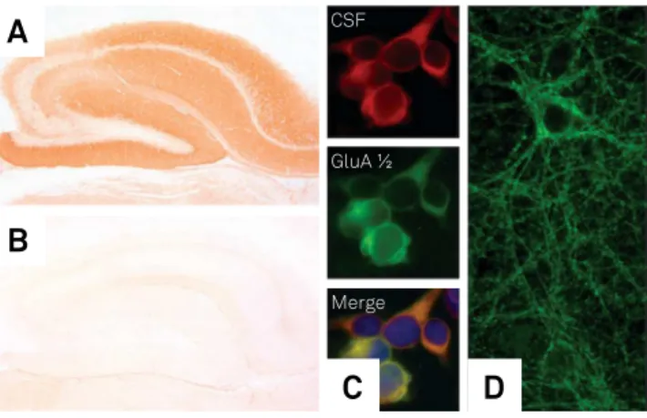
Figure 3. Techniques for the detection of anti-neuronal antibodies. Indirect immunohistochemistry on rat brain tissue with a serum of a patient with anti-NMDAR encephalitis shows strong labeling of hippocampal neurons (A) whereas a serum of a healthy individual is negative (B). In the cell-based assay, antibodies are identified on HEK293 cells transfected with related antigens. (C) red: CSF from a patient with AMPAR antibodies, green: commercial antibody against the GluA1/2 subunits of AMPARs). (D) Live hippocampal neuron culture; strong dot-like labeling of the neuronal membrane indicates antibodies against cell surface antigens.
C
D
A
B
CSF
GluA ½
Because antibodies may remain positive despite clinical fea-tures, clinicians should focus on patient treatment rather than antibody titers.
Differential diagnosis
Diferential diagnosis in anti-neuronal AIE includes
Hashimoto’s encephalopathy and other steroid-responsive encephalopathies, acute disseminated encephalomyeli-tis, neuromyelitis optica spectrum disorders, central ner-vous system vasculitis, neuropsychiatric lupus, angiocentric lymphoma, Rasmussen’s encephalitis and febrile infection-related epilepsy syndrome. It is important to rule out HIV
infection, syphilis, Creutzfeldt-Jakob disease, as well as
human herpes virus-6-associated encephalitis in immuno-compromised patients1,3,8,33.
Viral infections are known triggers for AIE3,4. It is believed
that virus-mediated cerebral tissue damage may lead to anti-gen exposition that triggers the development of anti-neuro-nal antibodies. Herpes viruses may trigger autoimmunity to NMDAR, dopamine D2 receptor, GABA-AR and other synap-tic proteins that have are not yet been characterized.
Tumor investigation
All patients with AIE should be screened for tumors at
disease onset. he nature of the antibody, and to a lesser
extent the clinical syndrome, determines the risk and type of an underlying malignancy47. Due to the low frequency of
tumor association in patients with anti-LGI1 and anti-GAD
antibodies, patient screening should be considered at disease onset, with no need for periodic screenings8. Tumor
treat-ment is essential for neurological improvetreat-ment25.
If initial tumor screening is negative but the patient has antibodies that are typically paraneoplastic (e.g.
anti-NMDAR in young adult women, anti-CASPR2, anti-AMPAR
and anti-GABA-BR), screening should be repeated after three to six months, followed by screenings every six months for four years48.
Computed tomography (CT) of the chest should be per
-formed; if negative, luorodeoxyglucose positron emission tomography (FDG-PET) is indicated as it increases cancer
detection by 20%49. Mammograms followed by MRI are used
for breast cancer screening. For the pelvic region and testes, ultrasound is the investigation of irst choice followed by pel
-vic CT47. It is important to note that ovarian teratomas are
not seen in FDG-PET scans.
Treatment and prognosis
Various treatment approaches including corticosteroids, intravenous immunoglobulin, plasma exchange, rituximab and cyclophosphamide are currently used13. Data on
treat-ment response and prognosis are mostly available for anti-NMDAR encephalitis50.
Evidence suggests that early immunotherapy improves outcome3, thus treatment for AIE should not be delayed.
Patients should receive either methylprednisolone 1 g IV for
3–5 days and intravenous immunoglobulin (0.4 g/kg/day for
ive days) or methylprednisolone and plasmapheresis. We
favor the use of plasmapheresis in patients with refractory seizures and severe dysautonomia, although there is no com-pelling evidence of superiority of any approach. If an associ-ated tumor is detected, oncologic management (chemother-apy or tumor resection) is important for improvement.
Autoimmune encephalitis patients who fail to improve after 10–14 days should receive second-line therapies such as rituximab or cyclophosphamide, or both3,13. Specialized
cen-ters usually prescribe rituximab (375 mg/m2) weekly for four
weeks and cyclophosphamide (750 mg/m2) for six months in
patients older than 16 years. Younger patients should receive rituximab alone3.
Better prognosis has been associated with early treat-ment, no requirement for intensive care admission and non-paraneoplastic AIE20. he treatment response and relapse
rate vary among patients with AIE. Half of the patients with anti-NMDAR encephalitis fail initial immunotherapy and may require second-line treatment options, with relapses occurring in 12%. Relapses may occur in 31% of patients with
anti-LGI1 encephalitis and 10% of those with anti-CASPR2 encephalitis, sometimes years after the irst episode. About 33% of the patients with anti-LGI1 encephalitis are left dis -abled, mostly due to memory problems.
FINAL REMARKS
Autoimmune encephalitis may present with a wide variety
of symptoms of either acute or subacute onset. he diagnosis
should not rely solely on antibody testing as patients with AIE
may be seronegative. Clinicians must be aware of the difer -ent laboratory assessm-ent methods available as well as proper
interpretation of results. Clinical presentation and physical examination are of extreme importance in AIE. Patients ben
-eit from early aggressive treatment and relapses may occur.
References
1. Granerod J, Ambrose HE, Davies NW, Clewley JP, Walsh AL, Morgan D et al. Causes of encephalitis and differences in their clinical presentations in England: A multicentre, population-based prospective study. Lancet Infect Dis. 2010;10(12):835-44. https://doi.org/10.1016/S1473-3099(10)70222-X
3. Graus F, Titulaer MJ, Balu R, Benseler S, Bien CG, Cellucci T et al. A clinical approach to diagnosis of autoimmune encephalitis. Lancet Neurol. 2016;15(4):391-404. https://doi.org/10.1016/S1474-4422(15)00401-9
4. Linnoila JJ, Binnicker MJ, Majed M, Klein CJ, McKeon A. CSF herpes virus and autoantibody profiles in the evaluation of encephalitis. Neurol - Neuroimmunol Neuroinflammation. 2016;3(4):e245. https://doi.org/10.1212/NXI.0000000000000245
5. Sabater L, Gaig C, Gelpi E, Bataller L, Lewerenz J, Torres-Vega E et al. A novel non-rapid-eye movement and rapid-eye-movement parasomnia with sleep breathing disorder associated with antibodies to IgLON5: a case series, characterisation of the antigen, and post-mortem study. Lancet Neurol. 2014;13(6):575-86. https://doi.org/10.1016/S1474-4422(14)70051-1
6. Lancaster E, Dalmau J. Neuronal autoantigens: pathogenesis, associated disorders and antibody testing. Nat Rev Neurol. 2012;8(7):380-90. https://doi.org/10.1038/nrneurol.2012.99 7. Coevorden-Hameete MH, de Graaff E, Titulaer MJ, Hoogenraad
CC, Sillevis Smitt PE. Molecular and cellular mechanisms underlying anti-neuronal antibody mediated disorders of the central nervous system. Autoimmun Rev. 2014;13(3):299-312. https://doi.org/10.1016/j.autrev.2013.10.016
8. Leypoldt F, Wandinger K-P, Bien CG, Dalmau J.
Autoimmune Encephalitis. Eur Neurol Rev. 2013;8(1):31-7. https://doi.org/10.17925/ENR.2013.08.01.31
9. Davis R, Dalmau J. Autoimmunity, seizures, and status epilepticus. Epilepsia. 2013;54(6 Suppl 6):46-9. https://doi.org/10.1111/epi.12276 10. Tobin WO, Lennon VA, Komorowski L, Probst C, Clardy SL, Aksamit
AJ et al. DPPX potassium channel antibody: frequency, clinical accompaniments, and outcomes in 20 patients. Neurology. 2014; 83(20):1797-803. https://doi.org/10.1212/WNL.0000000000000991 11. Dalmau J, Tüzün E, Wu HY, Masjuan J, Rossi JE, Voloschin A et al.
Paraneoplastic anti- N -methyl-D-aspartate receptor encephalitis associated with ovarian teratoma. Ann Neurol. 2007;61(1):25-36. https://doi.org/10.1002/ana.21050
12. Dalmau J, Lancaster E, Martinez-Hernandez E, Rosenfeld MR, Balice-Gordon R. Clinical experience and laboratory investigations in patients with anti-NMDAR encephalitis. Lancet Neurol. 2011;10(1):63-74. https://doi.org/10.1016/S1474-4422(10)70253-2PMID:21163445 13. Titulaer MJ, McCracken L, Gabilondo I, Armangué T, Glaser C,
Iizuka T et al. Treatment and prognostic factors for long-term outcome in patients with anti-NMDA receptor encephalitis: an observational cohort study. Lancet Neurol. 2013; 12(2):157-65. https://doi.org/10.1016/S1474-4422(12)70310-1
14. Schmitt SE, Pargeon K, Frechette ES, Hirsch LJ. Extreme delta brush: a unique EEG pattern in adults with anti-NMDA receptor encephalitis. Neurology. 2012;79:1094-100. https://doi.org/10.1212/WNL.0b013e3182698cd8 15. Höftberger R, Sonderen A, Leypoldt F, Houghton D,
Geschwind M, Gelfand J et al. Encephalitis and AMPA receptor antibodies: novel findings in a case series of 22 patients. Neurology. 2015;84(24):2403-12. https://doi.org/10.1212/WNL.0000000000001682
16. Petit-Pedrol M, Armangue T, Peng X, Bataller L, Cellucci T, Davis R et al. Encephalitis with refractory seizures, status epilepticus, and antibodies to the GABAA receptor: a case series, characterisation of the antigen, and analysis of the effects of antibodies. Lancet Neurol. 2014;13(3):276-86. https://doi.org/10.1016/S1474-4422(13)70299-0 17. Spatola M, Petit-Pedrol M, Simabukuro MM, Armangue T, Castro
FJ, Barcelo Artigues MI et al. Investigations in GABAA receptor antibody-associated encephalitis. Neurology. 2017;88 (11):1012-20. https://doi.org/10.1212/WNL.0000000000003713
18. Ohkawa T, Satake S, Yokoi N, Miyazaki Y, Ohshita T, Sobue G et al. Identification and characterization of GABA(A) receptor autoantibodies in autoimmune
encephalitis. J Neurosci. 2014;34(24):8151-63. https://doi.org/10.1523/JNEUROSCI.4415-13.2014 19. Lancaster E, Lai M, Peng X, Hughes E, Constantinescu R,
Raizer J et al. Antibodies to the GABA(B) receptor in limbic encephalitis with seizures: case series and characterisation of the antigen. Lancet Neurol. 2010;9(1):67-76.
https://doi.org/10.1016/S1474-4422(09)70324-2
20. Höftberger R, Titulaer MJ, Sabater L, Dome B, Rózsás A, Hegedus B et al. Encephalitis and GABAB receptor antibodies: novel findings in a new case series of 20 patients. Neurology. 2013;81(17):1500-6. https://doi.org/10.1212/WNL.0b013e3182a9585f
21. Lancaster E, Martinez-Hernandez E, Dalmau J. Encephalitis and antibodies to synaptic and neuronal cell surface proteins. Neurology. 2011;77(2):179-89. https://doi.org/10.1212/WNL.0b013e318224afde 22. Sonderen A, Schreurs MW, Bruijn MA, Boukhrissi S, Nagtzaam MM,
Hulsenboom ES et al. The relevance of VGKC positivity in the absence of LGI1 and Caspr2 antibodies. Neurology. 2016;86(18):1692-9. https://doi.org/10.1212/WNL.0000000000002637
23. Lilleker JB, Jones MS, Mohanraj R. VGKC complex antibodies in epilepsy: diagnostic yield and therapeutic implications. Seizure. 2013;22(9):776-9. https://doi.org/10.1016/j.seizure.2013.06.004 24. Irani SR, Pettingill P, Kleopa KA, Schiza N, Waters P, Mazia C et al.
Morvan syndrome: clinical and serological observations in 29 cases. Ann Neurol. 2012;72(2):241-55. https://doi.org/10.1002/ana.23577 25. van Sonderen A, Schreurs MWJ, Wirtz PW, Sillevis Smitt PAE, Titulaer
MJ. From VGKC to LGI1 and Caspr2 encephalitis: the evolution of a disease entity over time. Autoimmun Rev. 2016;15(10):970-4. https://doi.org/10.1016/j.autrev.2016.07.018
26. Lancaster E, Huijbers MG, Bar V, Boronat A, Wong A,
Martinez-Hernandez E et al. Investigations of caspr2, an autoantigen of encephalitis and neuromyotonia. Ann Neurol. 2011;69(2):303-11. https://doi.org/10.1002/ana.22297
27. Sonderen A, Thijs RD, Coenders EC, Jiskoot LC, Sanchez E, Bruijn MA et al. Anti-LGI1 encephalitis: clinical syndrome and long-term follow-up. Neurology. 2016;87(14):1449-56. https://doi.org/10.1212/WNL.0000000000003173
28. Poliak S, Salomon D, Elhanany H, Sabanay H, Kiernan B, Pevny L et al. Juxtaparanodal clustering of Shaker-like K+ channels in myelinated axons depends on Caspr2 and TAG-1. J Cell Biol. 2003;162(6):1149-60. https://doi.org/10.1083/jcb.200305018 29. Sonderen A, Petit-Pedrol M, Dalmau J, Titulaer MJ. The
value of LGI1, Caspr2 and voltage-gated potassium channel antibodies in encephalitis. Nat Rev Neurol. 2017;13(5):290-301. https://doi.org/10.1038/nrneurol.2017.43
30. Vale TC, Pedroso JL, Alquéres RA, Dutra LA, Barsottini OGP. Spontaneous downbeat nystagmus as a clue for the diagnosis of ataxia associated with anti-GAD antibodies. J Neurol Sci. 2015;359(1-2):21-3. https://doi.org/10.1016/j.jns.2015.10.024 31. Alexopoulos H, Akrivou S, Dalakas MC. Glycine receptor
antibodies in stiff-person syndrome and other GAD-positive CNS disorders. Neurology. 2013;81(22):1962-4.
https://doi.org/10.1212/01.wnl.0000436617.40779.65
32. Khawaja AM, Vines BL, Miller DW, Szaflarski JP, Amara AW. Refractory status epilepticus and glutamic acid decarboxylase antibodies in adults: presentation, treatment and outcomes. Epileptic Disord. 2016;18(1):34-43. https://doi.org/10.1684/epd.2016.0797 33. Saiz A, Blanco Y, Sabater L, González F, Bataller L, Casamitjana
R et al. Spectrum of neurological syndromes associated with glutamic acid decarboxylase antibodies: diagnostic clues for this association. Brain. 2008;131(10):2553-63. https://doi.org/10.1093/brain/awn183
35. Hutchinson M, Waters P, McHugh J, Gorman G, O’Riordan S, Connolly S et al. Progressive encephalomyelitis, rigidity, and myoclonus: a novel glycine receptor antibody. Neurology. 2008;71(16):1291-2. https://doi.org/10.1212/01.wnl.0000327606.50322.f0 36. McKeon A, Martinez-Hernandez E, Lancaster E, Matsumoto JY,
Harvey RJ, McEvoy KM et al. Glycine receptor autoimmune spectrum with stiff-man syndrome phenotype. JAMA Neurol. 2013;70(1):44-50. https://doi.org/10.1001/jamaneurol.2013.574
37. Ariño H, Gresa-Arribas N, Blanco Y, Martínez-Hernández E, Sabater L, Petit-Pedrol M et al. Cerebellar ataxia and glutamic acid decarboxylase antibodies. JAMA Neurol. 2014;71(8):1009-16. https://doi.org/10.1001/jamaneurol.2014.1011
38. Martinez-Hernandez E, Sepulveda M, Rostásy K, Höftberger R, Graus F, Harvey RJ et al. Antibodies to aquaporin 4, myelin-oligodendrocyte glycoprotein, and the glycine receptor α1 subunit in patients with isolated optic neuritis. JAMA Neurol. 2015;72(2):187-93. https://doi.org/10.1001/jamaneurol.2014.3602
39. Piepgras J, Höltje M, Michel K, Li Q, Otto C, Drenckhahn C et al. Anti-DPPX encephalitis: pathogenic effects of antibodies on gut and brain neurons. Neurology. 2015;85(10):890-7. https://doi.org/10.1212/WNL.0000000000001907
40. Boronat A, Gelfand JM, Gresa-Arribas N, Jeong HY, Walsh M, Roberts K et al. Encephalitis and antibodies to dipeptidyl-peptidase-like protein-6, a subunit of Kv4.2 potassium channels. Ann Neurol. 2013;73(1):120-8. https://doi.org/10.1002/ana.23756 41. Gelpi E, Höftberger R, Graus F, Ling H, Holton JL, Dawson
T et al. Neuropathological criteria of anti-IgLON5-related tauopathy. Acta Neuropathol. 2016;132(4):531-43. https://doi.org/10.1007/s00401-016-1591-8 42. Gaig C, Graus F, Compta Y, Högl B, Bataller L,
Brüggemann N et al. Clinical manifestations of the anti-IgLON5 disease. Neurology. 2017;88(18):1736-43. https://doi.org/10.1212/WNL.0000000000003887
43. Lopez-Chiriboga AS, Komorowski L, Kümpfel T, Probst C, Hinson SR, Pittock SJ et al. Metabotropic glutamate receptor type 1 autoimmunity. Neurology. 2016;86(11):1009-13. https://doi.org/10.1212/WNL.0000000000002476
44. Lancaster E, Martinez-Hernandez E, Titulaer MJ, Boulos M, Weaver S, Antoine JC et al. Antibodies to metabotropic glutamate receptor 5 in the Ophelia syndrome. Neurology. 2011;77(18):1698-701. https://doi.org/10.1212/WNL.0b013e3182364a44
45. Gresa-Arribas N, Titulaer MJ, Torrents A, Aguilar E, McCracken L, Leypoldt F et al. Antibody titres at diagnosis and during follow-up of anti-NMDA receptor encephalitis: a retrospective study. Lancet Neurol. 2014;13(2):167-77. https://doi.org/10.1016/S1474-4422(13)70282-5
46. Dahm L, Ott C, Steiner J, Stepniak B, Teegen B, Saschenbrecker S et al. Seroprevalence of autoantibodies against brain antigens in health and disease. Ann Neurol. 2014;76(1):82-94. https://doi.org/10.1002/ana.24189
47. Titulaer MJ, Soffietti R, Dalmau J, Gilhus NE, Giometto B, Graus F et al. Screening for tumours in paraneoplastic syndromes: report of an EFNS Task Force. Eur J Neurol. 2011;18(1):19-e3. https://doi.org/10.1111/j.1468-1331.2010.03220.x
48. Dalmau J, Rosenfeld MR. Paraneoplastic syndromes of the CNS. Lancet Neurol. 2008;7(4):327-40. https://doi.org/10.1016/S1474-4422(08)70060-7
49. McKeon A, Apiwattanakul M, Lachance DH, Lennon VA, Mandrekar JN, Boeve BF. Positron emission tomography-computed tomography in paraneoplastic neurologic disorders. Arch Neurol. 2010;67(3):322. https://doi.org/10.1001/archneurol.2009.336