O
r i g i n a la
rt i c l e3 8 2 Arq Bras Oftalmol. 2017;80(6):382-5 http://dx.doi.org/10.5935/0004-2749.20170093
INTRODUCTION
Glaucoma comprises a group of eye diseases that results in da mage to the optic nerve and permanent visual loss, and more than 60 million people globally are affected(1). Increased intraocular pressure (IOP) is the most important risk factor for the development of optic nerve damage and changes in the visual field. Therefore, the most effective treatment modality is the reduction of IOP to prevent glaucoma progression and optic nerve atrophy(2). Trabecu-lectomy and episcleral aqueous drainage implants are currently the most widely used surgical methods for reducing IOP and treating glaucoma. Unfortunately, these procedures are linked to potentially destructive intraoperative and postoperative complications, such as hypotony, choroidal effusion, blebitis, and endophthalmitis(3,4). Therefore, investigators have sought to develop new surgical proce-dures to avoid the complications arising from conventional surgical procedures. These new surgical methods, collectively called “mini-mally invasive glaucoma surgery” (MIGS), have some advantages
over conventional glaucoma surgical procedures, such as ab interno
applicability, the elimination of external intervention requirements, and the ease of performance alongside cataract surgery(5-8). These advantages allow MIGS methods to be safer and less invasive than other surgical methods.
The XEN gel stent (Allergan, Dublin, Ireland) is a recently develo-ped, permanent, ab interno collagen implant that reduces IOP by draining aqueous luid from the anterior chamber into the subcon-junctival space. The stent is a hydrophilic tube that is 6-mm long, and it is composed of porcine gelatin crosslinked with glutaraldehyde. The stent comes in three sizes, 45, 63, and 140 μm, all possessing diferent inner diameters. The XEN45 gel stent is CE-marked in the Eu-ropean Union, and it is indicated for the treatment of refractory glau-coma that has proven resistant to previous surgical treatment and for patients with primary open angle glaucoma (POAG), pseudoex-fo liative glaucoma, or pigmentary glaucoma that cannot be con-trolled with maximum tolerated medical therapy. It is also licensed for use in Canada, Switzerland, and Turkey. The XEN45 gel stent
An innovation in glaucoma surgery: XEN45 gel stent implantation
Uma inovação na cirurgia de glaucoma: implante de endoprótese de gel XEN45
Sadık altan Ozal1, OSman kaplaner2, BayBarS BarıS BaSar2, Hande Guclu1, ece Ozal3
Submitted for publication: March 22, 2017 Accepted for publication: July 17, 2017
1 Department of Ophthalmology, Trakya University Faculty of Medicine, Edirne, Turkey. 2 Department of Ophthalmology, Uzunkopru State Hospital, Edirne, Turkey. 3 Department of Ophthalmology, Edirne State Hospital, Edirne, Turkey.
Funding: No specific financial support was available for this study.
Disclosure of potential conflicts of interest: None of the authors has any potential conflicts of interest to disclose.
Corresponding author: Sadık Altan Ozal. Trakya University, Faculty of Medicine. Department of
Ophthalmology, Edirne-Turkey - E-mail: [email protected]
Approved by the following research ethics committee: Trakya University (# TUTF-BAEK 2016/213).
ABSTRACT
Purpose: To report follow-up data for patients who underwent XEN45 gel stent implantation, a new method of minimally invasive glaucoma surgery.
Methods: Fifteen eyes in fifteen patients who underwent XEN45 gel stent im-plantation surgery were investigated in the study. All patients were examined preoperatively and at the following postoperative time points: 1 day; 1 and 2 weeks; and 1, 2, 3, 6, and 12 months. Intraocular pressure (IOP) was measured via Goldmann applanation tonometry. Combined surgical procedures (XEN45 + phacoemulsification + intraocular lens) were performed in patients who that had cataracts in addition to glaucoma.
Results: The mean IOP values were significantly lower than the preoperative values at all postoperative visits (p<0.001). In two patients, the IOP exceeded 20 mmHg 12 months after surgery. These IOP increases were controlled by medical therapy, and none of the patients needed another surgical procedure. Conclusion: XEN45 gel stent implantation is a minimally invasive glaucoma surgery that ensures the effective reduction of IOP. This new treatment modality also avoids the destructive complications encountered in other invasive surgical procedures. However, further studies with greater numbers of patients and longer follow-up periods are needed to clarify certain points.
Keywords: Glaucoma open-angle/surgery; Trabeculectomy; Minimally invasive surgical procedure, stent
RESUMO
Objetivo: Relatar os dados de acompanhamento dos pacientes que apresentaram implante de endoprótese de gel XEN45, um novo método de cirurgia de glaucoma minimamente invasiva.
Métodos: Foram investigados quinze olhos de quinze pacientes que tiveram cirur-gia de implante de endoprótese de gel XEN45 no estudo. Todos os pacientes foram examinados no pré-operatório e nos seguintes pontos de tempo pós-operatório: 1 dia; 1 e 2 semanas; E 1, 2, 3, 6 e 12 meses. A pressão intraocular foi medida pela tonometria de aplanamento de Goldmann. Procedimentos cirúrgicos combinados (XEN 45 + facoemulsificação + lente intraocular) foram realizados nos casos que apresentavam catarata além do glaucoma.
Resultados: Os valores médios de PIO foram significativamente menores em todas as visitas pós-operatórias quando comparados aos valores pré-operatórios (p<0,001). Em 2 casos, os valores da pressão intraocular foram superiores a 20 mmHg aos 12 meses pós-operatório. Estes aumentos da pressão intraocular foram controlados por terapia médica, e nenhum dos pacientes necessitou de outro procedimento cirúrgico. Conclusão: Implantação de endoprótese de gel XEN45 é uma cirurgia de glaucoma minimamente invasiva que garante a redução efetiva da pressão intraocular. Esta nova modalidade de tratamento também evita as complicações destrutivas encontradas em outros procedimentos cirúrgicos invasivos. No entanto, estudos adicionais com um maior número de pacientes e períodos de acompanhamento mais longos são necessários para esclarecer certos pontos.
Oz a l Sa, e t a l.
3 8 3 Arq Bras Oftalmol. 2017;80(6):382-5 was recently approved by the US Food and Drug Administration(9-12).
Because it is a new device, some aspects of its eicacy and safety and of the proper surgical technique for its use remain unknown. This study aimed to report follow-up data after XEN45 gel stent implanta-tion surgery in patients with diferent types of glaucoma.
METHODS
This was a retrospective, consecutive case series study. Patients who underwent XEN45 gel stent implantation surgery between January 2015 and March 2016 were enrolled. Patients with refractory glaucoma that was resistant to previous surgical treatment; Those with POAG, pseudoexfoliative glaucoma, or pigmentary glaucoma in patients with uncontrolled IOP and progressive visual ield damage despite using the maximum tolerated dose of topical antiglaucoma-tous medications; and those who underwent prior glaucoma surgery were included. The exclusion criteria included primary closed-angle glaucoma, a history of uveitis, and the presence of corneal disease. Patients who did not attend follow-up visits were also excluded. All patients were informed about the treatment and potential compli-cations. Informed consent was obtained preoperatively. The proce-dures of the study were approved by the institutional review board of the hospital, and they adhered to the tenets of the Declaration of Helsinki. The study protocol was approved by the local ethics com-mittee (approval code: TUTF-BAEK 2016/213).
Before the XEN45 implantation, slit-lamp biomicroscopy and gonioscopic examination were performed. IOP was measured using Goldmann applanation tonometry (AT 900, Haag Strait, Bern, Ger-ma ny). A detailed fundus examination was performed using a 78-diopter non-contact lens after pupil dilatation induced by 1% Gutt tropicamide.
All surgical procedures were performed under sterile conditions by the same surgeon (OK). Irrigation of the eyelashes, eyelids, and periorbicular tissues was performed with antiseptic 10% povidone io-dine. While using an eye speculum, 5% povidone iodine was lushed onto the conjunctiva and fornix for 2 min, after which conjunctival irrigation was completed with saline. The surgery was performed under peribulbar anesthesia with 5 mL of prilocaine and 5 mL of bu-pivacaine. After peribulbar anesthesia, the superonasal quadrant of the conjunctiva was marked using blue surgical dye at a point 3 mm posterior to the limbus. Clear corneal incisions (main and side-port) were created in the inferotemporal and superotemporal quadrants. A highly cohesive viscoelastic was used to stabilize the anterior chamber during surgery. A 27-gauge pre-loaded injector was placed into the eye through the main clear corneal incision at the inferotemporal quadrant. The needle was directed toward the superonasal quadrant (Figure 1). One hand was used to manipulate the injector, and the other was used to ix and rotate the eye. The needle was pushed into the sclera and carried forward into the subconjunctival space. The surgery was performed under gonioscopic control. A 6-mm-long implant was positioned 2 mm into the subconjunctival space, 3 mm into the sclera, and 1 mm into the anterior chamber. Therefore, visua-lization of the tip of the needle exiting from the sclera indicated that an optimal 3-mm intrascleral tunnel had been formed. The XEN45 implant was deployed by rotating a dial on the inserter (Figure 2). Once bleb formation was observed, the needle was withdrawn (Figure 3). The viscoelastic material was aspirated, and then 0.1 mL of 1% cefuroxime was injected into the anterior chamber. Finally, the clear corneal incisions were hydrated with balanced salt so-lution. Mitomycin C was not injected into the conjunctiva at any stage of the surgery. Topical steroid and antibiotic treatments were applied after surgery. A topical steroid (prednisolone acetate 1%) was applied six or more times daily to control postoperative intraocular inflammation. Topical steroid treatment was stopped 1 month after surgery. Ocular massage for 1 min four times daily was recommended during the postoperative period. Needling was not performed at
any of the postoperative visits. Topical antiglaucomatous medications were added as needed according to the postoperative IOP levels. Follow-up visits were planned for 1 day; 1 and 2 weeks; and 1, 2, 3, 6, and 12 months after surgery.
Combined surgery (XEN45 + phacoemulsiication + intraocular lens) was performed in patients who had cataracts in addition to glau coma. In these cases, the XEN45 implantation was performed after phacoemulsiication, illing the anterior chamber with a highly cohesive viscoelastic material after implantation of the intraocular lens in the capsular bag.
All data were compiled into a computer ile for statistical analysis, and Statistical Package for Social Sciences for Windows 17.0 was used to perform the Wilcoxon signed rank test. A p value less than 0.05 was considered statistically signiicant.
Figure 1. The left panel shows an image from the surgical microscope. The right panel shows a schematic image of the direction of the needle (Vera V, Horvath C. Xen gel stent: the solution designed by AqueSys. In: Samples JR, Ahmed, IIK, editors. Surgical innovations in glaucoma. New York: Springer Science+Business Media; 2014. p. 189-98).
Figure 2. The left panel shows an image from the surgical microscope. The right side shows a schematic image of the XEN gel stent being deployed into position (Vera V, Horvath C. Xen gel stent: the solution designed by AqueSys. In: Samples JR, Ahmed, IIK, editors. Surgical innovations in glaucoma. New York: Springer Science+Business Media; 2014. p. 189-98).
An i n n o vAt i o ni ng l Au c o m As u r g e ry: X e n 4 5 g e l s t e n ti m p l A n tAt i o n
3 8 4 Arq Bras Oftalmol. 2017;80(6):382-5
RESULTS
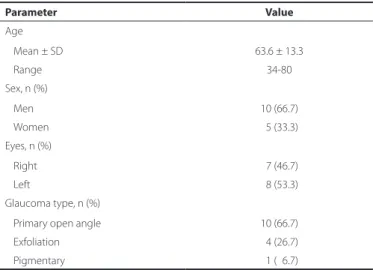
A total of 15 eyes of 15 patients were investigated in the study. Tables 1-2 show the baseline patient demographics, characteristics, and findings. Three patients (20%) previously underwent incisional glaucoma surgery. Of the 15 patients examined, nine (60%) underwent only XEN45 gel stent implantation surgery, with six (40%)undergoing XEN45 gel stent implantation combined with cataract surgery. There were no complications related to cataract surgery.
The mean IOP values were signiicantly lower than the preope-rative values at all postopepreope-rative visits (p<0.001; Figures 4-5). The mean number of antiglaucomatous medications was 3.60 ± 0.50 in the preoperative period; at 1 year after surgery, the mean number of antiglaucomatous medications had decreased to 0.33 ± 0.90 (p<0.001). At the end of the follow-up period, only two patients (13.3%) required antiglaucomatous treatment. In one patient, the stent was removed because it prolapsed into the anterior chamber, and antiglaucomatous treatment was started 12 months after sur-gery. None of the patients required further glaucoma sursur-gery.
Anterior chamber hemorrhage occurred in only one patient (6.6%) intraoperatively. This complication was easily resolved via an intraoperative viscoelastic injection into the anterior chamber. The vis -coelastic material was removed at the end of the operation. It did not cause any IOP increase in the postoperative period. None of the patients experienced any serious ocular complications (e.g., hy-potony, choroidal efusion, iritis, stent exposure, endophthalmitis) in the postoperative period.
DISCUSSION
In this study, implantation of an XEN45 gel stent signiicantly re duced IOP during the 12-month postoperative period. There are many advantages of this new surgical procedure; speciically, it is easy to learn and perform, and it allows for a short operating time with topical anesthesia. Another advantage is that this method can be easily performed in combination with cataract surgery. This new treatment modality also avoids the destructive complications encountered in other invasive surgical procedures. Because the XEN45 gel stent has a small internal lumen diameter (45 μm), it eli mi-nates postoperative hypotony by providing approximately 6-8 mmHg of outlow resistance(9-10,13).
The role of topical antiglaucomatous therapy in ocular surface disease has been demonstrated in several studies(14,15). Some patients with glaucoma cannot adapt to the use of topical antiglaucomatous drops, and thus, they discontinue the treatment. Therefore, XEN45 gel stent implantation should be considered to reduce the need for to-pical antiglaucomatous therapy and prevent ocular surface diseases. Because the XEN45 gel stent is a new device, there are few articles to date about its implantation in the literature(16-18). Previous authors(17) performed XEN45 gel stent implantation with mitomycin C injections combined with cataract surgery in 30 eyes. They achieved a signii-cant IOP reduction (29.34%) after 12 months of follow-up. At the end of the follow-up period, three patients required antiglaucomatous treatment. The most common complications were hemorrhage at the scleral exit point (90%) and minor intracameral hemorrhage (86%). No vision-threatening complications were observed.
Other authors(18) reported the results of XEN63 and XEN140 gel stent implantations, but without mitomycin C injections, combined with cataract surgery in 37 eyes. They achieved a signiicant IOP re-duction and a decreased number of antiglaucomatous medications
Table 2. Patient indings
Cases Sex Age Type of glaucoma Surgery Baseline IOP
Last visit IOP
Number of medications preoperatively
Number of medications
on last visit Complications
Case 01 M 57 POAG Xen 36 17 4 0 No
Case 02 M 71 POAG Xen+Phaco 35 15 4 0 No
Case 03 M 34 POAG Xen 39 16 3 0 No
Case 04 F 80 POAG Xen+Phaco 39 15 3 0 No
Case 05 M 69 PEXG Xen 32 12 4 0 No
Case 06 M 54 POAG Xen 39 25 4 2 No
Case 07 M 64 POAG Xen 34 24 4 3 Stent prolapsus
Case 08 M 78 POAG Xen 30 15 3 0 No
Case 09 F 76 POAG Xen+Phaco 34 18 3 0 No
Case 10 F 77 POAG Xen+Phaco 30 18 3 0 No
Case 11 M 40 POAG Xen 37 17 3 0 AC hemorrhage
Case 12 M 60 PG Xen 43 16 4 0 No
Case 13 F 62 PEXG Xen 40 16 4 0 No
Case 14 M 63 PEXG Xen+Phaco 38 15 4 0 No
Case 15 F 69 PEXG Xen+Phaco 35 12 4 0 No
POAG= primary open angle glaucoma; PG= pigmentary glaucoma; PEXG= pseudoexfoliative glaucoma; Phaco= phacoemulsiication; IOP= intraocular pressure. Table 1. Baseline patient demographics and characteristics
Parameter Value
Age
Mean ± SD 63.6 ± 13.3
Range 34-80
Sex, n (%)
Men 10 (66.7)
Women 05 (33.3)
Eyes, n (%)
Right 07 (46.7)
Left 08 (53.3)
Glaucoma type, n (%)
Primary open angle 10 (66.7)
Exfoliation 04 (26.7)
Oz a l Sa, e t a l.
3 8 5 Arq Bras Oftalmol. 2017;80(6):382-5
Figure 4. Changes in the mean intraocular pressure and percent reduction.
Figure 5. Changes in the intraocular pressure at 12 months after surgery compared to the preoperative value for each patient.
used after 12 months of follow-up. Twelve patients (32%) required a needling procedure with antiibrotic agents in the postoperative period. Hypotony was observed in the irst month after surgery, par-ticularly in the XEN140 group.
One study(19) presented the results of XEN45 gel stent implanta-tions with preoperative mitomycin C injecimplanta-tions in 71 eyes. The authors maintained a 36% decrease in the IOP level in 34 eyes at 12 months. The intraoperative complications were anterior camera bleeding in two patients, subconjunctival bleeding in one patient, and the preo-perative fracture of one implant. The postopreo-perative complications were a lat anterior chamber (1.4%), hyphema (4.1%), and hypotony below 6 mmHg on the irst day (12.6%).
Additionally, internal ostium occlusion of the XEN45 gel stent has been reported in the literature(20). The authors removed a blood clot occluding the internal ostium after XEN45 gel stent implantation, leading to a reduction in IOP.
In our study, the IOP reduction rate was higher at 12 months after surgery than reported in other studies in the literature. We suspect that this occurred because our baseline IOPs were higher than those in the other studies. Because mitomycin C may increase the incidence of late bleb-related complications, we did not inject this agent into the conjunctiva at any stage of the surgery. However, even without mitomycin C, we achieved a significant IOP reduction 12 months after the implantation of XEN45 gel stents.
There are some limitations to this study; in particular, our small number of patients and the lack of control group limit our
compari-sons. Thus, it has not been evaluated whether the eicacy of XEN45 gel implantation difers between patients according to prior glau-coma surgery. Furthermore, the mild IOP-reducing efect of phacoe-mulsiication alone was not evaluated. In addition, we could not eva-luate in which glaucoma type this surgical method is most efective. Clinical trials (NCT02006693, NCT02036541) are ongoing for XEN gel stents.(21) We think that after the results of these studies have been published, new aspects of XEN gel stent implantation will be revealed.
Consequently, the results of our study are promising, and XEN45 gel stent implantation presents a valuable contribution to glaucoma surgery. Using XEN45 gel stents, we reduced IOP and medication use without signiicant complications in patients with diferent forms of glaucoma. We believe that this new surgical procedure will play an important role in glaucoma surgery in the future. However, further studies with greater numbers of patients and longer follow-up periods are needed to clarify certain points.
REFERENCES
1. Tham Y-C, Li X, Wong TY, Quigley HA, Aung T, Cheng C-Y. Global prevalence of glau-coma and projections of glauglau-coma burden through 2040: A systematic review and meta-analysis. Ophthalmology. 2014;121(11):2081-90.
2. Heijl A, Leske MC, Bengtsson B, Hyman L, Bengtsson B, Hussein M. Reduction of intrao cular pressure and glaucoma progression: results from the Early Manifest Glaucoma Trial. Arch Ophthalmol. 2002;120(10):1268-79.
3. Nouri-Mahdavi K, Brigatti L, Weitzman M, Caprioli J. Outcomes of trabeculectomy for primary open-angle glaucoma. Ophthalmology. 1995;102(12):1760-9.
4. Landers J, Martin K, Sarkies N, Bourne R, Watson P. A twenty-year follow-up study of trabeculectomy: risk factors and outcomes. Ophthalmology. 2012;119(4):694-702. 5. Minckler D, Mosaed S, Dustin L, Ms BF. Trabectome (trabeculectomy-internal approach):
additional experience and extended follow-up. Trans Am Ophthalmol Soc. 2008;106: 149-59.
6. Mosaed S, Rhee DJ, Filippopoulos T, Tseng H, Deokule S, Weinreb RN. Trabectome outcomes in adult open-angle glaucoma patients: One-year follow-up. Clin Surg Ophthal mol. 2010;28(8-9):182-6.
7. Ting JLM, Damji KF, Stiles MC. Ab interno trabeculectomy: Outcomes in exfoliation versus primary open-angle glaucoma. J Cataract Refract Surg. 2012;38(2):315-23. 8. Bussell II, Kaplowitz K, Schuman JS, Loewen NA. Outcomes of ab interno
trabeculec-tomy with the trabectome by degree of angle opening. Br J Ophthalmol. 2015;99(7): 914-9.
9. Lewis RA. Ab interno approach to the subconjunctival space using a collagen glau-coma stent. J Cataract Refract Surg. 2014;40(8):1301-6.
10. Vera V, Horvath C. Xen gel stent: the solution designed by AqueSys. In: Samples JR, Ahmed, IIK, editors. Surgical innovations in glaucoma. New York: Springer Science+ Business Media; 2014. p. 189-98.
11. Richter GM, Coleman AL. Minimally invasive glaucoma surgery: current status and future prospects (review). Clin Ophthalmol. 2016;28(10):189-206.
12. Kerr NM, Wang J, Barton K. Minimally invasive glaucoma surgery as primary stand-alone surgery for glaucoma (review). Clin Exp Ophthalmol. 2016;45(4):393-400.
13. Sheybani A, Reitsamer H, Ahmed II. Fluid Dynamics of a novel micro-istula implant for the surgical treatment of glaucoma. Invest Ophthalmol Vis Sci. 2015;56(8):4789-95. 14. Garcia-Feijoo J, Sampaolesi JR. A multicenter evaulation of ocular surface disease
prevalence in patients with glaucoma. Clin Ophthalmol. 2012;6:441-6.
15. Valente C, Iester M, Corsi E, Rolando M. Symptoms and signs of tear ilm dysfunction in glaucomatous patients. J Ocul Pharmacol Ther. 2011;27(3):281-5.
16. Dupont G, Collignon N. New surgical approach in primary open-angle glaucoma: Xen Gel Stent a minimally invasive technique. Rev Med Liege. 2016;71(2):90-3. 17. Perez-Torregrosa VT, Olate-Perez A, Cerda-Ibanez M, Gargallo-Benedicto A, Osorio-Alayo
V, Barreiro-Rego A, et al. Combined phacoemulsuication and XEN45 surgery from a temporal approach and 2 incisions. Arch Soc Esp Oftalmol. 2016;91(9):415-21. 18. Sheybani A, Lenzhofer M, Hohensinn M, Reitsamer H, Ahmed IIK. Phacoemulsiication
combined with a new ab interno gel stent to treat open-angle glaucoma: pilot study. J Cataract Refract Surg. 2015;41(9):1905-9.
19. Lavin-Dapena C, Arteaga JV, Cervero MAC, Suarez LC, Segarra MAS, Mardero OD, et al. 12 month results from a minimally-invasive, 45μm lumen ab interno gelatin stent in combination with a preoperative mmc from a multicenter study conducted in Spain. Hong Kong: World Glaucoma Congress; 2015.
20. Pinto Ferreira N, Abegao Pinto L, Marques-Neves C. XEN gel stent ınternal ostium occlusion: Ab-ınterno revision. J Glaucoma. 2017;26(4):e150-e152.