In vivo OCT assessment of anterior segment central axial lengths with accommodation
Avaliação por OCT
in vivo
do comprimentos axiais centrais do segmento anterior
Daniel Monsálvez-RoMín1, aikateRini i. Moulakaki1, José J. esteve-taboaDa1, teResa FeRReR-blasco1, RobeRt Montés-Micó1
Submitted for publication: February 13, 2017 Accepted for publication: July 17, 2017
1 Optometry Research Group (GIO), Optics and Optometry and Vision Sciences Department, University
of Valencia, Valencia, Spain.
Funding: This study was supported by Ministerio de Economía y Competitividad (Research project SAF2013-44510-R with ERDF Funds from the European Union). Finantial support by the Daniel Monsálvez-Romín is a “Formación de Profesorado Universitario” Grantee (FPU13/05332, Minis-terio de Educación, Cultura y Deporte) and Aikaterini I. Moulakaki is a “Marie Curie Early Stage Researcher” from the AGEYE Initial Training Network Program FP7-People-2013-608049 Project.
Disclosure of potential conflicts of interest: None of the authors have any potential conflict of interests to disclose.
Corresponding author: José J. Esteve-Taboada. Optics and Optometry and Vision Sciences De-partment. University of Valencia - C/ Dr. Moliner, 50 - 46100 - Burjassot - Spain
Email: [email protected]
Approved by the following researchethics committee: Universitat de Valencia (# H146063858 4959).
ABSTRACT
Purpose: To assess changes in anatomic structures in the anterior eye segment in terms of axial lengths with accommodation via optical coherence tomography. Methods: In this observational study, 25 eyes of 25 healthy adults were examined using the Visante®omni optical coherence tomography system. Central corneal thickness, anterior chamber depth, central lens thickness, and anterior segment length were assessed. The evaluated parameters were obtained with accommoda-tion using different stimulus vergences, namely 0.0, -1.0, -2.0, and -3.0 D. Variaaccommoda-tion of these parameters was compared among different levels of accommodation. Results: Central corneal thickness was not altered at any stimulus vergence during accommodation (p>0.05). Conversely, anterior chamber depth was significantly reduced (p<0.05), whereas central lens thickness was significantly increased (p<0.05). Anterior segment length also increased with accommodation (p<0.05), indicating backward movement of the posterior pole.
Conclusions: There are significant variations in anterior segment lengths that occur with accommodation. Studying these changes will provide useful information regarding the accommodation mechanism that can improve our understanding of this process and facilitate clinical decision-making by practitioners.
Keywords: Anterior eye segment; Ocular accommodation; Optical coherence tomography
RESUMO
Objetivo: Avaliar as mudanças das estruturas anatômicas no segmento anterior do olho em termos de comprimentos axiais com acomodação por meio da tomografia de coerência óptica.
Métodos: Neste estudo observacional, foram incluídos 25 olhos de vinte e cinco adultos saudáveis e medidos com o sistema Visante® omni tomografia de coerência óptica. A espessura corneana central, a profundidade da câmara anterior, a espes-sura central da lente e o comprimento do segmento anterior foram avaliados. Os parâmetros avaliados foram obtidos com acomodação usando diferentes vergências de estímulo: 0,0, -1,0, -2,0 e -3,0 D. A variação desses parâmetros foi comparada para os diferentes níveis de acomodação.
Resultados: A espessura corneana central não foi alterada em nenhum estímulo durante a acomodação (p>0,05). A ACD mostrou uma redução significativa (p<0,05), enquanto a espessura central da lente foi significativamente aumentada (p<0,05). O comprimento do segmento anterior também aumentou com acomodação (p<0,05) indicando um movimento do polo posterior para trás.
Conclusões: Há variações significativas nos comprimentos do segmento anterior que ocorrem com acomodação. Estudar essas mudanças fornece informações úteis sobre o mecanismo de acomodação para os profissionais, a fim de obter uma melhor compreensão desse processo e ajudá-los a tomar suas decisões clínicas.
Descritores: Segmento anterior do olho; Acomodação ocular; Tomografia de coe-rência óptica
INTRODUCTION
Accommodation of the human eye is a dynamic process. Accor ding to Helmholtz’s theory, accommodation is a muscle-in duced activity. Specifically, the ciliary muscle contracts caused the release of the zonular fibers, leading to an increase of its refractive power to focus on close targets. The efficiency of this process declines with age, as the crystalline lens loses its elasticity and the activity of the ciliary muscle deteriorates, leading to presbyopia. Nevertheless, the in vivo crystalline lens response to ciliary muscle contraction remains under investigation, mainly due to visualization difficulties faced in recent decades(1).
Meanwhile, recent improvements in imaging technology have provided greater insights into the accommodation process in terms of biometrical changes that occur in the anterior segment of the
eye(2). At present, the presbyopic population must choose between
limited solutions of reading additions or implanting accommodative intraocular lenses (IOLs), among other techniques. However, it has also been reported in the literature that multiple companies are working to develop efficient accommodative IOLs, which rely on the action of the ciliary muscle. However, there is limited understanding of the presbyopic ciliary muscle function(3).
A number of studies have been conducted using different me-thods to visualize these structures such as magnetic resonance imaging, ultrasound biomicroscopy, and anterior segment optical
coherence tomography (OCT)(4,5). In the present study, the imaging
capabilities of the Visante®omni OCT system were used to measure
the changes that occur in the anterior segment at different accommo-dative demands.
such as blurred vision, headache, ocular discomfort, and loss of con-centration throughout a task(6,7). Thus, it is essential to visualize and evaluate the changes in the anterior segment to provide a better understanding of the processes occurring during accommodation to identify new solutions to correct presbyopia or other types of accommodative dysfunction.
METHODS
S
UBJECTSTwenty-five right eyes of healthy adults aged 20-45 years (29.5 ± 6.7 years) were included in this study. The refractive error of the patients was obtained via noncycloplegic autorefraction, and the value averaged -0.77 ± 1.90 diopters (D). The subjects had sufficient amplitude of accommodation (at least -3.00 D), which was evaluated
monocularly using Donders’ method(8). Their best-corrected visual
acuity evaluated using the ETDRS chart (Precision Vision, USA) was at least 20/20 in Snellen equivalents. The subjects had no ocular abnormality or systemic condition and no histories of ocular surgery, and they all presented clear intraocular media. The study followed the Declaration of Helsinki, and it was approved by the Ethics Committee of our research institution. The patients were informed about the details of the study, and each provided written consent after receiving written and verbal explanation of the implications.
M
EASUREMENTDEVICEANDPARAMETERSThe measurement device was the Visante®omni system (Carl Zeiss
AG, Oberkochen, Germany). This device combines OCT techno logy with Placido disk topography to obtain advanced corneal and ante-rior segment measurements. The noncontact diagnostic instrument acquires and analyzes detailed cross-sectional tomographic images of the anterior segment. It uses low-coherence interferometry to obtain the captures. Infrared light of 1310 nm is sent along an optical path that reaches the eye together with another reference path of the interferometer. Both paths are then combined at the photodetector to determine the axial depth of the tissue via the reflectivity signal.
The device allows the examiner to adjust the vergence of the visual target using a set of internal lenses, which is useful for evalua-ting the changes occurring with accommodation. Thus, the refractive error of the participants was corrected with the internal lenses, and all measurements were taken for the stimulus presented at 0.0 D relative to the patient’s far point (henceforth referred as the “unaccommoda-ted state” on) and for the accommoda“unaccommoda-ted eye with increasing stimuli at -1.0, -2.0, and -3.0 D.
The software of the Visante® system (version 3.0) disposes of a
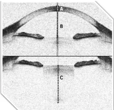
set of different scan types, two of which were used in this study: the “enhanced anterior segment single” and “raw image” mode. The “enhanced anterior segment single” mode catches four different tomograms composed of 256 A-scan primitive lines that represent a total area of 16 mm in width and 6 mm in depth and provides an average. This mode requires the examiner to take the capture centered on the cornea. It then processes the final image to detect the anterior and posterior corneal surfaces and fits two limiting lines matching these surfaces. This adjustment is useful for reshaping the tomograms and obtaining corrected distances. This mode was used to capture the central corneal thickness (CCT) and central anterior chamber depth (ACD).
The “raw image” mode was also used in this study. With the “raw image” mode, obtaining images deep into the eye is possible, allowing a full assessment of the lens thickness. This mode consists of 256 primitive A-scans that also represent a total area of 16 mm in width and 6 mm in depth. This mode was used to capture the axial central lens thickness (CLT). Additionally, the anterior segment length (ASL) was included as a calculated parameter by adding ACD and CLT. Normally, the ASL is defined as the distance from the central anterior
corneal surface to the posterior surface of the lens. In this work, this measurement was considered from the posterior surface of the cornea (endothelium) to the posterior surface of the lens to evaluate the change in position of the posterior pole of the crystalline lens without adding extra variability due to the corneal measurements.
All captured images were exported from the Visante® software
to external software. The measurements were extracted by using ImageJ, a public domain software for image processing and analysis developed by Wayne Rasband (National Institutes of Health, USA). All measurements were obtained in pixels in the first instance. The CCT was measured from the central corneal anterior surface or epithelium to the central posterior surface or endothelium. The ACD was measu-red from the central corneal endothelium to the anterior surface of the lens. The CLT was extracted from the central anterior surface to its central posterior surface (Figure 1).
The measurements were obtained in pixels and then converted to real physical measurements prior to analyzing the differences among accommodative stimuli. For this purpose, the measurements in pixels were transformed considering the refractive indices that the software assigns, as well as the total depth of the image yielded by the instrument with these modes (6 mm). The software only uses two different refractive indices for ocular media, namely those of the cornea (1.388) and aqueous humor (1.343)(9). To correct the crystalline lens measurements, the corneal index was used as an approximation because it is closer to the refractive index of the crystalline lens.
E
XPERIMENTALPROCEDUREThe patients were requested to maintain their fixation on the vi-sual target of the instrument before image capture. The patients were also requested to blink before starting the exam and to open their eyes wide until the measurements were finished to avoid affecting the measurement during the acquisition process. All measurements were obtained during a single session.
S
TATISTICALANALYSISStatistical analysis was performed using SPSS software (version 22.0, SPSS Inc., Chicago, IL, USA). Each measurement was extracted
three times per image, and a mean value was calculated. Repea-ted-measures analysis of variance (ANOVA) was performed to investigate significant differences among stimulus vergences for each parameter. The normality of datasets was assessed using the Shapiro-Wilk test. The ANOVA procedure based on the F statistic is robust under the breach of the normality assumption, provided that the data samples have no important asymmetries or similar
distri-bution shapes(10). Prior to ANOVA, the sphericity assumption was
checked using Mauchly’s sphericity test. The Greenhouse-Geisser correction was applied in cases in which the sphericity was
statisti-cally significant(11). The Bonferroni method was used as a post-hoc
test for comparisons among groups when ANOVA revealed signifi-cant differences between measurements. This procedure provides the significance level for paired differences between the individual conditions. The statistical significance limit was defined as p<0.05.
RESULTS
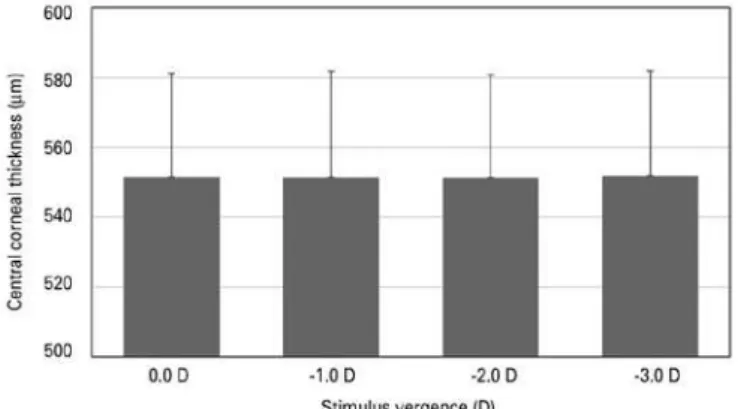
Regarding the central axial measurements that were analyzed, the variations of the CCT were not statistically significant among any of the considered pair comparisons, i.e., any accommodation stimulus (-1.0, -2.0, or -3.0 D) -induced change in this parameter with respect to the unaccommodated state or between accommodative states (p>0.05) (Figure 2). When considering the ACD measurements, the accommodation involved a significant reduction (p<0.05) for every pair comparison. More specifically, the ACD decreased with increasing accommodative stimulus (Figure 3). Moving deeper into the eye, the CLT also significantly increased with accommodation at
any stimulus vergence comparison. The lens increased its thickness with increasing stimulus size (Figure 4). Concerning the ASL, statisti-cally significant differences were also found for every pair comparison (Figure 5).
Table 1 shows the p-values of all stimulus comparisons for every studied parameter and thus all of the statistically significant differences that were found. Table 2 shows all of the results (mean ± standard deviation) extracted from the images of the anterior eye
segment using Visante®omni at all stimulus vergences, namely 0.0,
-1.0, -2.0, and -3.0 D.
DISCUSSION
Evaluating the anterior eye segment and variations of the ana-tomic structures is feasible thanks to the noninvasive technique
Figure 2. Central corneal thickness with accommodation at stimulus vergences of 0.0 (unaccommodated eye), -1.0, -2.0, and -3.0 D (accommodated eye).
Figure 3. Anterior chamber depth with accommodation at stimulus vergences of 0.0 (unaccommodated eye), -1.0, -2.0, and -3.0 D (accommodated eye).
Figure 4. Central lens thickness with accommodation at stimulus vergences of 0.0 (unaccommodated eye), -1.0, -2.0, and -3.0 D (accommodated eye).
Figure 5. Anterior segment length with accommodation at stimulus vergences of 0.0 (unaccommodated eye), -1.0, -2.0, and -3.0 D (accommodated eye).
Table 1. Pair comparisons
Pair comparison CCT ACD CLT ASL
-0.0 to -1.0 D 0.90 * * *
-0.0 to -2.0 D 0.83 * * *
-0.0 to -3.0 D 0.90 * * *
-1.0 to -2.0 D 0.91 * * *
-1.0 to -3.0 D 0.78 * * *
-2.0 to -3.0 D 0.80 * * *
termed anterior segment OCT. In this sense, this technique provides a useful tool for clinicians, as better comprehension of the mecha-nisms that involve the eye structures can be achieved(12–14). Since its introduction into clinical practice, it has been used to analyze structu-res such as the cornea, anterior chamber, and crystalline lens. This has crucial importance for making decisions such as special contact lens fittings such as those needed for keratoconus or post-LASIK corneas, as well as for cataract surgery and planning a pseudopha-kic or phapseudopha-kic IOL implantation(15,16). Opposed to just considering the eye as a static organ, the constant adjustment of its refractive power throughout the day to focus on objects at different distances must be considered. This is known as accommodation, and during this
process, the anatomical configuration of the eye changes(17). The
crystalline lens plays the main role in this process. It adjusts its refractive power as a response to a close stimulus to obtain a clear image in the retina. This involves secondary anatomical changes in the anterior segment of the eye(18-21).
In this research, we evaluated a set of central axial measurements and the effect of accommodation on these measurements. Specifi-cally, the CCT, ACD, CLT, and ASL were evaluated at different stimulus vergences corresponding to 0.0, -1.0, -2.0, and -3.0 D. The CCT was not altered during accommodation at any stimulus (p>0.05). Nevertheless, although the CCT appears unaffected, other changes in the shape of the cornea have been investigated in the presence of accommoda-tion. Prior studies evaluated the changes in its curvature with accom-modation using corneal topographers. In this regard, Yasuda et al. found statistically significant differences in the maximum K-values for the central 3.0, 5.0, and 7.0 mm by a mean of 0.62 D(22). Similarly, He et al.(23) found changes in the corneal surface with a Placido-based videoke-ratographer during accommodation that suggested an increase in the peripheral curvature with flattening at the vertex.
Concerning ACD measurements, statistically significant diffe-rences were noted for every pair comparison. The ACD was reduced with accommodation, and this reduction increased with increasing stimulus size. A pilot study performed by Del Águila et al.(24) analyzed the changes in the ACD at different parts of the anterior chamber using a Scheimpflug camera rotating system. They analyzed the variations with accommodation for the ACD at the central position, as well as at the superior-inferior and temporal-inferior pairs 2 mm away from the center. They also found a reduction of the mean ACD value with accommodation at all positions, with the extent of the reduction increasing with greater stimulus size up to -4.0 D. The reduction of the ACD with accommodation in a given patient is an important parameter to consider when an anterior chamber phakic IOL is to be implanted into the eye, as its proximity to the corneal en-dothelium and the push-up of the ocular structures with
accommo-dation could induce cell loss due to peripheral contact(25,26). The
anterior segment OCT device used in this study includes a set of embedded functions that allow preoperative simulation of the fit of a phakic IOL into the patient’s eye. This can be simulated at diffe-rent accommodative stimuli, which helps practitioners observe the changes of the ACD and assess the feasibility of this procedure(16).
Regarding the CLT, we found statistically significant changes for every group. The CLT increased with increasing accommodative sti-mulus up to the maximum vergence studied in this work. This beha-vior is well known as one the main implications during the process of accommodation. Other publications evaluated the change in the thickness of the crystalline lens, with the findings aligning with our
reported data(19,27). Furthermore, the ASL, as previously mentioned,
was calculated from the corneal endothelium to the posterior surface of the lens as the sum of the ACD and CLT. The point of including this measurement was assessing the change in position of the posterior pole of the crystalline lens. The change in position was proportional to the distance. The change of the anterior surface of the lens is directly related to the change in the ACD, as the anterior chamber is reduced or expanded proportionally to the displacement of its ante-rior surface, assuming that the cornea does not change its position during accommodation.
The ASL significantly changed with accommodation for all com-parisons in our study. This indicates that the posterior surface of the lens is moving backwards with accommodation. This is in accordance
with the results of Dubbelman et al.(28), who used a Scheimpflug
device and also found that the increase in the lens thickness with accommodation is larger than the decrease in the ACD. However, as the ASL has a smaller absolute variation than the ACD, this means that this posterior movement is smaller than that of the anterior lens surface, which carries a more significant accommodative component by moving closer to the cornea. In fact, in addition to increasing its thickness, the crystalline lens moves forward, which plays an important role in increasing the refractive power of the accommodated eye(29).
Nevertheless, it must be considered that the accommodation provided by the crystalline lens is due to the increasing thickness of its structure or the change in the distance to the cornea based on its movement, but a change in shape that increases its power
by steepening the curvature radii also occurs. Dubbleman et al.(28)
reported that both the anterior and posterior lens curvatures in-crease during accommodation, although the change in the latter is much smaller. Regarding the change in curvature of the 3-mm central zone, they found that both surfaces steepen, indicating that the lens power increases due to the more convex shape of the lens (approximately 64% steepening of the anterior curvature and 36% steepening of the posterior curvature). They also studied the change in the conic constant during accommodation of the anterior lens surface, determining that this value decreases with accommodation. This fact indicates that the lens surface becomes more curved and hyperbolic, in addition to moving toward the cornea. Additionally, it must be considered that the ciliary muscle plays a primary role in accommodation, although little is known about its exact behavior during contraction and there are some
limitations with its imaging and measurement(30). It is known that
the ciliary muscle relaxes or contracts to enable the lens to change its shape for focusing. Lossing et al.(1) analyzed changes in the ciliary muscle thickness with accommodation in young adults. Using their method, they confirmed that it is possible to examine the action
of the ciliary muscle with accommodation using the Visante® OCT
system and observed possible thickening of the anterior portion and thinning of the posterior portion of the ciliary muscle. However, the methods for measuring the ciliary muscle must be improved, and the results should be considered carefully.
Our findings provide additional useful information regarding the accommodation mechanism that can increase our understanding of this process and help practitioners with clinical decision-making. In our study, we considered different vergences, comparing the va-riations between them and with respect to the nonaccommodative state of the eye. This also helps reinforce the usefulness of the device
(Visante®omni system) used for the measurements, as many other
studies employed different systems. In addition, our study is a first step for further investigating this subject to compare the results at
Table 2. Variables extracted from the images of the anterior eye seg-ment using the Visante® omni system at various stimulus vergences
Vergence (D) CCT (µm) ACD (mm) CLT (mm) ASL (mm)
diffe rent conditions considering other factors, such as age, light con-ditions, level of ametropia, or nonsurgical treatments for presbyopia. In this sense, our study confirms previous findings and permits further research. Moreover, further clarification of this mechanism will fa-cilitate the design of new strategies for providing optical solutions for correcting presbyopia. Recent and future advances in imaging technology will improve the examination of changes of the ocular anatomy with accommodation.
REFERENCES
1. Lossing LA, Sinnott LT, Kao C-Y, Richdale K, Bailey MD. Measuring changes in ciliary muscle thickness with accommodation in young adults. Optom Vis Sci. 2012;89(5): 719-26.
2. Leung CKS, Chan W-M, Ko CY, Chui SI, Woo J, Tsang M-K, et al. Visualization of anterior chamber angle dynamics using optical coherence tomography. Ophthalmology. 2005; 112(6):980-4.
3. Glasser A. Restoration of accommodation: surgical options for correction of presbyo-pia. Clin Exp Optom. 2008;91(3):279-95.
4. Stachs O, Martin H, Kirchhoff A, Stave J, Terwee T, Guthoff R. Monitoring accommo-dative ciliary muscle function under three-dimensional ultrasound. Graefes Arch Clin Exp Ophthalmol. 2002;240(11):906-12.
5. Strenk SA, Strenk LM, Guo S. Magnetic resonance imaging of aging, accommodating, phakic, and pseudophakic ciliary muscle diameters. J Catarct Refract Surg. 2006;32(11): 1792-8.
6. Hokoda SC. General binocular dysfunctions in an urban optometry clinic. J Am Optom Assoc. 1985;56(7):560-2.
7. Mutti DO, Mitchell GL, Hayes JR, Jones LA, Moeschberger ML, Cotter SA, et al. Accommo-dative lag before and after the onset of myopia. Invest Ophthalmol Vis Sci. 2006; 47(3):837-46.
8. Donders F. On the anomalies of the accommodation and refraction of the eye. [edition translated from the author’s manuscript by William Daniel Moore]. UK: The New Sydenham Society; 1864. p.207-9.
9. Lehman BM, Berntsen DA, Bailey MD, Zadnik K. Validation of optical coherence tomography-based crystalline lens thickness measurements in children. Optom Vis Sci. 2009;86(3):181-7.
10. Tan WY. Sampling distributions and robustness of t, F and variance-ratio in two samples and ANOVA models with respect to departure from normality. Communication in statistics–Theory and Methods. 1982;11:486-511.
11. Box GEP. Some theorems on quadratic forms applied in the study of analysis of variance problems. II: Effects of inequality of variance and of correlation between errors in the two-way classification. Ann Mathemat Stat. 1965;25:484-98.
12. Ramos JL, Li Y, Huang D. Clinical and research applications of anterior segment optical coherence tomography - a review. Clin Experiment Ophthalmol. 2009;37(1):81-9.
13. Farouk MM, Naito T, Shinomiya K, Eguchi H, Sayed KM, Nagasawa T, et al. Optical coherence tomography reveals new insights into the accommodation mechanism. J Ophthalmol. 2015;2015.
14. Radhakrishnan S, Rollins a M, Roth JE, Yazdanfar S, Westphal V, Bardenstein DS, et al. Real-time optical coherence tomography of the anterior segment at 1310 nm. Arch Ophthalmol. 2001;119(8):1179-85.
15. Doors M, Berendschot TT, de Brabander J, Webers CAB, Nuijts RM, Huang D, et al. Value of optical coherence tomography for anterior segment surgery. J Cataract Refract Surg. 2010;36(7):1213-29.
16. Doors M, Berendschot TT, Hendrikse F, Webers CA, Nuijts RM. Value of preoperative phakic intraocular lens simulation using optical coherence tomography. J Cataract Refract Surg. 2009;35(3):438-43
17. Baikoff G, Lutun E, Ferraz C, Wei J. Static and dynamic analysis of the anterior segment with optical coherence tomography. J Cataract Refract Surg. 2004;30(9):1843-50. 18. Domínguez-Vicent A, Monsálvez-Romín D, Del Águila-Carrasco AJ, Ferrer-Blasco T,
Montés-Micó R. Changes in the anterior chamber during accommodation assessed with a Scheimpflug system. J Cataract Refract Surg. 2014;40(11):1790-7
19. Richdale K, Bullimore MA, Zadnik K. Lens thickness with age and accommodation by optical coherence tomography. Ophthalmic Physiol Opt. 2008;28(5):441-7. 20. Malyugin BE, Shpak AA, Pokrovskiy DF. Accommodative changes in anterior chamber
depth in patients with high myopia. J Cataract Refract Surg. 2012;38(8):1403-7. 21. Ciuffreda KJ. Chapter 4 - Accommodation, the pupil, and presbyopia. In: Benjamin
Consultant WJ, Borish IM, editors. Borish’s clinical refraction. 2nd ed. Saint Louis: Butterworth-Heinemann; 2006. p.93-144.
22. Yasuda A, Yamaguchi T, Ohkoshi K. Changes in corneal curvature in accommodation. J Cataract Refract Surg. 2003;29(7):1297-301.
23. He JC, Gwiazda J, Thorn F, Held R, Huang W. Change in corneal shape and corneal wave-front aberrations with accommodation. J Vis. 2003;3(7):456-63.
24. Del Águila-Carrasco AJ, Domínguez-Vicent A, Monsálvez-Romín D, Pérez-Vives C, Sanchis V. Changes in anterior chamber depth during accommodation: a pilot study. J Emmetropia, 2013;87-91.
25. Coullet J, Mahieu L, Malecaze F, Fournié P, Leparmentier A, Moalic S, et al. Severe endothelial cell loss following uneventful angle-supported phakic intraocular lens implantation for high myopia. J Cataract Refract Surg. 2007;33(8):1477-81. 26. Kim M, Kim JK, Lee HK. Corneal endothelial decompensation after iris-claw phakic
intraocular lens implantation. J Cataract Refract Surg. 2008;34(3):517-9.
27. Dubbelman M, Van Der Heijde GL, Weeber HA, Vrensen GFJM. Changes in the internal structure of the human crystalline lens with age and accommodation. Vision Res. 2003;43(22):2363-75.
28. Dubbelman M, Van Der Heijde GL, Weeber HA. Change in shape of the aging human crystalline lens with accommodation. Vision Res. 2005;45(1):117-32.
29. He JC, Wang J, Wei M, Mao J. Forward movement of the crystalline lens plays an important role in accommodation. Invest Ophthalmol Vis Sci. 2014;55(13):3779. 30. Bailey MD. How should we measure the ciliary muscle? Invest Ophthalmol Vis Sci.