O
r i g i n a la
rt i c l e3 7 8 Arq Bras Oftalmol. 2017;80(6):378-81 http://dx.doi.org/10.5935/0004-2749.20170092
INTRODUCTION
Optical coherence tomography (OCT) is a well-known tool for the high-resolution assessment of retinal pathologies. Spectral-domain OCT (SD-OCT) is a more recent technique that allows the imaging of ocular structures with faster scan rates than OCT(1-2). The evaluation of intraretinal layer thickness is becoming increasingly important for the diagnosis and monitoring of various ocular diseases(3). However, few studies have assessed the repeatability and reproducibility of automated total retinal thickness measurements using diferent SD-OCT instruments in healthy individuals(4,5) or those with ocular pathology(6-11).
ABSTRACT
Purpose: To evaluate the repeatability and reproducibility of automatic seg-mentation in healthy subjects using a Spectralis optical coherence tomography (OCT) system.
Methods: A total of 60 eyes from 60 patients were included in this prospective study. Spectral-domain optical coherence tomography images were generated using the Spectralis OCT system. An automated algorithm was used to segment the macular retina into nine layers and evaluate the thickness of each layer in the foveal, inner, and outer Early Treatment Diabetic Retinopathy Study (ETDRS) subield rings. The eyes were imaged three times by an examiner to assess intraobserver repeatability and imaged once by a second examiner to assess interobserver re-producibility. The first scan was used for reference, whereas the second and third scans were collected using the device’s follow-up mode. Intraclass correlation coefficients (ICCs) of repeatability and reproducibility were analyzed.
Results: The examiners achieved high repeatability and reproducibility for all parameters. Good agreement was found for all parameters in all ETDRS subdivi-sions with an ICC of >0.96 for all measurements.
Conclusion: It is possible to obtain cross-sections from the same location using the device’s follow-up mode, making it virtually impossible to distinguish between repeated measurements taken with the device by different examiners.
Keywords: Retina; Macula lutea; Tomography, optical coherence; Reproducibility of results; Image processing, Computer-assisted
RESUMO
Objetivo: Avaliar a repetibilidade e a reprodutibilidade da segmentação auto-má tica com tomografia de coerência óptica Spectralis em indivíduos saudáveis.
Métodos: Foram incluídos neste estudo prospectivo um total de 60 olhos de 60 pa-cientes. As imagens de tomografia de coerência óptica de domínio espectral (SD-OCT) foram geradas com Spectralis OCT (Heidelberg Engineering, Heidelberg, Alemanha). Um algoritmo automatizado foi utilizado para segmentar a retina macular em nove camadas e quantificar a espessura de cada camada em anéis de subcampo ETDRS fovetais, internos e externos. Os olhos de cada paciente foram imaginados três vezes pelo primeiro examinador para avaliar a repetibilidade intra-observador e uma vez pelo segundo examinador para avaliar a reprodutibilidade entre observadores. A primeira verificação foi definida como a varredura de referência, enquanto a segunda e a terceira varredura foram as varreduras de seguimento e foram realizadas com o uso do modo de acompanhamento, respectivamente. O coeficiente de correlação intraclasse (ICC) de repetibilidade e reprodutibilidade foi analisado. Repetibilidade e reprodutibilidade das medidas obtidas foram analisadas utilizando o coeficiente de correlação intraclasse.
Resultados: Os examinadores alcançaram alta repetibilidade e reprodutibilidade para todos os parâmetros. Foi encontrado um bom acordo para todos os parâmetros em todas as subdivisões de ETDRS com um coeficientes de correlação intraclasse superior a 0,96 para todas as medições.
Conclusão: É possível obter seções transversais do mesmo local usando o modo de acompanhamento, o que torna praticamente impossível distinguir entre medições repetidas do dispositivo independentemente do examinador.
Descritores: Retina; Mácula lútea; Tomografia de coerência óptica; Reprodutibilidade dos testes; Processamento de imagem assistida por computador
The aim of this study was to investigate the repeatability and reproducibility of automatic segmentation of nine intraretinal layers in healthy subjects using a SD-OCT system.
METHODS
S
UBJECTSThis study followed the tenets of the Declaration of Helsinki and was approved by the Ethics Committee of Afyon Kocatepe Uni-versity. Healthy subjects (28 males, 32 females) who were admitted to the ophthalmology department for regular ophthalmological
Repeatability and reproducibility of automatic segmentation of retinal layers in
healthy subjects using Spectralis optical coherence tomography
Repetibilidade e reprodutibilidade da segmentação automática de camadas retinianas em
sujeitos saudáveis com tomograia de coerência óptica Spectralis
Ersan ÇEtinkaya1, rahmi Duman2, Reşat Duman2, mEhmEt CEm sabanEr2
Submitted for publication: April 25, 2017 Accepted for publication: July 17, 2017
1 Department of Ophthalmology, Manisa State Hospital, Manisa, Turkey.
2 Department of Ophthalmology, School of Medicine, Afyon Kocatepe University, Afyonkarahisar,
Turkey.
Funding: No specific financial support was available for this study.
Disclosure of potential conflicts of interest: None of the authors have any potential conflict of interest to disclose.
Corresponding author: Ersan Çetinkaya. Department of Ophthalmology. Manisa State Hospital. Manisa, Turkey - E-mail:[email protected]
Çe t i n kaya e, e t a l.
3 7 9 Arq Bras Oftalmol. 2017;80(6):378-81 exa minations with no history of ocular disease except for refractive
errors were included, and each signed an informed consent form. The exclusion criteria were ocular surface disorder, corneal disease, previous corneal surgery, contact lens wear, history of ocular trauma or inlammation, and systemic disease.
M
EASUREMENTDEVICERetinal OCT imaging was performed using the Spectralis OCT sys-tem (Heidelberg Engineering GmbH, Heidelberg, Germany). Segmen-tation of the retinal layers from each SD-OCT scan was performed using the built-in Spectralis mapping software the Heidel berg Eye Explorer (version 6.0). Spectralis segmentation software was used to obtain the following thickness measurements: total retinal thickness (Retina), retinal nerve iber layer (RNFL), ganglion cell layer (GCL), inner plexiform layer (IPL), inner nuclear layer (INL), outer plexiform layer (OPL), outer nuclear layer (ONL), and retinal pigment epithelium (RPE). The automatic segmentation tool of the posterior pole scan also provides the thickness of the inner retinal layer (IRL), measured from the internal limiting membrane to the external limiting mem-brane (ELM), and that of the outer retinal layer (ORL), measured from the ELM to the Bruch membrane (Figure 1). The Spectralis mapping software generates automated retinal thickness measurements based on analyses of the central, inner ring, and outer ring subields as deined by the Early Treatment Diabetic Retinopathy Study (ETDRS)(12).
M
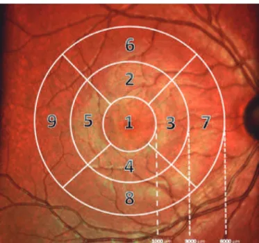
EASUREMENTTECHNIQUEMeasurements were taken according to the guidelines of the respective device manufacturers. Before imaging, all eyes were subjected to an ocular examination including visual acuity testing, auto-refraction, intraocular pressure, and ophthalmoscopic exami-nation. After enrollment, all eyes were imaged without mydriasis. Three repeated measurements were performed within a short pe-riod of time on the same day by a single examiner (EC) to test the in traobserver repeatability. The first examination was marked as the reference in the follow-up mode, a special feature of the Heidelberg SD-OCT system that allows scanning of the same part of the retina during follow-up visits. SD-OCT imaging was also performed once by another examiner (MCS) on the same day to test interobserver re-producibility. The thicknesses of all layers within the central ETDRS zones of 0-1000 and 1000-3000 μm (inner ring) and 3000-6000 μm diameter (outer ring) were recorded for each scan (Figure 2).
S
TATISTICALANALYSISAll of the statistical analyses were performed by Statistical Package for the Social Sciences software (version 17.0 for Windows; SPSS Inc., Chicago, IL, USA). Descriptive statistics are shown as mean ± standard deviation. The intraobserver repeatability was measured with three OCT images obtained by the same operator, while the interobserver reproducibility was measured with two OCT images obtained by two diferent operators. The overall mean thickness and intraclass correla-tion coeicients (ICCs) were calculated to evaluate the repea tability
and reproducibility of the thickness measurements. The overall mean thickness of the nine intraretinal layers along the central macular 6-mm scan length was determined as the mean of the three mea-surements by the same examiner. The paired ICCs were analyzed to evaluate the agreement of the thickness measurements between the two SD-OCT instruments.
RESULTS
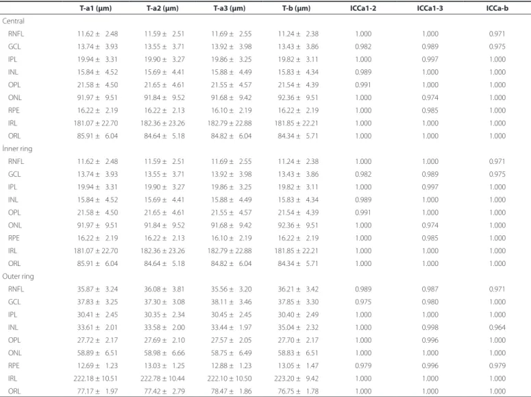
Sixty volunteers (28 men, 32 women) aged 20-40 years (mean age, 30.26 ± 6.31 years) participated in this study. Table 1 shows the repeatability and reproducibility of the thickness measurements for intraretinal layers measured using the Spectralis OCT system. There were no signiicant diferences among the three thickness measu-rements obtained by the same examiner. ICCs obtained for the in-traobserver repeatability and interobserver reproducibility tests were >0.964 for the entire retina and for all of the intraretinal layers in all of the subields (Table 1).
DISCUSSION
Repeatability and reproducibility are very important for assessing measurement luctuations for an instrument and when an instru-ment is used by multiple operators. Measureinstru-ments of the intraretinal layers are signiicant morphometric parameters in the diagnosis of retinal and neurological diseases as well as for monitoring disease progression(13). In this study, we evaluated the repeatability and re-producibility of measurements for nine intraretinal layers (RNFL, GCL, IPL, INL, OPL, ONL, RPE, IRL, ORL) determined by an automated algo-rithm applied to images obtained using the SD-OCT system with the follow-up mode. The follow-up mode is also called AutoRescan and implements active eye tracking to perform OCT scans automatically on the retina at the same point as in the previous review(14). In our study, for both intraobserver and interobserver comparisons, ICCs were high for all layers. The intraobserver and interobserver test results indicated that the SD-OCT system produced excellent repeatable and reproducible measurements for all of the intraretinal layers. The repeatability of measurements in the present study is consistent with those in previous reports involving diferent OCT devices.
Figure 1. Segmented view of the retinal layers created using the Heidelberg Spectralis
Re p e ata b i l i t ya n dR e p R o d u c i b i l i t yo fau t o m at i cs e g m e n tat i o no fR e t i n a ll ay e R si nh e a lt h ys u b j e c t su s i n g sp e c t R a l i so p t i c a lc o h e R e n c et o m o g R a p h y
3 8 0 Arq Bras Oftalmol. 2017;80(6):378-81
Table 1. Intraobserver and interobserver agreement for retinal layer thickness
T-a1 (μm) T-a2 (μm) T-a3 (μm) T-b (μm) ICCa1-2 ICCa1-3 ICCa-b
Central
RNFL 011.62 ± 02.48 011.59 ± 02.51 011.69 ± 02.55 011.24 ± 02.38 1.000 1.000 0.971
GCL 013.74 ± 03.93 013.55 ± 03.71 013.92 ± 03.98 013.43 ± 03.86 0.982 0.989 0.975
IPL 019.94 ± 03.31 019.90 ± 03.27 019.86 ± 03.25 019.82 ± 03.11 1.000 0.997 1.000
INL 015.84 ± 04.52 015.69 ± 04.41 015.88 ± 04.49 015.83 ± 04.34 0.989 1.000 1.000
OPL 021.58 ± 04.50 021.65 ± 04.61 021.55 ± 04.57 021.54 ± 04.39 0.991 1.000 1.000
ONL 091.97 ± 09.51 091.84 ± 09.52 091.68 ± 09.42 092.36 ± 09.51 1.000 0.974 1.000
RPE 016.22 ± 02.19 016.22 ± 02.13 016.10 ± 02.19 016.22 ± 02.19 1.000 0.985 1.000
IRL 181.07 ± 22.70 182.36 ± 23.26 182.79 ± 22.88 181.85 ± 22.21 1.000 1.000 1.000
ORL 085.91 ± 06.04 084.64 ± 05.18 084.82 ± 06.04 084.34 ± 05.71 1.000 1.000 1.000
İnner ring
RNFL 011.62 ± 02.48 011.59 ± 02.51 011.69 ± 02.55 011.24 ± 02.38 1.000 1.000 0.971
GCL 013.74 ± 03.93 013.55 ± 03.71 013.92 ± 03.98 013.43 ± 03.86 0.982 0.989 0.975
IPL 019.94 ± 03.31 019.90 ±0 3.27 019.86 ± 03.25 019.82 ± 03.11 1.000 0.997 1.000
INL 015.84 ± 04.52 015.69 ± 04.41 015.88 ± 04.49 015.83 ± 04.34 0.989 1.000 1.000
OPL 021.58 ± 04.50 021.65 ± 04.61 021.55 ± 04.57 021.54 ± 04.39 0.991 1.000 1.000
ONL 091.97 ± 09.51 091.84 ± 09.52 091.68 ± 09.42 092.36 ± 09.51 1.000 0.974 1.000
RPE 016.22 ± 02.19 016.22 ± 02.13 016.10 ± 02.19 016.22 ± 02.19 1.000 0.985 1.000
IRL 181.07 ± 22.70 182.36 ± 23.26 182.79 ± 22.88 181.85 ± 22.21 1.000 1.000 1.000
ORL 085.91 ± 06.04 084.64 ± 05.18 084.82 ± 06.04 084.34 ± 05.71 1.000 1.000 1.000
Outer ring
RNFL 035.87 ± 03.24 036.08 ± 03.81 035.56 ± 03.20 036.21 ± 03.42 0.989 0.987 0.971
GCL 037.83 ± 03.25 037.30 ± 03.08 038.11 ± 03.46 037.85 ± 03.30 0.975 0.980 1.000
IPL 030.41 ± 02.45 030.35 ± 02.34 030.45 ± 02.45 030.40 ± 02.49 1.000 1.000 1.000
INL 033.61 ± 02.01 033.58 ± 02.00 033.44 ± 01.97 035.04 ± 02.32 1.000 0.998 0.964
OPL 027.72 ± 02.17 027.69 ± 02.10 027.57 ± 02.05 027.70 ± 02.17 1.000 0.996 1.000
ONL 058.89 ± 06.51 058.98 ± 06.66 058.75 ± 06.49 058.83 ± 06.51 1.000 1.000 1.000
RPE 012.69 ± 01.23 013.03 ± 01.25 012.88 ± 01.23 013.05 ± 01.47 0.979 0.996 0.979
IRL 222.18 ± 10.51 222.78 ± 10.44 222.10 ± 10.50 223.20 ± 09.42 1.000 1.000 1.000
ORL 077.17 ± 01.97 077.42 ± 02.79 078.47 ± 01.86 076.75 ± 01.78 1.000 1.000 1.000
T-a1= mean thickness of irst measurement by examiner 1; T-a2= mean thickness of second measurement by examiner 1; T-a3= mean thickness of third measurement by examiner 1; T-b= mean thickness of irst measurement by examiner 2; ICC a1-2= intraclass correlation coeicients of repeatability (T-a1-T-a2); ICC a1-3= intraclass correlation coeicients of repea-tability (T-a1-T-a3); ICCa-b= intraclass correlation coeicients of reproducibility; RNFL= retinal nerve iber layer; GCL= ganglion cell layer IPL; inner plexiform layer; INL= inner nuclear layer; OPL= outer plexiform layer; ONL= outer nuclear layer; RPE= retinal pigment epithelium; IRL= inner retinal layer; ORL= outer retinal layer.
Previous studies reported a higher degree of repeatability for ma -cular thickness with Spectralis OCT measurements. Fiore et al. de-monstrated that the eye tracking mechanism and follow-up function improved reproducibility, ensuring the same scanning location was selected on following visits and that moving artifacts were redu-ced(15). Ctori et al. reported excellent repeatability and reproducibility for the thickness of each of eight individual retinal layers located centrally or at 2° or 5° eccentricity away from the foveal measure-ments obtained using the SD-OCT segmentation software in a young healthy cohort(16). What diferentiates our study from previous studies is its assessment of repeatability and reproducibility in all subdivisions (wide area) of ETDRS. Debuc et al. reported the repeatability and reproducibility of thickness measurements for six intraretinal layers using automated custom software developed for the Stratus OCT with healthy subjects. They found that ICCs were >0.75 for all layers except for the OPL and OS/RPE(17). Wang et al. reported the repeatabi-lity of thickness measurements for nine intraretinal layers determined by manual segmentation of ultra-high-resolution OCT (UHR-OCT) images with an axial resolution of approximately 2 μm with healthy subjects. The ICC was >0.90 in most of the intraretinal layers(18). Liu
et al. reported the repeatability and reproducibility of thickness mea-surements for eight-retinal layers using a custom-built UHR-OCT instrument (~3 μm resolution) and a commercial RTVue100 OCT (~5 μm resolution) instrument with 20 normal subjects. They found ICCs of >0.80 for all layers except for the inner and outer segments(13). Our results are consistent with those of these studies, showing that the thickness measurements of higher-axial-resolution OCT instru-ments have better repeatability for all retinal layers.
There are some limitations to the present study. First, it included only healthy subjects. Diseased retinal structures may vary subs-tantially among patients, which is likely to increase the frequency of segmentation errors. Thus, the repeatability and reproducibility values may be reduced in diseased retinas. In future studies, we will apply our new method to evaluate a variety of retinal diseases. However, the purpose of the current study was to determine the clinical signiicance of any changes in the repeatability and repro-ducibility of the intraretinal measurements.
Çe t i n kaya e, e t a l.
3 8 1 Arq Bras Oftalmol. 2017;80(6):378-81
REFERENCES
1. Nassif N, Cense B, Park B, Pierce M, Yun S, Bouma B, et al. In vivo high resolution vi-deo-rate spectral-domain optical coherence tomography of the human retina and optic nerve. Opt Express. 2004;12(3):367-76.
2. Wojtkowski M, Bajraszewski T, GorczyÅska I, Targowski P, Kowalczyk A, Wasilewski W, et al. Ophthalmic imaging by spectral optical coherence tomography. Am J Ophthalmol. 2004;138(3):412-9.
3. Cesareo M, Ciufoletti E, Martucci A, Balducci C, Cusumano A, Ricci F, et al. Automatic segmentation of posterior pole retinal layers ın patients with early stage glaucoma using spectral domain optical coherence tomography. J Clin Exp Ophthalmol. 2016; 7:538. doi: 10.4172/2155-9570.1000538.
4. Tan CS, Li KZ, Lim TH. A novel technique of adjusting segmentation boundary layers to achieve comparability of retinal thickness and volumes between spectral domain and time domain optical coherence tomography. Invest Ophthalmol Vis Sci. 2012; 53(9):5515-9.
5. Wolf-Schnurrbusch UE, Ceklic L, Brinkmann CK, Iliev ME, Frey M, Rothenbuehler SP, et al. Macular thickness measurements in healthy eyes using six diferent optical coherence tomography instruments. Invest Ophthalmol Vis Sci. 2009;50(7):3432-7. 6. Krebs I, Smretschnig E, Moussa S, Brannath W, Womastek I, Binder S. Quality and
re producibility of retinal thickness measurements in two spectral-domain optical coherence tomography machines. Invest Ophthalmol Vis Sci. 2011;52(9):6925-33. 7. Comyn O, Heng LZ, Ikeji F, Bibi K, Hykin PG, Bainbridge JW, et al. Repeatability of
Spectralis OCT measurements of macular thickness and volume in diabetic macular edema. Invest Ophthalmol Vis Sci. 2012;53(12):7754-9.
8. Patel PJ, Chen FK, Ikeji F, Xing W, Bunce C, Da Cruz L, et al. Repeatability of stratus optical coherence tomography measures in neovascular age-related macular dege-neration. Invest Ophthalmol Vis Sci. 2008;49(3):1084-8.
9. Patel PJ, Chen FK, Ikeji F, Tufail A. Intersession repeatability of optical coherence to-mography measures of retinal thickness in early age-related macular degeneration. Acta Ophthalmol. 2011;89(3):229-34.
10. Pinilla IM, Garcia-Martin EM, Fernandez-Larripa SM, Fuentes-Broto LP, Sanchez-Cano AI, Abecia EM. Reproducibility and repeatability of cirrus and spectralis fourier-domain optical coherence tomography of healthy and epiretinal membrane eyes. Retina. 2013; 33(7):1448-55.
11. Diabetic Retinopathy Clinical Research Network Writing Committee, Bressler SB, Edwards AR, Chalam KV, Bressler NM, Glassman AR, Jafe GJ, Melia M, Saggau DD, Plous OZ. Reproducibility of spectral-domain optical coherence tomography retinal thickness measurements and conversion to equivalent time-domain metrics in diabetic macular edema. JAMA Ophthalmol. 2014;132(9):1113-22. doi: 10.1001/ jamaophthalmol.2014.1698.
12. Photocoagulation for diabetic macular edema. Early Treatment Diabetic Retinopathy Study Report No.1. 1. Early Treatment Diabetic Retinopathy Study research group. Arch Ophthalmol. 1985;103(12):1796-806.
13. Liu X, Shen M, Huang S, Leng L, Zhu D, Lu F. Repeatability and reproducibility of eight macular intra-retinal layer thicknesses determined by an automated segmentation algorithm using two SD-OCT instruments. PLoS One. 2014;9(2):e87996.
14. Heidelberg Engineering. Spectralis HRA + OCT User manual software version 6.0. Heidelberg: Germany; 2015.
15. Fiore T, Lupidi M, Androudi S, Giansanti F, Fruttini D, Cagini C. Repeatability of retinal macular thickness measurements in healthy subjects and diabetic patients with clinically signiicant macular edema: Evaluation of the follow-up system of Spectralis optical coherence tomography. Ophthalmologica. 2015;233(3-4):186-91.
16. Ctori I, Huntjens B. Repeatability of foveal measurements using spectralis optical coherence tomography segmentation software. PLoS One. 2015;10(6):e0129005. doi: 10.1371/journal.pone.0129005.
17. DeBuc DC, Somfai GM, Ranganathan S, Tátrai E, Ferencz M, Puliaito CA. Reliability and reproducibility of macular segmentation using a custom-built optical coherence tomography retinal image analysis software. J Biomed Opt. 2009;14(6):064023. doi: 10.1117/1.3268773.