623
Arq Neuropsiquiatr 2010;68(4):623-626
Article
Suboccipital craniectomy with
or without duraplasty
What is the best choice in patients
with Chiari type 1 malformation?
Flávio Ramalho Romero1, Clemente Augusto de Brito Pereira2
ABSTRACT
The best surgical treatment for Chiari malformation is unclear, especially in patients with syringomyelia. We reviewed the records of 16 patients who underwent suboccipital craniectomy at our institution between 2005 and 2008. Of the six patients who did not undergo duraplasty, four showed improvement postoperatively. Two patients without syringomyelia showed improvement postoperatively. Of the four patients with syringomyelia, three showed improvement, including two with a decrease in the cavity size. One patient showed improvement in symptoms but the syringomyelia was unchanged. The cavity size increased in the one patient who did not show improvement. Among the 10 patients who underwent duraplasty, improvements were noted in four of the five patients without syringomyelia and in all of the five with syringomyelia. There is a suggestion that patients with syringomyelia may have a higher likelihood of improvement after undergoing duraplasty.
Key words: Chiari type 1, syringomyelia, suboccipital craniectomy, duraplasty.
Craniectomia suboccipital com ou sem duroplastia: qual a melhor escolha em pacientes com malformação de Chiari tipo 1?
RESUMO
A melhor opção de tratamento cirúrgico na malformação de Chiari é desconhecido, especialmente em paciente com siringomielia. Nós revisamos casos de 16 pacientes submetidos à craniectomia suboccipital em nossa instituição de 2005 à 2008. Dos 6 pacientes que não foram submetidos à duroplastia, quatro tiveram melhora pós operatória. Dois pacientes sem siringomielia tiveram melhora pós operatória. Dos quatro pacientes com siringomielia, três tiveram melhora, incluindo dois com diminuição do tamanho da cavidade. Um paciente teve melhora da sintomatologia mas a siringomielia não se modificou. Dez pacientes foram submetidos à duroplastia. Melhora foi observada em 4 de 5 pacientes sem siringomielia e 5 de 5 casos com siringomielia. Há uma sugestão de que pacientes com siringomielia possam ter uma maior taxa de melhora após serem submetidos à duroplastia.
Palavras-chave: Chiari tipo 1, siringomielia, craniectomia suboccipital, duroplastia.
Correspondence Flávio Ramalho Romero Rua Pascoal Vita 366 / 94 05445-000 São Paulo SP - Brazil E-mail: [email protected] [email protected]
Received 26 October 2009
Received in final form 20 January 2010 Accepted 2 February 2010
Neurosurgery Service of Hospital Heliópolis - SUS, São Paulo SP, Brazil: 1Former Resident at Hospital Heliópolis - SUS.
Neurosurgeon at Hospital Ipiranga, Hospital São Camilo, and Hospital São Luiz, São Paulo SP, Brazil; 2Former head of
Neurosurgery Division at Hospital Heliópolis - SUS. Neurosurgeon at Hospital Santa Catarina, São Paulo SP, Brazil. Chiari malformation type I (CM1)
con-sists of protrusion of the cerebellar ton-sils through the foramen magnum, de-fined radiologically as tonsillar descent of 5 mm or more1-5. Although many indi-viduals with CM1 are asymptomatic, the malformation can cause headaches,
inves-Arq Neuropsiquiatr 2010;68(4)
624
Chiari type 1 malformation: suboccipital craniectomy Romero et al.
tigation of brain magnetic resonance images (MRIs) re-ported that the prevalence of CM1 was one case in 1,280 individuals8.
CM1 has been associated with syringomyelia in 30 to 70% of patients4,5. he best surgical treatment for Chiari malformation is unclear, especially in patients with sy-ringomyelia1-5. he various surgical approaches attempt-ed have includattempt-ed suboccipital craniectomy, syringostomy, obex plugging, syringosubarachnoid shunting, ventricu-loperitoneal shunting and fourth ventriculosubarachnoid shunting1-3,7,9-12. Posterior craniocervical decompression is the procedure currently most used for treating CM1 (alone or in association with syringomyelia, in the ab-sence of hydrocephalus).
Patients with syringomyelia have a poorer out-come with surgery, compared with those without syringomyelia3,6,13-15. Craniocervical junction decompres-sion in hindbrain-related syringomyelia achieves an im-provement in symptoms in approximately two-thirds of patients14. In a review of patients who underwent Chiari decompression with or without duraplasty, Matsumoto and Symon15 noted no diference in the reduction of sy-ringomyelia and Munshi et al.15 showed that regarding the improvement in symptoms, patients without duraplasty had a signiicantly worse outcome, compared with those who underwent duraplasty.
In the present report, we review our experience with the treatment of CM1 in patients who underwent decom-pression with or without duraplasty.
METHOD
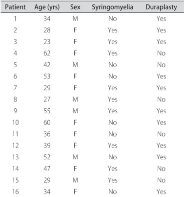
We reviewed the records of 16 patients who under-went Chiari decompressions at our institution between 2005 and 2008 (Table 1). heir histories were obtained from clinical charts, and the symptoms and their duration were determined. Preoperative magnetic resonance imag-ing scans were examined for the presence of syrimag-ingomy- syringomy-elia. he diagnosis of Chiari malformation and syringo-myelia was made exclusively by means of MRI in this se-ries. he diameter of the syringomyelic cavity was mea-sured in relation to the diameter of the spinal cord, as pro-posed by Fujii et al.11. he indications for surgery includ-ed but were not limitinclud-ed to progressive or disabling symp-toms, such as headache or tussive headache; drop attacks; neck, arm, or back pain; swallowing diiculties; upper-extremity numbness or tingling; or progressive scoliosis. Postoperative clinical improvement was assessed from clinical notes and generally relected subjective reports of improvement in symptoms, return to work and decreas-es in pain medication in relation to preoperative dosag-es. Radiological improvement was deined as any demon-strable decrease in maximum syrinx diameter, as seen on postoperative MRI scans.
he speciic surgical procedure, i.e. non-duraplasty (without durotomy) or duraplasty, was chosen by each surgeon on the basis of training and personal prefer-ence. All patients underwent decompressive suboccipi-tal craniectomy extending at least 2 cm above the fora-men magnum, with bilateral removal of the C1 laminae. Six of the patients then underwent removal of all dural scarring or bands on the outside of the dura, as described by Isu et al.16. Ten patients underwent bone removal and dural grafting using the fascia lata.
Follow-up was performed postoperatively at one, three and six months. Postoperative improvement or worsening of symptoms was determined and noted in the chart. For patients with postoperative MRI scans, the change in the size of the syrinx cavity was classiied as im-proved (decreased maximum diameter), unchanged or increased. his study was approved by the local ethics committee.
RESULTS
he patient population ranged in age from 23 to 62 years (mean 40.625) and included six men and ten wom-en. he follow-up period ranged from nine months to two years (Table 1).
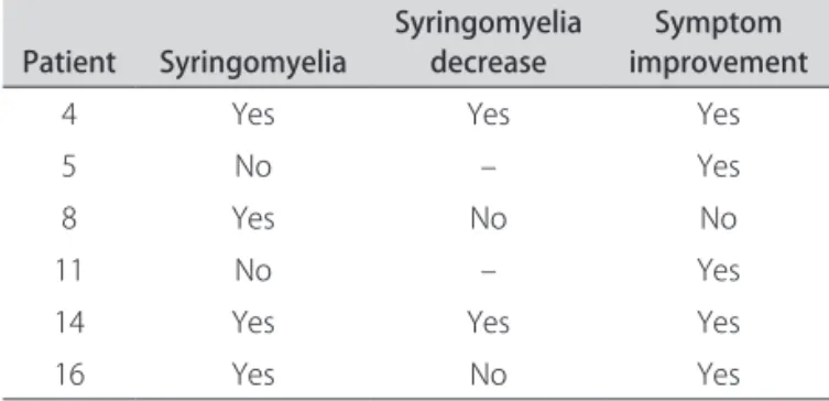
Of the six patients who did not undergo duraplasty, four showed improvement postoperatively. Two patients without syringomyelia showed improvement postoper-atively. Of the four patients with syringomyelia, three showed improvement, including two with a decrease in the cavity size. One patient showed improvement in
Table 1. Clinical and radiological summary of the patients.
Patient Age (yrs) Sex Syringomyelia Duraplasty
1 34 M No Yes
2 28 F Yes Yes
3 23 F Yes Yes
4 62 F Yes No
5 42 M No No
6 53 F No Yes
7 29 F Yes Yes
8 27 M Yes No
9 55 M Yes Yes
10 60 F No Yes
11 36 F No No
12 39 F Yes Yes
13 52 M No Yes
14 47 F Yes No
15 29 M Yes No
16 34 F No Yes
Arq Neuropsiquiatr 2010;68(4)
625
Chiari type 1 malformation: suboccipital craniectomy Romero et al.
symptoms but the syringomyelia was unchanged. The cavity size increased in the one patient who did not show improvement (Table 2).
Among the 10 patients who underwent duraplasty, improvement was noted in four of the ive patients with-out syringomyelia and in all of the ive with syringomyelia. All of the patients with syringomyelia underwent postop-erative MRI; all showed a decrease in the volume of their syringomyelia and clinical improvement (Table 3).
When the dura was opened, the surgical complica-tions included two cases of cerebrospinal luid (CSF) leaks associated with aseptic meningitis in one patient; one case of subgaleal CSF or seroma collection that subsequent-ly resolved with conservative treatment onsubsequent-ly; one case of supericial wound infections; and postoperative occipi-tal nerve pain in one patient. When the dura was not opened, the only complication was a supericial wound infection that resolved.
DISCUSSION
Chiari type 1 malformations occur in the region where the brain and the spinal cord join1,4,15-17. In this disorder, the portions of the brain called the cerebellum and/or brainstem lie lower than usual. Often, a portion of the
cerebellum called the cerebellar tonsils protrudes out of the base of the skull into the spinal canal. his protrusion causes pressure in the brain, thus contributing towards the symptoms that such individuals experience. he cause of CM1 is not known. Some CM1 cases are believed to be present at birth16.
The posterior fossa is smaller and shallower in pa-tients with Chiari malformation, compared with that in normal individuals17. he cisterna magna is also small or absent in patients with CM118-20. herefore, enlargement of the foramen magnum or creation of a cisterna magna is one goal of surgery20-23. Furthermore, cardiac-gated dy-namic MRI scans have demonstrated obstruction of CSF low at the craniocervical junction in these patients22-24. he postoperative low velocity of CSF is improved in the majority of these patients25-30. his low change may un-derlie the change in syringomyelia size observed post-operatively.
Several mechanisms for the pathogenesis of syringo-myelia have been proposed. According to the theory of Gardner and Angel 25, the obstruction of CSF low at the craniocervical junction causes CSF to enter the cervical central canal. However, no such communication exists in the majority of these patients18,24 and therefore this the-ory cannot explain the formation of the cavity. Oldield et al.18 proposed that CSF enters the spinal cord direct-ly via the perivascular spaces, or so-called Virchow-Rob-in spaces, by means of arterial pulsations. his is consis-tent with studies that show movement of water-soluble contrast media from the subarachnoid space to the cavi-ty. Aboulker1 considered that an increase in venous pres-sure in the epidural veins was the reason for the increase in luid in the spinal cord, with subsequent hydromyelia. Recently, Stoodley et al.23 showed that, under normal con-ditions in a sheep model, CSF lows rapidly from the sub-arachnoid space to the perivascular spaces, and that this low is dependent on arterial pulsations. With any ob-struction to the low of CSF in the central canal, such as at the craniocervical junction, a cavity can form that could subsequently enlarge.
Although the exact mechanism for the formation of the hydromyelia in Chiari malformation is still contro-versial, there is general agreement on the importance of decompressing the craniocervical junction in treatments for Chiari malformation and hydromyelia. Suboccipital decompression, with or without duraplasty, serves to di-rectly relieve the bony compression at the craniocervical junction. However, most authors difer on the usefulness and safety of additional procedures, such as duraplasty, syringosubarachnoid shunting or obex plugging.
If the purpose of surgery for CM1 is enlargement of the cisterna magna19, thereby allowing improved CSF low, adequate bone removal must be obtained in all patients.
Table 2. Clinical summary of patients who did not undergo duraplasty.
Patient Syringomyelia Syringomyelia decrease improvementSymptom
4 Yes Yes Yes
5 No – Yes
8 Yes No No
11 No – Yes
14 Yes Yes Yes
16 Yes No Yes
Table 3. Clinical summary of patients who underwent duraplasty.
Patient Syringomyelia Syringomyelia decrease improvementSymptom
1 No – Yes
2 Yes Yes Yes
3 Yes Yes Yes
6 No – No
7 Yes Yes Yes
9 Yes Yes Yes
10 No – Yes
12 Yes Yes Yes
13 No – Yes
Arq Neuropsiquiatr 2010;68(4)
626
Chiari type 1 malformation: suboccipital craniectomy Romero et al.
hree of our four patients with syringomyelia showed im-provement with bone removal only, and two (50%) of the four showed a decrease in the size of the syringomyelia. he two patients with a decrease in cavity size showed an increase in CSF space behind the cerebellum. he patient with no change in the cavity size showed no change in the CSF space behind the cerebellum. It is also possible that this patient harbored or developed arachnoid scarring or subarachnoid adhesions.
All of the ive patients with syringomyelia who under-went duraplasty showed improvement and all ive (100%) showed a decrease in the size of the cavity. Four of the ive patients without syringomyelia who underwent du-raplasty showed clinical improvement. Four of our six pa-tients showed a decrease in syringomyelia, with an im-provement in symptoms, through bone removal alone. his improvement was associated with an increase in the size of the cisterna magna, presumably allowing improved CSF low, thereby leading to resolution of the syringomy-elia. It therefore seems that duraplasty provides a better chance of enlarging the size of the cisterna magna.
Complications such as electrolyte imbalances, tran-sient postoperative swallowing problems and cer-ebellar infarctions have been reported after Chiari decompression1,3,17,21. CSF leaks occurred in two of our patients. he wounds were resutured, thus resolving the leaks. he subgaleal luid collections that developed after surgery were also resolved, by means of tapping. Occipital nerve pain, as seen in one patient, is a well-known com-plication following posterior fossa surgery20. hese mi-nor complications would not ordinarily warrant concern. However, the fact that they occurred predominantly in the patients undergoing duraplasty is of some signiicance.
It is diicult to draw conclusions from a series of lim-ited size; however, there is a suggestion that patients with syringomyelia may have a higher likelihood of improve-ment after undergoing duraplasty. Nevertheless, some pa-tients showed a decrease in syringomyelia, with an im-provement in symptoms, through bone removal alone. his improvement was associated with an increase in the size of the cisterna magna, presumably allowing improved CSF low and thereby leading to resolution of the syringo-myelia. It therefore seems that duraplasty provides a bet-ter chance of enlarging the size of the cisbet-terna magna.
Performing duraplasty to treat Chiari I malforma-tion may lead to a greater decrease in concurrent syrin-gomyelia. However, a subset of patients whose syringo-myelia will decrease through bone removal alone still ex-ists. Further studies are needed in order to better char-acterize these patients and to determine which patients with Chiari I malformation are better served with bone decompression alone, and which patients will require du-raplasty to resolve their syringomyelia.
REFERENCES
Dauser RC, DiPietro MA, Venus JL. Symptomatic Chiari I malformation in 1.
childhood: a report of 7 cases. Pediatr Neurosci 1988;14:184-190. Duddy MJ, Williams B. Hindbrain migration after decompression for hindbrain 2.
hernia: a quantitative assessment using MRI. Br J Neurosurg 1991;5:141-152. Gonçalves da Silva JA, Leiros da Costa MD, Holanda MMA, et al. Impacted 3.
cisterna magna without syringomyelia associated with spastic paraparesis: case report. Arq Neuropsiquiatr 2006;64:672-675.
Isu T, Iwasaki Y, Akino M, Abe H. Hydrosyringomyelia associated with a Chiari 4.
I malformation in children and adolescents. Neurosurgery 1990;26:591-596. Quencer RM, Post MJD, Hinks RS. Cine MRI in the evaluation of normal and 5.
abnormal CSF low: intracranial and intraspinal studies. Neuroradiology 1990; 32:371-391.
Aboulker J. Syringomyelia and intra-rachidian luids. Parts I-XIII. Neurochir-6.
urgie 1979;25:1-144.
Armonda RA, Citrin CM, Foley KT, Ellenbogen RG. Quantitative cine-mode 7.
magnetic resonance imaging of Chiari I malformation: an analysis of cere-brospinal luid dynamics. Neurosurgery 1994;35:214-224.
Stover LJ, Bergan U, Nilsen G, Sjaastad O. Posterior cranial fossa dimensions 8.
in the Chiari I malformation: relation to pathogenesis and clinical presenta-tion. Neuroradiology 1993;35:113-118.
Batzdorf U. Chiari I malformation with syringomyelia: evaluation of surgical 9.
therapy by magnetic resonance imaging. J Neurosurg 1988;68:726-730. Blagodatsky MD, Larionov SN. Surgical treatment of “hindbrain related” syrin-10.
gomyelia: long-term results. Acta Neurochir (Wien) 1993;123:209-210. Nohria V, Oakes WJ. Chiari I malformation: a review of 43 patients. Pediatr 11.
Neurosurg 1990;16:222-227.
Fujii K, Natori Y, Nakagaki H, Fukui M. Management of syringomyelia associat-12.
ed with Chiari malformation: comparative study of syrinx size and symptoms by magnetic resonance imaging. Surg Neurol 1991;38:161-162.
Barbaro NM, Wilson CB, Gutin PH, Edwards MS. Surgical treatment of syrin-13.
gomyelia: favorable results with syringo-peritoneal shunting. J Neurosurg 1984;61:531-538.
Matsumoto T, Symon L. Surgical management of syringomyelia: current re-14.
sults. Surg Neurol 1989;32:253-256.
Wan MJ, Hiroshi N, Tator, CH. Conversion to symptomatic Chiari I malforma-15.
tion after minor head or neck trauma. Neurosurgery 2008;63:748-753. Cahan LD, Bentson JR. Considerations in the diagnosis and treatment of sy-16.
ringomyelia and the Chiari malformation. J Neurosurg 1982;57:24-31. Nagib MG. An approach to symptomatic children (age 4-14 years) with Chiari 17.
type 1 malformation. Pediatr Neurosurg 1994;21:31-35.
Oldield EH, Muraszk K, Shawker TH. Pathophysiology of syringomyelia as-18.
sociated with Chiari I malformation of the cerebellar tonsils: implications for diagnosis and treatment. J Neurosurg 1994;80:3-15.
McGirt MJ, Nimjee SM, Floyd J, Bulsara KR, George TM.
19. Correlation of
cere-brospinal luid low dynamics and headache in Chiari I malformation. Neu-rosurgery 2005;56:716-721.
Menezes AH. Chiari I malformations and hydromyelia: complications. Pediatr 20.
Neurosurg 1991;17:146-154.
Newman PK, Tereny TR, Foster JB. Some observations on the pathogenesis of 21.
syringomyelia. J Neurol Neurosurg Psychiatry 1981;44:964-969.
Taricco MA, Melo LRS. Retrospective study of patients with Chiari: malforma-22.
tion submitted to surgical treatment. Arq Neuropsiquiatr 2008;66:184-188. Samii M, Klekamp A, Sepehrnia A, et al. Syringomyelia associated with Arnold 23.
Chiari malformation. Acta Neurochir (Wien) 1993;123:195. Munshi I
24. , Frim D, Stine-Reyes R, Weir BK, Hekmatpanah J, Brown F. Efects of posterior fossa decompression with and without duraplasty on Chiari malfor-mation-associated hydromyelia. Neurosurgery 2000;46:1384-1390. Gonçalves da Silva JA, Holanda MMA, Leiros da Costa MD, et al. Basilar im-25.
pression associated with impacted cisterna magna, spastic paraparesis and distress of balance: case report. Arq Neuropsiquiatr 2006;64:668-671. Dyste GN, Menezes AH. Presentation and management of pediatric Chiari 26.
malformation without myelodysplasia. Neurosurgery 1988;23:589-597. Gardner WJ, Angel J. The cause of syringomyelia and its surgical treatment. 27.
Cleve Clin Q 1958;25:4-8.
Imae S. Clinical evaluation on etiology and surgical outcome in syringo-28.
myelia associated with Chiari type I malformation. No To Shinkei 1997;49: 1131-1138.
Isu T, Sasaki H, Takamura H, Kobayashi N. Foramen magnum decompression 29.
with removal of the outer layer of the dura as treatment for syringomyelia oc-curring with Chiari I malformation. Neurosurgery 1993;33:844-849. Thorn K, Quigley K, Huang MC, Myseros JS, Yaun A, Keating RF.
30. Chiari 1.5