LANGUAGE BRAIN DOMINANCE IN PATIENTS
WITH REFRACTORY TEMPORAL LOBE EPILEPSY
A comparative study between functional magnetic
resonance imaging and dichotic listening test
Denise Ren da Fontoura
1, Daniel de Moraes Branco
2, Mauricio Anés
3,
Jaderson Costa da Costa
4, Mirna Wetters Portuguez
5Abstract – Purpose: To identify brain dominance for language functions with DLT and correlate these results with those obtained from fMRI in patients suffering from intractable temporal lobe epilepsy. Method: This study reports on 13 patients who underwent pre-surgical epileptic evaluation between April and October 2004 at the Epilepsy Surgery Program, Hospital Sao Lucas, PUCRS. In DLT, dominance was assessed through a consonant-vowel task, whereas in fMRI patients performed a verb generation task. Results: Our results identified a correlation between the fMRI lateralization index and the DLT ear predominance index and reply difference index (r=0.6, p=0.02; Pearson Correlation Coefficient), showing positive correlation between results obtained from fMRI and DLT. Conclusion: DLT was found to significantly correlate with fMRI. These findings indicate that DLT (a non-invasive procedure) could be a useful tool to evaluate language brain dominance in pre-surgical epileptic patients as it is cheaper to perform than fMRI.
KEy wORDS: dichotic listening test, functional magnetic resonance imaging, refractory temporal lobe epilepsy, brain dominance, language.
Determinação de dominância cerebral para linguagem em pacientes com epilepsia refratária de lobo temporal: estudo comparativo entre ressonância magnética funcional e teste de escuta dic�ticateste de escuta dic�tica
Resumo – Objetivo: Identificar a dominância cerebral para funções de linguagem através do teste de escuta dicótica (TED) e correlacionar com os resultados de ressonância magnética funcional (RMf) em pacientes com epilepsia refratária de lobo temporal. Método: Foram estudados 13 pacientes com epilepsia refratária de lobo temporal, que realizaram avaliações pré-cirúrgicas no período de abril a outubro de 2004 no Programa de Cirurgia de Epilepsia do Hospital São Lucas da PUCRS. Realizada investigação da dominância hemisférica para linguagem através do TED Consoante-Vogal e da RMf pela geração de verbos. Resultados: Verificou-se a existência de correlação entre os índices de lateralidade (RMf) e os índices de predomínio de orelha e de diferença de resposta (TED) (r=0,6, p=0,02). Conclusão: Existe correlação entre os resultados obtidos através da RMf (índice de lateralidade) e do TED (índice de predomínio de orelha e índice de diferença de resposta) em pacientes com epilepsia refratária de lobo temporal.
PALAVRAS-CHAVE: teste de escuta dicótica, ressonância magnética funcional, epilepsia temporal refratária, dominância cerebral, linguagem.
Serviço de Neurologia do Hospital São Lucas da PUCRS, Programa de Cirurgia de Epilepsia, Porto Alegre RS, Brazil: 1Fonoaudióloga, Mestre do Programa
de Pós-Graduação em Medicina e Ciências da Saúde/Neurociências, Faculdade de Medicina da PUCRS, Doutoranda em Ciências da Linguagem e da Comunicação pela Universidade Nova de Lisboa; 2Médico Neurologista, Doutor em Medicina e Ciências da Saúde/Neurociências pela PUCRS, Post-doc
pela Harvard Medical School, HMS; 3Físico Médico do Hospital de Clínicas de Porto Alegre; 4Professor Titular de Neurologia da Faculdade de Medicina da
PUCRS; 5Professora Adjunta de Neurologia e da Pós-Graduação em Medicina e Ciências da Saúde/Neurociências, Faculdade de Medicina da PUCRS .
Received 30 July 2007, received in inal form 8 October 2007. Accepted 20 November 2007.
Dra. Denise Ren da Fontoura – Rua Dr. Prudente de Moraes 349 - 91330-170 Porto Alegre RS - Brasil. E-mail: [email protected]
Epilepsy is a consequence of altered cerebral func-tions caused by various pathologic processes. Depending on the localization of the epileptogenic discharges, the disease can assume different forms, which may be
asso-ciated with several distinct neurological impairments1-4.
arrest. Consequently, assessment of cerebral dominance, particularly for language function, is of fundamental im-portance in the evaluation of candidate patients to refrac-tory epilepsy surgery. In addition, the presence of epilep-tic discharges since childhood can cause an atypical reor-ganization of cortical functions, resulting in migration of language to other areas of the same hemisphere or even to the contra-lateral (usually right) hemisphere. Accord-ing to Portuguez5, “what is most important and necessary
is mapping the language cortex and identifying memory dominance in the brain hemispheres of patients who will undergo epilepsy surgery, to guide the neurosurgeon and minimize the deicits in the post-surgical period.”
Functional magnetic resonance imaging (fMRI) cur-rently is one of the most important and used techniques for brain mapping. It has been employed by many epilepsy centers to replace the sodium amobarbital test (SAT) and electrical cortical stimulations (ECS), and is regarded as a non-invasive procedure with minimal risks for patients5-7.
As the SAT procedure is not presently performed in Bra-zil due to dificulties in obtaining the barbiturate (whose import is forbidden), we employed the dichotic listen-ing test (DLT) to determine cerebral dominance of lan-guage functions. This is a non-invasive alternative tech-nique, which consists of verbal stimuli simultaneously de-livered to both ears of the patient8.
This study aimed at identifying brain dominance of language through the dichotic listening test and correlat-ing the results to those obtained with functional magnet-ic resonance imaging in order to assess the validity of DLS as a pre surgical planning tool.
METHOD
A transversal study was performed with patients from the Epilepsy Surgery Program of the Hospital São Lucas da PUCRS (PCE-HSL-PUCRS). All individuals were enrolled between April and October 2004. fMRI and DLT were performed in all of them to establish brain dominance of language. The results were then analyzed in terms of left or mixed hemisphere language domi-nance and the correlation between both tests.
Patients
Male and female adult patients (older than 16) were enrolled. The sample consisted of 13 patients (seven men and six women) between 17 and 48 years old (±33,4 years old), all with an estab-lished diagnosis of refractory temporal lobe epilepsy.
Patients with previous brain surgery, presenting other neu-rological and/or psychiatric disorders or loss of audition in the audiometric examination, as well as patients with altered lan-guage understanding (worse than a small alteration as assessed through the Token Test), or with IQ values below the inferior av-erage, were excluded from the study.
The patients of this study were included after receive an
in-formed agreement that was previously obtained by HSL-PUCRS and sign by the patients or responsibles.
Procedures
Examinations were performed when the patients were clin-ically stable and able to keep the level of attention required for the neuropsychological and language tests. These included lan-guage understanding with the Token Test, Estimated IQ through the wAIS-R (vocabulary and cubes) subtests and a hearing test with an air conduction audiometer, performed in a soundproof cabinet.
After the hearing evaluation, the Dichotic Consonant-Vow-el Listening Test was performed in an attention-free situation to analyze the perception asymmetry of language stimulation, which provided results on cerebral dominance for language functions.
The test consisted of a total of 12 pairs of different sylla-bles with the “a” vowel (“ta”, “da”, “ga”, “ca”, “ba”, “pa”), presented in a synchronized way and with the same intensity (50 dBNA), applied to both ears in an acoustically isolated room to avoid the interference of external noise. In the attention-free test, the patients were instructed to repeat one of the syllables they heard (the one that appeared to be loudest), while the examin-er wrote down the vexamin-erbalized syllable on the answexamin-er sheet. The same procedure was then repeated after inversion of the head-phones. The number of repeated syllables and the mistakes ob-served were recorded for posterior analysis. The prevalence of correct answers for one of the ears as compared to the other was called the “ear predominance index” (EPI), and the differ-ence between correct answers derived from the right and the left ears was called the “reply difference index” (RDI)7.
During the fMRI exam, patients performed a verb-gener-ation task. They heard concrete words through the scanner headphones and were asked to think of their utility (e.g. pen-cil >> write), without verbalizing of making any facial or tongue movements, keeping silent and with eyes closed. The patients were previously instructed about the proposed task. we used a blocked paradigm and the control blocks (rest condition) con-sisted simply of absence of verbal stimuli, when they should keep their eyes closed and think of a white screen. In each run X xx-seconds blocks were interleaved with X rest blocks (TR=4.5s).
Functional T2*-weighed images were realigned, normalized to MNI space, and smoothed with the SPM package (wellcome Department of Imaging Neuroscience, London, UK).
we took the whole hemisphere bilaterally (excluding cere-bellum) as two distinct regions of interest (ROI). The number of activated voxels in each ROI (L and R) was then counted, and a lateralization index (LI) was calculated as follows9-16. The number
Statistical analysis was performed by a Pearson correlation coeficient to establish a correlation between the results from the Dichotic Listening Test and the fMRI.
RESULTS
Language hemisphere dominance: DLT and fMRI Results relative to language hemisphere dominance will be presented through DLT, according to ear predomi-nance (EPI) and reply difference (RDI) indices and through
Table 1. Correlation between side index (fMRI) and ear predom-inance and reply difference indices (DLT).
Characteristic r p Test SI x EPI 0.62 0.02 Pearson Correlation SI x RDI 0.6 0.02 Pearson Correlation
EPI, Ear predominance indice; RDI: Reply difference indice; SI, Side index.
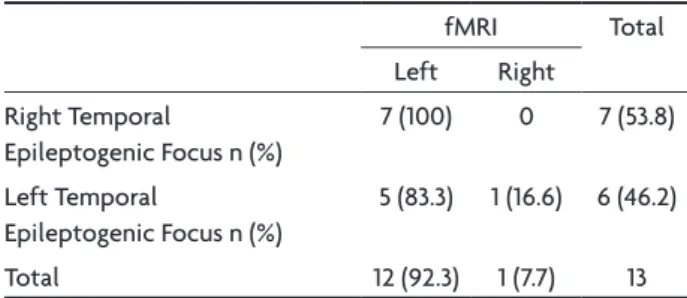
Table 2. Comparison between lateralization of the temporal ep-ileptogenic focus and language hemisphere dominance tested by fMRI: Kappa’s Concordance Coeficient and Fisher Exact Test.
fMRI Total Left Right
Right Temporal
Epileptogenic Focus n (%)
7 (100) 0 7 (53.8)
Left Temporal
Epileptogenic Focus n (%)
5 (83.3) 1 (16.6) 6 (46.2)
Total 12 (92.3) 1 (7.7) 13
Fisher Exact Test, p=0.26; Kappa=0.17, p=0.13; fMRI, functional magnet-ic resonance image.
Table 3. Comparison between lateralization of the temporal ep-ileptogenic focus and language hemisphere dominance tested by DLT: Kappa’s Concordance Coeficient and Fisher Exact Test.
DLT Total Left Right
Right Temporal
Epileptogenic Focus n (%)
7 (100) 0 7 (53.8)
Left Temporal
Epileptogenic Focus n (%)
4 (66.6) 2 (33.3) 6 (46.2)
Total 11 (84.6) 2 (15.4) 13
Fisher Exact Test, p=0,19; Kappa=0.68, p=0.005; DLT, dichotic listening test.
Table 4. Comparison between age of beginning of the epileptogenic crises and language hemi-sphere dominance testes by fMRI: Kappa’s Concordance Coeficient and Fisher Exact Test.
fMRI Total Mix Left
Age of beginning of the epileptogenic crises
Before 5 years old n (%) 1 (50) 1 (50) 2 (15.4) After 5 years old n (%) 0 11 (100) 11 (84.6) Total 1 (7.7) 12 (92.3) 13 (100)
Fisher Exact Test, p=0.15; Kappa=0.63, p=0.007; fMRI, functional magnetic resonance image.
fMRI, according to the side index (SI). For EPI, the domi-nant language hemisphere is the one contralateral to the predominant ear.
The determination of language hemisphere domi-nance by fMRI and DLT showed that all left-handed pa-tients presented left hemisphere language dominance (DLT and fMRI). Among the right-handed patients, 81.8% showed left hemisphere language and 18.8% mixed lan-guage function, as detected by DLT; fMRI results showed that 90.9% of them presented left hemisphere language and 9.1% mixed language function. No cases of right hemi-sphere language dominance were observed.
The analysis of the values obtained by DLT (ear pre-dominance and reply difference indices) and by fMRI (lat-erality index) showed a relationship between the lat(lat-erality indices (fMRI) and the ear predominance and reply differ-ence indices (DLT). Table 1 shows that, in the comparison of LI with EPI and LI with RDI, the Pearson test with r of 0.62 and 0.6 respectively presented a signiicant result (p=0.02).
Language hemisphere dominance and lateralization of the temporal epileptogenic focus
Tables 2 and 3 present the relationship between the side of the temporal epileptogenic focus and language brain dominance as assessed by fMRI and DLT. Analysis by the Fisher Exact Test did not show correlation between the temporal epileptogenic focus and language lateraliza-tion, as tested by fMRI or DLT. Concordance between the temporal epileptogenic focus and language lateralization as analyzed by DLT was however apparent.
Language hemisphere dominance and age of beginning of the epileptogenic crises
Patients were divided in two groups according to the age of beginning of the crises: those in whom the epi-leptic crises began before ive years of age, and those in whom they began later on.
one of them presented mixed language, whereas the oth-er ten patients presented left hemisphoth-ere language dom-inance (Table 5).
DISCUSSION
Language hemisphere dominance: DLT and fMRI DLT and fMRI are widely employed for the study of language brain dominance, but no studies were found in which results relative to the ear predominance index ob-tained with those tests were analyzed for their correla-tion with the reply difference index (DLT) and with the laterality index (fMRI). On the other hand, most of the researches does the relationship between the fMRI and the DLT has been very scarcely studied.
The present study showed that 92.3% of the patients analyzed by fMRI presented left hemisphere language and 7.7% presented mixed language function, supporting the results reported by Springer et al.10, who however
inves-tigated healthy individuals. In that work, language brain dominance was investigated by fMRI in normal and epi-leptic individuals, showing that 94% of the normal per-sons presented left hemisphere dominance for language, whereas 6% presented bilateral language. Among epileptic patients, 78% presented left hemisphere dominance, 16% bilateral, and 6%, right hemisphere dominance. The report by Springer et al.10 included, contrary to the present work,
not only temporal but also extratemporal epilepsies. It is believed that the frequency of left hemisphere dominance for language is higher among the patients studied in the present work, since all of them presented hippocampal sclerosis, limited to mesial structures of the temporal lobe, as the only lesion.
The analysis of brain dominance through DLT showed that 84% of the patients presented left hemisphere lan-guage and 15.4% mixed lanlan-guage function, with no cases presenting right hemisphere language dominance. These results are in accordance with reports by Geffen and Cau-drey17, Zatorre18, and Hugdahl et al.19, who compared DLT
and the Sodium Amobarbital Test (SAT) and concluded that most of the individuals presented left hemisphere language dominance.
According to Strauss et al.20, however, a predominant ear
was not found in the case of patients with right hemisphere language function, but only in patients with left hemisphere dominance for language, with a predominance of the right ear in 100% of them. The results obtained in present work, similarly, do not allow the conclusion that DLT is eficient in the detection of hemisphere dominance in patients with right hemisphere language function, since no patient with this characteristic was present in the sample group.
The analysis of correlation (Pearson’s Correlation Coef-icient) between the two tests employed in this work (DLT and fMRI), based on the quantitative results of EPI, RDI and LI, showed a positive association. DLT is correlated to fMRI, with a simultaneous increase in the values result-ing from both examinations. A number of other studies have already compared DLT with SAT17-21, and fMRI with
SAT9,11,13,15,16,22, showing a positive relationship and conirming
the eficiency of the two tests. The relationship between DLT and fMRI, however has been scarcely studied. we can cite the studies of Fernandes et al.23 and Bethmann et al.17.
Fernandes et al.23, investigating the DLT and the fMRI
in 14 children with epilepsy, veriied that it does exist con-cordance between both tests, and the DLT can be used to determine the dominant language hemisphere in pre-sur-gical patients. The DLT results showed that 6 children had mixed language (both ears response), 8 left hemisphere dominance (right ear dominance), and no one of the pa-tients had right hemisphere language dominance.
Bethmann et al.24 made a comparison between these
2 tests in 30 healthy people. The authors went to a con-clusion that there was short difference between the ears, suggesting that the DLT can not be used with the aim of determine a laterality of the language, on the opposite of the fMRI, that eficiently identiied the dominant hemi-sphere of the participants.
Language hemisphere dominance and lateralization of the temporal epileptogenic focus
Bilateral or right hemisphere language representation is uncommon in the populations, since in most people
Table 5. Comparison between age of beginning of the epileptogenic crises and language hemi-sphere dominance testes by DLT: Kappa’s Concordance Coeficient and Fisher Exact Test.
DLT Total Mix Left
Age of beginning of the epileptogenic crises
Before 5 years old n (%) 1 (50) 1 (50) 2 (15.4) After 5 years old n (%) 1 (9.1) 10 (90.9) 11 (84.6) Total 2 (15.4) 11 (84.6) 13 (100)
the left hemisphere is dominant for language functions. In case of neurological lesions with injury on the left hemi-sphere, there is an increased probability of language trans-fer to other regions. Loring et al.25 observed that language
transfer to the right hemisphere is more related to exten-sive lesions in areas beyond the temporal lobe. Chugani et al.26, furthermore, pointed out that small neocortical
lesions are associated to compensation of ipsilateral brain regions, whereas more extensive lesions induce compen-satory modiications on the contralateral hemisphere.
The results observed in the present work support those studies, since no correlation was observed between the brain hemisphere affected by epilepsy and language brain dominance, as tested by DLT or fMRI. The patients analyzed presented lesions limited to mesial (hippocam-pus) structures, with no evidence of extensive brain le-sions. In the three cases in which mixed language domi-nance was observed after individualized analysis, however, bilateral language localization occurred only in patients with left temporal epilepsy, with a partial brain reorgani-zation in the contralateral hemisphere. Furthermore, con-cordance between the temporal epileptogenic focus and language lateralization as evaluated by DLT was apparent, since the two patients with mixed language function had left epileptogenic foci.
Knecht27 observed, in a study with fMRI, results similar
to the three cases described above. The author concluded that, in cases in which the lesions were located in or near the Broca’s area, language brain lateralization to the right hemisphere did not occur; lesion on the left hippocam-pus, parahippocampus or on the inferior temporal pole, however, resulted in right or bilateral lateralization of lan-guage functions.
Language hemisphere dominance and age at epilepsy onset
It is well known that the sooner the brain lesions oc-cur, the higher are the probabilities of recovering the damaged functions, due to the increased brain plasticity in that life period. According to Springer et al.10 and
Jan-szky et al.28, modiications of the left hemisphere result in
an interhemispheric reconstitution of language functions to the right hemisphere. The same authors observed that after ive years of age, when language becomes gradually more lateralized, the contralateral reorganization of lan-guage after a brain seizure occurs less frequently. For this reason, the sample was separated in two groups in the present work: patients with onset of the epileptic crises before 5 years old, and after patients in whom epilepsy onset occurred later on.
Although the sample is not large, with only two pa-tients presenting epileptogenic crises before 5 years old, the results of fMRI showed that in all patients in whom the crises began after 5 years old language was on the left brain hemisphere, with a concordance among the results.
One of the two patients in whom the crises began before 5 years of age presented mixed language as tested on fMRI; this patient present left hemisphere temporal epilepsy, began before one year old. These results sup-port the theory of brain reorganization (neuronal plastic-ity) mentioned by Springer et al.10 and Janszky et al.28, as
well as the theory proposed by Duchowny et al.29, stating
that only damages on the irst years of life result in con-sistent contralateral reorganization of language. Risse et al.30 emphasize that the incidence of atypical language
is much more frequent in patients with a history of left hemisphere early lesions (from childhood). Springer et al.10
showed that all the cases of atypical language were asso-ciated to onset of epileptic crises in childhood.
In a study with refractory epilepsy patients, Duchowny et al.29 concluded that language transfer to the right
hemi-sphere happens only in cases of lesions occurring before ive years of age and in which the language cortex is de-stroyed, and not in cases of development lesions and epi-leptogenic discharges.
In relation to the patients investigated through fMRI, all those in whom the crises began after ive years of age presented left hemisphere language dominance, without brain reorganization in those patients with left temporal epilepsy, probably due to a late crisis onset when neuro-nal plasticity is smaller. According to Lent31,
neuroplastic-ity is higher during development, decreasing gradually in adult life without however disappearing.
In DLT, two of the patients, both with left temporal epilepsy, showed mixed language representation; however, in one of them the crises began before age 5 while in the other one they began later on. The irst case suggests the occurrence of brain reorganization due to neuronal plas-ticity, but modiication exclusively of pathways involved in the central hearing processing can also be suggested since the patient showed left hemisphere language domi-nance in the fMRI test. According to Ortiz et al.32, the
pro-cessing of hearing information during childhood may be modiied if any predisposition factor is present during de-velopment, and neurological alterations represent one of the risk factors for dysfunctions of the hearing processing.
Related to the second case, which crises began after 5 years old, the fact of has been noticed the mixed language in DLT agrees with the neuroplasticity theories, although been smaller, it also occurs in adults as well27,28,31.
re-habilitation, mainly the linguistic one, can be successfully achieved in cases of people with neuronal damage.
In conclusion, considering the observation conditions and the sample selected in the present study, the results indicate a correlation between the results obtained by fMRI and the DLT, allowing its application in the evalua-tion of language lateralizaevalua-tion in patients with refractory temporal lobe epilepsy. Left hemisphere dominance for language is more frequent, as shown by DLT as well as by fMRI, independent on hand dominance. No correlation was observed between the hemisphere affected by the epileptogenic focus and language brain dominance, em-ploying fMRI and DLT. The comparison of age crises onset and language lateralization showed that all the patients in whom the crises began after ive years of age present left brain hemisphere language dominance, as studied by fMRI. These results do not represent a suggestion for the replacement of fMRI by DLT, since the two exams, in spite of having the same objectives, represent different procedures. The irst one is an image test, more speciic and capable to inform about speciic regions for language activation in the brain; the second informs about the brain hemisphere which is responsible for language functions. DLT, however, besides being an exam of low cost and easy performance, represents an alternative in centers where SAT and fMRI are not available.
REFERENCES
1. Caner-Cukiert AR. Avaliação neuropsicológica das epilepsias refra-tárias. In Cukiert A. Tratamento clínico e cirúrgico das epilepsias de difícil controle. São Paulo: Lemos Editorial, 2002:189-199.
2. Cendes F, Kobayashi E. Epilepsia de lobo temporal. In Guerreiro CA, Guerreiro MM, Cendes F, Lopes-Cendes I. Epilepsia. São Paulo: Lemos Editorial, 2000:201-213.
3. Costa JC, Palmini A. Epilepsias refratárias em crianças. In Costa JC, Pal-In Costa JC, Pal-mini A, Yacubian EMT, Cavalleiro EA. Fundamentos neurobiológicosFundamentos neurobiológicos das epilepsias: aspectos clínicos e cirúrgicos. Vol 2. São Paulo: Lemos Editorial, 1998:817-829.
4. Yacubian EMT. Epilepsias refratárias em adultos. In Costa JC, Palmi-ni A, Yacubian EMT, Cavalleiro EA. Fundamentos neurobiológicos das epilepsias: aspectos clínicos e cirúrgicos. Vol 2. São Paulo: Lemos Edi-torial, 1998:807-816.
5. Portuguez MW. Ressonância magnética funcional. In Nunes M, Mar-rone. Semiologia neurológica. Porto Alegre: EDIPUCRS, 2002. 6. Palmini A, Costa JC, Calcagnotto ME, Martinez JVL. Avaliação
pré-cirúrgica de pacientes com epilepsia parcial refratária. In Costa JC, Pal-Costa JC, Pal-mini A, Yacubian EMT, Cavalleiro EA. Fundamentos neurobiológicosFundamentos neurobiológicos das epilepsias: aspectos clínicos e cirúrgicos. Vol 2. São Paulo: Lemos Editorial, 1998:857-878.
7. Portuguez MW, Veras JM. Teste do amital sódico: avaliação pré-cirúr-gica. In Guerreiro CA, Guerreiro MM, Cendes F, Lopes-Cendes I. Epi-lepsia. São Paulo: Lemos Editorial, 2000:119-130.
8. Tedesco ML. Consoante-vogal de escuta direcionada. In Pereira LD, Schochat E. Processamento auditivo central. São Paulo: Lovise, 1997: 139-145.
9. Binder JR, Swanson TA, Hammeke GL, et al. Determination of lan-guage dominance using functional MRI: a comparison with the Wada test. Neurology 1996;46:978-984.
10. Springer JA, Binder JR, Hammeke TA, Swanson SJ, Frost JA, Bellgowan PSF, et al. Language dominance in neurologically normal and epilepsy subjects: a functional MRI study. Brain 1999;122:2033-2045.
11. Benson RR, Fitzgeralg DB, Lesueur LL, Kennedy DN, Kwong KK, Buch-binder BR, et al. Language dominance determined by whole brain func-tional MRI in patients with brain lesions. Neurology 1999;52:798-809. 12. Lurito JT, Dzemidzic M. Determination of cerebral hemisphere lan-guage dominance with functional magnetic resonance imaging. Neu-roimaging Clin N Am 2001;11:355-363.
13. Rutten GJM, Ramsey NF, Van Rijen PC, Alpherts WC, Van Veelen CWM. fMRI-Determined language lateralization in patients with unilateral or mixed language dominance according to Wada test. Neuroimage 2002; 17:447-460.
14. Deblaere K, Backes WH, Holfman P, et al. Developing a comprehensive presurgical functional MRI protocol for patients with intractable tem-poral lobe epilepsy: a pilot study. Neuroradiology 2002;44:667-673. 15. Sabbah P, Chassoux F, Leveque C, et al. Functional MR imaging in
assessment of language dominance in epileptic patients. NeuroImage 2003;18:460-467.
16. Adcock JE, Wise RG, Oxbury JM, Oxbury SM, Matthews PM. Quanti-tative fMRI assesment of the diferences in lateralization of language-related brain activation in patients with temporal lobe epilepsy. Neu-roImage 2003;18:423-438.
17. Geffen G, CaudreyD. Reliability and validity of the dichotic monitor-ing test for language laterality. Neuropsychologia 1981;19:413-423. 18. Zatorre RJ. Perceptual assimetry on the dichotic fused words test and
cerebral speech lateralization determined by the carotid sodium amytal test. Neuropsychologia 1989;27:1207-1219.
19. Hugdahl K, Carlsson G, UvebrantP, Lundervold AJ. Dichotic-listen-ing performance and intracarotid injections of amobarbital in children and adolescents: pre-operative post-operative comparisons. Arch Neu-rol 1997;54:1494-1500.
20. Strauss E, GaddeS WH, Wada J. Performance on free-recall verbal dich-otic listening task and cerebral dominance determined by the carotid amytal test. Neuropsychologia 1987;25:747-753.
21. Caner-Cukiert AR, Cukiert A. Cerebral dominance as determined by the intracarotid amytal procedure and the dichotic words listening test. Epilepsia 1995;Suppl 4:129 (abstract).
22. Santos F, Costa JC. A ressonância magnética funcional na avaliação dacional na avaliação da dominância hemisférica para linguagem em pacientes portadores de epilepsia refratárica. Dissertação de Mestrado em Neurociências, Pon-tifícia Universidade Católica do Rio Grande do Sul, Porto Alegre, 2001. 23. Fernandes MA, Smith ML, Logan W, Crawley A, McAndrews MP. Com-paring language lateralization determined by dichotic listening and fMRI activation in frontal and temporal lobes in children with epilep-sy. Brain Lang 2006;96:106-114.
24. Bethmann A, Tempelmann C, De Bleser R, Scheich H, Brechmann A. Determining language laterality by fMRI and dichotic listening. Brain Res 2007;16;1133:145-157.
25. Loring DW, Strauss E, Herman BP, et al. Effects of anomalous language representation on neuropsychological performance in temporal lobe ep-ilepsy. Neurology 1999;53:260-264.
26. Chugani HT, Müller RA, Chugani DC. Functional brain reorganization in children. Brain Develop 1996;18:347-356.
27. Knecht S. Editorial. Does language lateralization depend on the hippo-campus? Brain 2004;127:1217-1218 (editorial).
28. Janszky J, Jokeit H, Heinemann D, Schulz R, Woermann FG, Ebner A. Epileptic activity inluences the speech organization in medial tempo -ral lobe epilepsy. Brain 2003;26:2043-2051.
29. Duchowny M, Jayacas P, Harvey S, et al. Language cortex representa-tion: effects of developmental versus acquired pathology. Ann Neurol 1996;40:3-38.
30. Risse GL, Gates JR, Fangman MC. A reconsideration of bilateral repre-sentation based on the intracarotid amobarbital procedure. Brain CognBrain Cogn 1997;33:118-132.
31. Lent R. Cem bilhões de neurônios: conceitos fundamentai de neuro-ciência. São Paulo: Editora Atheneu, 2001.São Paulo: Editora Atheneu, 2001.