AR
TIGO ORIGINAL / ORIGINAL AR
TICLE
INTRODUCTION
Gastrointestinal stromal tumors or GISTs are uncommon abdominal neoplasias, but that represent the most frequent mesenchymal tumors of the diges-tive tract (80%) and 5% of all sarcomas(1). The disease can affect any portion of the gastrointestinal tract, being the stomach the most affected organ (45% to 65%), followed by the small intestine (15% to 25%), colon (5% to 10%) and other regions of the abdominal cavity (5%)(6).
Approximately 4,500 to 6,000 cases of GIST are diagnosed annually in the USA, with an incidence pick between the sixth and seventh decades of life, representing around 1.0% to 3.0% of gastric tumors, 20% of the small intestine tumors, and 0.2%-0.1% of colorectal tumors(2, 26).
Although this mesenchymal neoplasia has been
TOMOGRAPHIC FINDINGS OF GASTRIC
GASTROINTESTINAL STROMAL TUMOR AND
CORRELATION WITH THE MITOTIC INDEX
Gustavo Lemos
PELANDRÉ
1, Maria Célia
DJAHJAH
2,
Emerson Leandro
GASPARETTO
2, Marcelo Souto
NACIF
3,
Edson
MARCHIORI
2and Eduardo Linhares Riello de
MELLO
4ABSTRACT – Context – Gastrointestinal stromal tumors are uncommon abdominal neoplasms and can affect any portion of the
gas-trointestinal tract. Objectives - Describe the tomographic indings of the gastrointestinal stromal tumor of gastric origin, correlating it with the mitotic index. Methods - Twenty-one patients were selected within the period of January 2000 and 2008, with histo-pathological and immunohistochemical diagnosis of gastric gastrointestinal stromal tumors, who presented computed tomography done before the treatment. The tomographic variables analyzed were lesion topography, dimensions, contours, morphology, pattern and intensity enhancement through venous contrast, growth pattern, invasion of adjacent organs, presence of ulceration, istula, calciications, iniltration of mesenteric fat, lymphadenopathy and metastasis. The mitotic index was determined through optic microscopy, counting the number of mitosis igures in 50 high power ields. Results - The tumors were located in the body (66.7%)
or gastric fundus (33.3%), with dimensions varying between 4.2 and 21.2 cm (average of 10.5 cm). The growth was predominantly extraluminal (47.6%) or intra/extra luminal (28.6%). The enhancement by venous contrast was heterogeneous in 66.7%. The statistical analysis showed that irregular morphology (P = 0.027) and iniltration of mesenteric fat (P = 0.012) presented correlation with the
high mitotic index. Conclusions - In the present study, most part of the tumors were located in the gastric body, with average size of
10.5 cm, presenting central hypo dense area, heterogeneous enhancement through contrast and predominantly extra luminal growth. Irregular morphology and iniltration of mesenteric fat present statistical correlation with high mitotic level.
HEADINGS - Gastrointestinal stromal tumors. Spiral computed tomography. Mitotic index.
known for decades, recent indings have allowed a better knowledge of its cellular origin, as well as the molecular events involved in the lesion(6). For about 20 years, we believed that most mesechymal tumors were originated in the smooth muscles, being called “leyo-miomas” and “leyomiossarcomas”. The utilization of electronic microscopy and immunohistochemistry, however, showed that only some of these tumors pre-sented smooth muscle characteristics(24). Afterwards, some authors demonstrated that these tumors also presented characteristics of neuronal differentiation, denominating them “plexossarcomas” and “gastro-intestinal tumors of the autonomic nerve”(19). Just recently it was clariied that this neoplasia constitutes one well deined entity denominated GIST, through the discovery of its origin from Cajal’s interstitial cells(17), and the expression of the kit protein(12). Kit is a transmembrane receptor with tyrosine kinase
Declared conflict of interest of all authors: none
1 Colégio Brasileiro de Radiologia (CBR), São Paulo, SP, Brasil; 2 Departamento de Radiologia da Universidade Federal do Rio de Janeiro (UFRJ), RJ, Brasil; 3
Depar-tamento de Radiologia da Universidade Federal Fluminense (UFF), Niterói, RJ, Brasil; 4 Departamento de Cirurgia Oncológica do Instituto Nacional de Câncer (INCA),
Rio de Janeiro, RJ, Brasil.
Research performed at: Hospital do Câncer I, Instituto Nacional de Câncer, Rio de Janeiro, RJ, Brasil.
activity, responsible for various cellular functions, such as adhesion, apoptosis, and differentiation. In the GIST, the mutation of the gene kit is responsible for the constitutive activation in the kit protein, which causes stimulus without the opposition for cell proliferation(14).
Computed tomography is the most important image mo-dality to detect and characterize the GIST, once it provides information about the size of the tumor, anatomic, growth pattern, necrosis evidence, invasion of adjacent organs, and metastasis, also allowing treatment monitoring and the as-sessment of the disease progression(5, 32). Some authors still describe the association among tomography variables and the high tumor degree, however these inding are conlicting and there is no consensus, which makes the prediction of the biological behavior of the tumor through image characteris-tics problematic(13, 16, 21, 26, 32-34). In recent years, the malignity potential has been classiied according to the tumor site, size, and mitotic index(26).
The authors aim at describing the tomography aspects of a group of 21 patients with gastric GIST, as well as to determine its importance for the prediction of the behavior of the tumor, through the correlation with the mitotic index.
METHODS
We reviewed the clinical, pathological and radiological indings for 39 patients with histopathological diagnosis of GIST who were treated in our institution between January 2000 and December 2008. From those, 18 patients who did not present abdominal computed tomography performed before the oncologic treatment or whose exams were incom-plete or were not found, were excluded.
The study population consisted of 14 (66.7%) female pa-tients and 7 (33.3%) male papa-tients. The average age observed was 61.6 ± 16 years old (average ± standard deviation).
The most common presenting features were pain or ab-dominal discomfort (n = 13), weight loss (n = 6), abab-dominal mass (n = 6), hematemesis (n = 3), melena (n = 2), vomiting (n =2), fever (n = 1), constipation (n = 1), asthenia (n = 1) and anemia (n = 1). One patient was asymptomatic and the tumor was incidentally found on abdominal ultrasound.
Thirteen patients had computed tomography in our in-stitution, in helical device Shimadzu SCT-7000 TS, through axial acquisitions collimation of 5 mm and reconstruction interval of 7 mm, pitch of 1.5, 120 KV and 130 mA. For the opaciication of the bowels, 1,000 mL iodized ionic contrast solution was administrated orally, one hour before each exam (meglumine diatrizoate 2%). The images were obtained through two volumetric acquisitions, performed before and after the administration of 100 mL of iodized non-ionic venous contrast (ioexol 300 mgI/mI, 2 mL/s, 55 seconds after the beginning of the infusion). Eight patients had the computed tomography performed in other institutions, with varied techniques; all of them utilized oral contrast and six used venous contrast.
Three radiologists evaluated all the scans independently. In discordant cases, the inal decision was deined by consen-sus. The assessed data were: lesion topography, size, contours, morphology, pattern and intensity of enhancement through venous contrast, growth pattern, invasion of adjacent or-gans, presence of ulceration, istula, calciications, central hypodensity, mesenteric fat iniltration, lymphadenopathy and metastasis.
In relation to topography, the lesions were classiied ac-cording to the segment gastric of origin. The contours were classiied as regular and irregular. The lesion morphology was deined as round/oval or irregular. The enhancement intensity through venous contrast was compared to the hepatic and abdominal muscles density: mild enhancement if density is equal or inferior to the muscle’s enhancement, moderate if density is superior to the muscle and inferior to the liver’s enhancement, and marked enhancement if density is superior to the liver’s one. The growth pattern was classiied accord-ing to the predominant component: intra and extraluminal. The cases which presented both components, none of them being predominant, were classiied as intra/extraluminal. Lesion dimensions were measured through the measuring of orthogonal plans.
Histopathological indings were revised by a pathologist physician. Most patients, 17 (80.9%) presented tumors with fusiform cells, 3 (14.3%) with epithelial cells and 1 (4.8%) with mixed cells. All cases had immunohistochemistry conirma-tion with positivity for c-kit (CD 117).
Mitotic index was determined through optical micros-copy, counting the number of mitosis igures in 50 high power ields (HPF). According to the number of mitosis by 50 HPF, the patients were classiied into 2 groups: ≤5 mitosis per 50 HPF or >5 mitosis per 50 HPF.
All patients were submitted to surgical treatment: stom-ach segmental resection in 13 (61.9%) cases, total gastrectomy in 6 (28.6%), and subtotal gastrectomy in 2 (9.5%).
Statistical analysis was performed utilizing Student’s t test, Fisher test and chi-square test. Prevalence reason and conidence interval of 95% were calculated to describe the association between tomography variables and mitotic in-dex. P-values smaller than 0.05 were considered statistically signiicant.
The study was approved by the Human Research Ethics Committee of our institution (project 065/07).
RESULTS
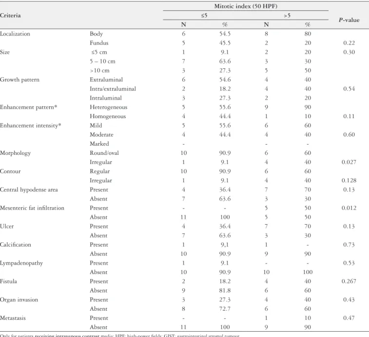
The main tomography indings of the 21 patients with gastric GIST, as well as the mitotic index found are dem-onstrated in Table 1. The tumors were located in the body (n = 14; 66.7%) and gastric fundus (n = 7; 33.3%).
TABLE 1. Computed tomographic indings of gastric GIST in 21 patients
Criteria
Mitotic index (50 HPF)
≤5 >5
P-value
N % N %
Localization Body 6 54.5 8 80
Fundus 5 45.5 2 20 0.22
Size ≤5 cm 1 9.1 2 20 0.30
5 – 10 cm 7 63.6 3 30
>10 cm 3 27.3 5 50
Growth pattern Extraluminal 6 54.6 4 40
Intra/extraluminal 2 18.2 4 40 0.54
Intraluminal 3 27.3 2 20
Enhancement pattern* Heterogeneous 5 55.6 9 90
Homogeneous 4 44.4 1 10 0.11
Enhancement intensity* Mild 5 55.6 6 60
Moderate 4 44.4 4 40 0.60
Marked - -
-Morphology Round/oval 10 90.9 6 60
Irregular 1 9.1 4 40 0.027
Contour Regular 10 90.9 6 60
Irregular 1 9.1 4 40 0.128
Central hypodense area Present 4 36.4 7 70 0.13
Absent 7 63.6 3 30
Mesenteric fat iniltration Present - - 5 50 0.012
Absent 11 100 5 50
Ulcer Present 4 36.4 7 70 0.13
Absent 7 63.6 3 30
Calciication Present 1 9,1 1 - 0.73
Absent 10 90.9 9 90
Lympadenopathy Present 1 9.1 - - 0.53
Absent 10 90.9 10 100
Fistula Present 2 18.2 4 40 0.267
Absent 9 81.8 6 60
Organ invasion Present 3 27.3 4 40 0.43
Absent 8 72.7 6 60
Metastasis Present - - 1 10 0.47
Absent 11 100 9 90
*Only for patients receiving intravenous contrast media; HPF: high-power ields; GIST: gastrointestinal stromal tumour
FIGURE 1. A 71 years old male patient, presenting abdominal pain
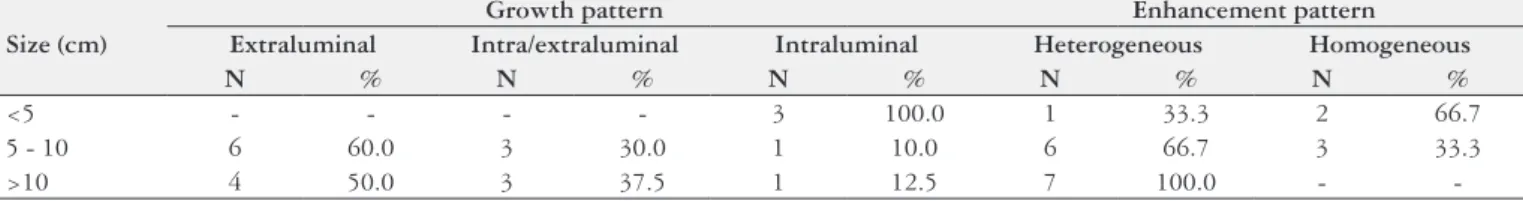
Lesion growth was predominantly extraluminal in 10 (47.6%) cases (Figure 2), intra/extra luminal in 6 (28.6%)
(Figure 3) and intraluminal in 5 (23.8%) (Figure 4). Among the tumors bigger than 10 cm, 50% presented extraluminal growth (Table 2), 37.5% presented intra/extra luminal growth, and 12.5% intraluminal. Among the tumors smaller than 5 cm, 100% presented intra luminal growth. Extraluminal tumors presented average size of 12.0 ± 5.7 cm and the in-traluminal tumors 6.6 ± 3.0 cm.
Enhancement through venous contrast was heterogene-ous in 14 (66.7%) patients and homogeneheterogene-ous in 5 (23.8%). In two (9.5%) patients, venous contrast was not utilized. Among the tumors bigger than 10 cm, 100% presented het-erogeneous enhancement, while among tumors smaller than 5.0 cm, 66.7% presented homogeneous enhancement (Table 2). Heterogeneous tumors presented average size of 11.9% ± 5.3 cm and the homogeneous 6.9 ± 2.2 cm.
The tumors presented oval/round morphology in 16 (76.8%) cases and irregular in 5 (23.8%). The presence of irregular morphology was observed in 40% of patients with high mitotic index (Figure 3) and 10% of the patients with reduced mitotic index.
Mesenteric fat iniltration was observed in 5 (23.8%) pa-tients, corresponding to 50% of the group with high mitotic level (Figure 3). No patient with low mitotic index presented iniltration of mesenteric fat.
Most patients still presented lesion with regular contours (76.8%), central hypodensity area (52.4%) and ulceration (52.4%). The presence of istula was observed in six (28.6%) patients, intratumoral calciication in two (9.5%) and adja-cent lymphadenopathy in just one (4.8%) patient.
FIGURE 2. A 61 years old female patient, presenting abdominal pain A and B: computed tomography with venous contrast showing expansive heterogeneous mass, predominantly extraluminal, located in the posterior wall of the gastric body (arrow); C: upper digestive endoscopy showing intraluminal component of the tumor; D and E: gross pathologic speci-men shows predominantly extraluminal growth (D) and the intraluminal aspect of the lesion (E).
FIGURE 3. A 51 years old female patient, presenting abdominal pain A and B: computed tomography with oral and venous contrast showing expansive heterogeneous mass, with central hypodense area and intra/ extraluminal growth, located in the anterior wall of the greater gastric curvature, presenting irregular morphology (arrow); B: hyperattenuation of adjacent mesenteric fat (*), characterizing tumor iniltration.
FIGURE 4. A 77 years old female patient, presenting upper digestive hemorrhage
A: computed tomography with oral and venous contrast showing intralu-minal vegetative lesion in the upper third of the greater curvature (white arrow), forming a istulous tract into the gastric lumen (black arrow); B: presence of gas and oral contrast in the lesion (arrow); C: intraoperatory aspect of the lesion (arrow); D: gross pathologic specimen showing intra-luminal lesion in the greater curvature (arrow)
TABLE 2 - Correlation between tumor size, growth pattern and enhancement pattern*
Size (cm)
Growth pattern Enhancement pattern Extraluminal Intra/extraluminal Intraluminal Heterogeneous Homogeneous
N % N % N % N % N %
<5 - - - - 3 100.0 1 33.3 2 66.7
5 - 10 6 60.0 3 30.0 1 10.0 6 66.7 3 33.3
>10 4 50.0 3 37.5 1 12.5 7 100.0 -
Invasion of adjacent organs was observed in 7 (33.3%) cases, the most affected being diaphragm, spleen, and pan-creas (n = 3; 14.3%). In only one case (4.8%), with high mitotic level, hepatic metastasis were identiied.
Statistical analysis showed that irregular morphology (P = 0.027) and iniltration of mensenteric fat (P = 0.012) correlated with high mitotic index.
DISCUSSION
Although this is an uncommon tumor, GIST is the most frequent mesenchymal neoplasia of the digestive tract. The disease is more common in the stomach and in individuals who are over 50 years old, with average age varying between 55 and 67(29, 31). Clinical manifestations are unspeciic and de-pend on the lesion site. Digestive hemorrhage and abdominal pain are the most frequent symptoms found(16, 21, 23, 30, 32). Our case comprised 14 (66.7%) female patients and 7 (33.3%) male patients, with average age of 61.6 years old, presenting pain or abdominal discomfort as the most frequent symptom.
Histopathological classiication of the tumor is based on the predominant cellular type (fusiform, epithelioid or mixed cells), and the diagnosis through immunohistochemistry as-sessment, which is based on the expression of the kit protein (CD 117)(7). GISTs are positive CD117 tumors (95%) and positive CD34 (30%-40%)(7). Some studies have yet dem-onstrated one alternative route in the disease pathogenesis, characterized by mutation in another receptor tyrosine kinase with activity similar to kit (Platelet-derived growth factor receptor – PDGFRA)(11).
Many factors are identiied in the literature as variables capable of predicting the evolution of the GIST, such as size, mitotic index, the presence of tumor necrosis, cellular proliferation markers, and tumor site(8). Whereupon, terms like “benign” or “malign” have been avoided, and GIST has been classiied according to the potential malignity based on the most relevant prognostic factors recognized in the literature (tumor site, size, and mitotic index)(26). Gastric tumors can then be classiied as high risk, intermediate risk, low risk or very low risk. We consider a tumor as a high risk one if it is bigger than 10 cm, if it presents more than 10 mitosis per HPF, or if bigger than 5 cm with more than ive mitosis per HPF. Intermediate risk tumors are smaller than 5 cm with 6 to 10 mitosis per HPF or its measure is 5 to 10 cm with less than ive mitosis per HPF. Low risk if its measure is between 2 and 5 cm with less than ive mitosis per HPF; very low risk if its measure is less than 2 cm with less than ive mitosis per HPF(6, 7).
Computed tomography is the most important imaging modality in the characterization of the GIST, as well as in the evaluation of adjacent organs invasion, abdominal me-tastasis, and response to treatment(1, 3, 10). Technological im-provement of this method has allowed a better assessment of large exophytic tumors and the relations of the gastric lesion with adjacent structures, and allows the characterization of tumors in speciic circumstances such as masses of unknown origin or originated from sites inaccessible to endoscopy(4, 18).
In the review of literature, the gastric body was the segment most affected by GIST (38%-75%)(16, 21, 23), with average size varying between 5.4 and 13.0 cm(2, 20), similarly to what was found in the present study. Tumors smaller than 5 cm present, still, predominantly intraluminal growth and homogeneous enhancement by venous contrast, while tumors bigger than 10 cm present extraluminal component and heterogeneous enhancement in most cases (3, 13, 16, 20, 27, 32). This trend was also observed in our results, where there was a bigger predominance of extraluminal growth among the tumors bigger than 10 cm (50%) and intraluminal growth in all tumors smaller than 5.0 cm. The same way, we found heterogeneous enhancement in 100% of the tumors bigger than 10 cm and homogeneous enhancement in 66.7% of the ones smaller than 5.0 cm.
Other characteristics can still be found especially in tumors of high histological degree, such as mucosa ulcera-tion (3%-88%), cavitaulcera-tion and areas of central hypodensity (20%-49%) which can correspond to cystic degeneration, hemorrhage or necrosis(9, 16, 23, 29, 32). Presence of gas or con-trast in the interior of the lesion can suggest presence of mucosa ulceration and istula formation(18). In our study, we found area of central hypodensity in 52.4% of the patients, mucosa ulceration in 52.4% and istulas in 28.6%. In both three groups, the percentage of patients with high mitotic level was, respectively, 63.7%, 63.7%, and 66.7%.
The presence of lymphadenopathy is very rare in pa-tients with GIST and its inding can cogitate the hypothesis of alternative diagnosis(28). Many series do not present any case with this inding(3, 22, 29, 30, 32), which is described in up to 2.5% of cases(16). In our cases, we found one case presenting lymphadenopathy, with mitotic index lower than ive mitosis per HPF.
Invasion of adjacent organs can be found in 6.2% to 20% of cases(16, 22, 33) and the presence of metastasis reaches up to 60% of cases in some series(2, 5). Metastatic potential seems to be related to the expression of the immunohistochemis-try marker CD34(25), being the liver and the peritoneum the most common sites of occurrence(29). In the present study, we found seven (33.3%) patients with invasion of adjacent organs, 57.2% of them presented high mitotic index and only 1 (4.8%) patient with hepatic metastasis, also presenting mitotic index superior to ive mitosis per HPF.
During the latest years, many authors have studied the tomography aspects of the GIST, trying to establish cor-relations between imaging indings and malignity potential. Kim et al.(16) observed that the presence of ulcer, mesenteric iniltration, invasion of adjacent organs, and presence of metastasis were characteristics more frequent in patients with high mitotic index. However, in the multi-varied analysis, just the size was the predictor for the high mitotic index. Similar results were described by Yang et al.(34). They found correlation between tumors bigger than 5.0 cm and malignity potential.
inva-sion, hepatic metastasis, and heterogeneous enhancement. Similarly, Ulusan et al.(33) have compared tomography vari-ables and mitotic index. The group with mitotic index higher than ive mitosis per 50 HPF presented positive correlation with homogeneous enhancement, tumor size, gastric location, presence of cyst-necrotic component, and presence of metas-tasis. Other series, however, did not ind statistical correlation between imaging characteristics and malignity potential(13, 15, 21).
In our case, some characteristics were more frequent in the group with high mitotic level, such as size bigger than 10 cm, heterogeneous enhancement, irregular morphology, irregular contour, central hypodensity, mesenteric iniltra-tion, ulcerainiltra-tion, istula, invasion of adjacent organs, and metastasis. However, only irregular morphology and mesen-teric iniltration presented statistically signiicant correlation. Irregular tumor morphology was observed in ive (23.8%) cases and 80% of them presented high mitotic index. Only one (9.1%) case in the group with mitotic index inferior to 5 presented this characteristic. Jeon et al.(15) found distorted morphology in 20.8% of the cases, being the frequency of this inding higher in the high-risk group (67%), if compared to the low-risk group (7%).
Mesenteric infiltration was also found in 5 (23.8%) patients of the present study, all with high mitotic index, corresponding to 50% of the patients of this group. Kim et al.(16) found mesenteric iniltration in 9.9% of the cases, all with high potential of malignity. Similarly, Ulusan et al.(33) found 23% of the cases with mesenteric iniltration, corresponding to 35.3% of the patients with high mitotic
level. However, no statistically signiicant correlation was established in these studies.
The reviewed literature presents bigger series with radi-ology assessment of the GIST. But a few studies correlate imaging indings with malignity potential based on histo-pathological data, with special interest for gastric tumors. Our series, although small, is concentrated in tumors of the stomach, which may have inluenced the results, as many authors have observed different predictions for lesions in other locations(5, 7, 16, 25, 26, 31, 34).
This study presents some limitations. It is a retrospective analysis, with a small number of cases, reviewed in many years in a tertiary institution. Nevertheless, some uncommon indings were observed, such as the statistical correlation between morphology, mesenteric iniltration and high mitotic index, what had not been described so far. These indings if conirmed in bigger series, can help in the identiication of the malignity potential of gastric GIST, through imaging characteristics.
CONCLUSIONS
In the present study, most gastric GISTs were located in the gastric body, with average size of 10.5 cm, presenting round/oval morphology, central hypodense area, ulceration, heterogeneous enhancement and predominantly extraluminal growth. Irregular morphology (P = 0.027) and iniltration of mesenteric fat (P = 0.012) presented correlated with high mitotic index.
Pelandré GL, Djahjah MC, Gaspareto EL, Nacif MS, Marchiori E, Mello ELR. Aspectos tomográicos do tumor estromal gastrointestinal de origem gástrica e correlação com índice mitótico. Arq Gastroenterol. 2013,50(4):244-50.
RESUMO – Contexto - Tumores estromais gastrointestinais são neoplasias raras e podem acometer qualquer segmento do trato gastrointestinal. A tomograia computadorizada é o método de imagem mais importante na detecção e caracterização do tumor. - Objetivos - Descrever os achados
tomográicos do tumor estromal gastrointestinal de origem gástrica, correlacionando com o índice mitótico. Métodos - No período de janeiro de
2000 a dezembro de 2008, foram selecionados 21 pacientes com diagnóstico histopatológico e imunohistoquímico de tumor estromal gastrointestinal, que apresentavam tomograia computadorizada realizada anteriormente ao tratamento. As variáveis tomográicas analisadas foram topograia da lesão, dimensões, contornos, morfologia, padrão e intensidade do realce pelo meio de contraste venoso, padrão de crescimento, invasão de órgãos adjacentes, presença de ulceração, fístula, calciicações, iniltração da gordura mesentérica, linfonodomegalias e metástases. O índice mitótico foi determinado através de microscopia óptica, com contagem do número de iguras de mitoses em 50 campos de grande aumento. Resultados - Os
tu-mores foram localizados no corpo (66,7%) ou fundo gástrico (33,3%), com dimensões variando entre 4,2 e 21,2 cm (média de 10,5 cm). O crescimento foi predominantemente extraluminal (47,6%) ou intra/extraluminal (28,6%). O realce pelo contraste venoso foi heterogêneo em 66.7%. A análise estatística mostrou que morfologia irregular (P = 0.027) e iniltração da gordura mesentérica (P = 0,012) apresentaram correlação com índice mitótico
elevado. Conclusões - No presente estudo, a maioria dos tumores localizava-se no corpo gástrico, com tamanho médio de 10.5 cm, apresentando área
hipodensa central, realce heterogêneo pelo meio de contraste e crescimento predominantemente extraluminal. Morfologia irregular e iniltração da gordura mesentérica apresentaram correlação estatística com índice mitótico elevado.
REFERENCES
1. Blay JY, Bonvalot S, Casali P, Choi H, Debiec-Richter M, Dei Tos AP, Emile JF, Gronchi A, Hogendoorn PC, Joensuu H, Le Cesne A, McClure J, Maurel J, Nupponen N, Ray-Coquard I, Reichardt P, Sciot R, Stroobants S, van Glabbeke M, van Oosterom A, Demetri GD;.GIST consensus meeting panelists. Consensus meeting for the management of gastrointestinal stromal tumors. Report of the GIST Consensus Conference of 20-21 March 2004, under the auspices of ESMO. Ann Oncol. 2005;16:566-78.
2. Burkill GJ, Badran M, Al-Muderis O, Meirion Thomas J, Judson IR, Fisher C, Moskovic EC. Malignant gastrointestinal stromal tumor: distribution, imaging features, and pattern of metastatic spread. Radiology. 2003;226:527-32. 3. Chourmouzi D, Sinakos E, Papalavrentios L, Akriviadis E, Drevelegas A.
Gastrointestinal stromal tumors: a pictorial review. J Gastrointestin Liver Dis. 2009;18:379-83.
4. Da Ronch T, Modesto A, Bazzocchi M. Gastrointestinal stromal tumour: spiral computed tomography features and pathologic correlation. Radiol Med. 2006;111:661-73.
5. DeMatteo RP, Lewis JJ, Leung D, Mudan SS, Woodruff JM, Brennan MF. Two hundred gastrointestinal stromal tumors: recurrence patterns and prognostic factors for survival. Ann Surg. 2000;231:51-8.
6. Efron DT, Lillemoe KD. The current management of gastrointestinal stromal tumors. Adv Surg. 2005;39:193-221.
7. Fletcher CD, Berman JJ, Corless C, Gorstein F, Lasota J, Longley BJ, Miettinen M, O’Leary TJ, Remotti H, Rubin BP, Shmookler B, Sobin LH, Weiss SW. Di-agnosis of gastrointestinal stromal tumors: a consensus approach. Hum Pathol. 2002;33:459-65.
8. Fujimoto Y, Nakanishi Y, Yoshimura K, Shimoda T. Clinicopathologic study of primary malignant gastrointestinal stromal tumor of the stomach, with special reference to prognostic factors: analysis of results in 140 surgically resected patients. Gastric Cancer. 2003;6:39-48.
9. Ghanem N, Altehoefer C, Furtwangler A, Winterer J, Schafer O, Springer O, Kotter E, Langer M. Computed tomography in gastrointestinal stromal tumors. Eur Radiol. 2003;13:1669-78.
10. Gong J, Kang W, Zhu J, Xu J. CT and MR imaging of gastrointestinal stromal tumor of stomach: a pictorial review. Quant Imaging Med Surg. 2012;2:274-9. 11. Heinrich MC, Corless CL, Duensing A, McGreevey L, Chen CJ, Joseph N,
Singer S, Grifith DJ, Haley A, Town A, Demetri GD, Fletcher CD, Fletcher JA. PDGFRA activating mutations in gastrointestinal stromal tumors. Science. 2003;299:708-10.
12. Hirota S, Isozaki K, Moriyama Y, Hashimoto K, Nishida T, Ishiguro S, Kawa-no K, Hanada M, Kurata A, Takeda M, Muhammad Tunio G, Matsuzawa Y, Kanakura Y, Shinomura Y, Kitamura Y. Gain-of-function mutations of c-kit in human gastrointestinal stromal tumors. Science. 1998;279:577-80.
13. Horton KM, Juluru K, Montogomery E, Fishman EK. Computed tomography imaging of gastrointestinal stromal tumors with pathology correlation. J Comput Assist Tomogr. 2004;28:811-7.
14. Huizinga JD, Thuneberg L, Kluppel M, Malysz J, Mikkelsen HB, Bernstein A. W/kit gene required for interstitial cells of Cajal and for intestinal pacemaker activity. Nature. 1995;373:347-9.
15. Jeon SW, Park YD, Chung YJ, Cho CM, Tak WY, Kweon YO, Kim SK, Choi YH. Gastrointestinal stromal tumors of the stomach: endosonographic differentiation in relation to histological risk. J Gastroenterol Hepatol. 2007;22:2069-75.
16. Kim HC, Lee JM, Kim KW, Park SH, Kim SH, Lee JY, Han JK, Choi BI. Gastrointestinal stromal tumors of the stomach: CT indings and prediction of malignancy. AJR Am J Roentgenol. 2004;183:893-8.
17. Kindblom LG, Remotti HE, Aldenborg F, Meis-Kindblom JM. Gastrointestinal pacemaker cell tumor (GIPACT): gastrointestinal stromal tumors show phenotyp-ic characteristphenotyp-ics of the interstitial cells of Cajal. Am J Pathol. 1998;152:1259-69. 18. Lau S, Tam KF, Kam CK, Lui CY, Siu CW, Lam HS, Mak KL. Imaging of
gastrointestinal stromal tumour (GIST). Clin Radiol. 2004;59:487-98. 19. Lauwers GY, Erlandson RA, Casper ES, Brennan MF, Woodruff JM.
Gastroin-testinal autonomic nerve tumors. A clinicopathological, immunohistochemical, and ultrastructural study of 12 cases. Am J Surg Pathol. 1993;17:887-97. 20. Lee CM, Chen HC, Leung TK, Chen YY. Gastrointestinal stromal tumor:
Com-puted tomographic features. World J Gastroenterol. 2004;10:2417-8.
21. Levy AD, Remotti HE, Thompson WM, Sobin LH, Miettinen M. Gastrointestinal stromal tumors: radiologic features with pathologic correlation. Radiographics. 2003;23:283-304.
22. Lupescu IG, Grasu M, Boros M, Gheorghe C, Ionescu M, Popescu I, Herlea V, Georgescu SA. Gastrointestinal stromal tumors: retrospective analysis of the computer-tomographic aspects. J Gastrointestin Liver Dis. 2007;16:147-51. 23. Martín-Lorenzo JG, Aguayo-Albasini JL, Torralba-Martínez JA, Lirón-Ruiz R,
Giménez-Bascuñana A, Miquel-Perelló J, Moreno-Egea A, Carrasco-González L. [Gastrointestinal stromal tumors. Diagnosis, prognosis and current surgical treatment. Follow-up of 18 treated patients]. Cir Esp. 2006;79:22-7.
24. Mazur MT, Clark HB. Gastric stromal tumors. Reappraisal of histogenesis. Am J Surg Pathol. 1983;7:507-19.
25. Miettinen M, Lasota J. Gastrointestinal stromal tumors--deinition, clinical, his-tological, immunohistochemical, and molecular genetic features and differential diagnosis. Virchows Arch. 2001;438:1-12.
26. Miettinen M, El-Rifai W, Sobin L, Lasota J. Evaluation of malignancy and prog-nosis of gastrointestinal stromal tumors: a review. Hum Pathol. 2002;33:478-83. 27. Nishida T, Kumano S, Sugiura T, Ikushima H, Nishikawa K, Ito T, Matsuda
H. Multidetector CT of high-risk patients with occult gastrointestinal stromal tumors. AJR Am J Roentgenol. 2003;180:185-9.
28. Rimondini A, Belgrano M, Favretto G, Spivach A, Sartori A, Zanconati F, Cova MA. Contribution of CT to treatment planning in patients with GIST. Radiol Med. 2007;112:691-702.
29. Sandrasegaran K, Rajesh A, Rushing DA, Rydberg J, Akisik FM, Henley JD. Gas-trointestinal stromal tumors: CT and MRI indings. Eur Radiol. 2005;15:1407-14. 30. Sandrasegaran K, Rajesh A, Rydberg J, Rushing DA, Akisik FM, Henley JD.
Gastrointestinal stromal tumors: clinical, radiologic, and pathologic features. AJR Am J Roentgenol. 2005;184:803-11.
31. Strickland L, Letson GD, Muro-Cacho CA. Gastrointestinal stromal tumors. Cancer Control. 2001;8:252-61.
32. Tateishi U, Hasegawa T, Satake M, Moriyama N. Gastrointestinal stromal tumor. Correlation of computed tomography indings with tumor grade and mortality. J Comput Assist Tomogr. 2003;27(5):792-8.
33. Ulusan S, Koc Z, Kayaselcuk F. Gastrointestinal stromal tumours: CT indings. Br J Radiol. 2008;81:618-23.
34. Yang TH, Hwang JI, Yang MS, Hung SW, Chan SW, Wang J, Tyan YS. Gas-trointestinal stromal tumors: computed tomographic features and prediction of malignant risk from computed tomographic imaging. J Chin Med Assoc. 2007;70:367-73.