OPTIMIZED HYPERVENTILATION PRESERVES
2,3-DIPHOSPHOGLYCERATE IN SEVERE
TRAUMATIC BRAIN INJURY
Rayne Borges Torres
1, Renato Giuseppe Giovanni Terzi
2, Antônio Luís Eiras Falcão
3,
Nelci Fenalti Höehr
4, Venâncio Pereira Dantas Filho
5ABSTRAT - Introduction: The concentration of 2,3-diphosphoglycerate (2,3-DPG/Hct) increases as a phys-iological occurrence to pH increase and hyperventilation. This response was tested in patients with se-vere traumatic brain injury (TBI). Method: The concentration of 2,3-DPG/Hct was measured daily for six days in eleven patients with severe TBI in need of optimized hyperventilation because of intracranial hypertension. Results: There was correlation between pH and the concentration of DPG/Hct. The concen-tration of 2,3-DPG/Hct remained predominantly within normal levels with slight increase in the sixth day of the study. The concentration of 2,3-DPG/Hct correlated significantly with measured partial pressure of oxy-gen that saturates 50% the hemoglobin of the blood (P50st), confirming the consistency of our data. Con-clusion: The expected physiological response of a progressive increase in concentration of 2,3-DPG/Hct to hyperventilation was not observed. This fact may be explained by the intermittent and not sustained hy-perventilation as dictated by the protocol of optimized ventilation.
KEY WORDS: 2,3-diphosphoglycerate, brain injury, traumatic brain injury, hyperventilation, mechanical ventilation.
A hiperventilação otimizada preserva o 2,3-difosfoglicerato no traumatismo craniencefálico grave
RESUMO - Introdução: A concentração de 2,3-difosfoglicerato nos eritrócitos (2,3-DPG/Hct) aumenta como ocorrência fisiológica ao aumento do pH e à hiperventilação. Esta resposta foi testada em pacientes com traumatismo craniencefálico (TCE) grave. Método: A concentração de 2,3-DPG/Hct foi medida diariamen-te, durante seis dias, em 11 pacientes com TCE grave necessitando de hiperventilação otimizada por cau-sa da hipertensão intracraniana. Resultados: Houve correlação entre o pH e a concentração de 2,3-DPG/ Hct. A concentração de 2,3-DPG/Hct permaneceu predominantemente dentro da normalidade, com ligei-ra tendência à elevação no sexto dia de estudo. Houve correlação entre a concentligei-ração de 2,3-DPG/Hct e a pressão parcial de oxigênio que satura 50% da hemoglobina (P50st), confirmando a consistência dos dados. Conclusão: A esperada resposta fisiológica de aumento progressivo da concentração de 2,3-DPG/ Hct não foi observada. Este fato pode ser explicado pela aplicação intermitente e não sustentada de hiper-ventilação, como ditado pelo protocolo clínico de ventilação otimizada.
PALAVRAS-CHAVE: 2,3-difosfoglicerato, traumatismo cerebral, lesão cerebral traumática, hiperventilação, ventilação mecânica.
Campinas State University (UNICAMP) Campinas, São Paulo, Brazil (FCM/UNICAMP): 1Physical Therapist and Master of Science. Direc-tor, Graduate Course of Phisiotherapy, Faculdades Integradas de Patos, Patos PB, Brazil; 2Full Professor of Surgery, FCM/UNICAMP; 3
Assistant Professor, FCM/UNICAMP and Head UTI - Hospital das Clínicas - UNICAMP; 4
Assistant Professor, Clinical Pathology Depart-ment, FCM/UNICAMP; 5Assistant Professor, Bioethics and Neurosurgery, FCM and Hospital das Clínicas da UNICAMP. Neurosurgeon Hospital das Clínicas - UNICAMP. This Project was partially funded by FAPESP Grant 99/12920-8 and Grant 00/02498-6.
Received 5 March 2007, received in final form 22 May 2007. Accepted 28 June 2007.
Dr. Rayne Borges Torres - Rua Bossuet Wanderley 685 - 58700-410 Patos PB - Brasil. E-mail: [email protected]
The organic phosphate present in the red blood cell - 2,3-diphosphoglycerate (2,3-DPG) - exerts im-portant function in oxygen transport. The increase of this phosphate in the red blood cell reduces the hemoglobin oxygen affinity, facilitating oxygen de-livery to the tissues. On the other hand, a reduction of 2,3-DPG in blood promotes an opposite effect1,2
. The main factors that regulate the concentration of 2,3-DPG in the red blood cell are changes in red
cell pH and hypoxia3
. The arterial blood carbon di-oxide (PaCO2) and, consequently, pH are altered by mechanical ventilation which has been used in some situations, such as the control of intracranial hyper-tension in patients with severe traumatic brain inju-ry (TBI).
The fall in PaCO2 (hypocapnia) reduces cerebral
blood flow and intracranial pressure4. Currently,
pa-tients with TBI has been questioned5, but it has been
used, in a more restricted way, monitoring the global brain metabolism. This has been done calculating the cerebral extraction of oxygen through continuous or intermittent sampling of the venous oxygen satura-tion of the jugular bulb vein. The coupling between the PaCO2 and the cerebral extraction of oxygen has
been called optimized ventilation6,7
.
It is known that the reduction of PaCO2 and, con-sequently, the increase in arterial pH, increases the affinity of hemoglobin for oxygen, a possible factor interfering with oxygen delivery to the tissues of the injured brain. The hypothesis of the present study was that an increase in the concentration of 2,3-DPG in the red blood cell must occur as a physiological re-sponse to increased arterial pH induced by mechani-cal ventilation. Therefore, the objective was to mea-sure PaCO2, arterial pH, the concentration of 2,3-DPG
and to calculate the partial pressure of oxygen that saturates 50% the hemoglobin of the blood (P50) of patients that suffered severe TBI and submitted to a protocol of optimized ventilation.
METHOD
The present study was carried out in the Intensive Care Unit of the Hospital das Clínicas da Universidade Estadu-al de Campinas, UNICAMP. The study was approved by the Hospital Research Ethics Committee and informed consent was obtained from the patient’s legal representatives.
Eleven patients were included. Gender, age, Glasgow Coma Scale (GCS) on admission8, the cause of the head trau-ma, the presence or absence of associated lesions, the evalu-ation of the computerized tomography by the Marshall
clas-sification9 and the APACHE II score as well as the APACHE II calculated risk of death are displayed on Table.
All patients exhibiting focal lesion as well as those with gunshot injuries underwent surgery. All patients were in-tubated and on mechanical ventilation. Intracranial pres-sure was continuously monitored as well as respiratory mechanics, capnography and pulse oximetry. The pressure transducer of titanium tip CODMAN® (Johnson & Johnson, Raynham, Me - USA) was placed in the brain parenchyma and was used to monitor intracranial pressure. Respirato-ry variables, capnography and oximetRespirato-ry were continuously recorded with a respiratory profile monitor CO2SMOPlus® (Respironics/Novametrix/Dixtal, São Paulo - Brazil). The he-moglobin saturation of jugular bulb blood was continu-ously monitored with a 4F fiber optic oxymetric catheter BAXTER® coupled to a VIGIILANCE® monitor (Baxter Health Care Corporation, Irvine, CA - USA) and/or in an intermit-tent way by hemo-oximetry measured in a blood sample collected from the same catheter, or from a 16G catheter INTRACATH®. A central venous catheter (superior vein ca-va) and a urinary bladder catheter were inserted. The cor-rect location of the venous catheters was confirmed by ra-diography.
The patients were kept supine with the head elevated to 30 degrees and the cervical spine maintained rectified to prevent compression of the jugular vein.
It was attempted to keep the cerebral perfusion pressure (mean arterial pressure less intracranial pressure) always above 70 mmHg, with volume replacement and/or admin-istered norepinephrine. Sedation was started with fentanyl associated to midazolam (0.5 mg of fentanyl and 75 mg of midazolam in 250 mL of 5% glucose. Dripping started at 10 to 20 microdrops/min, up to a sufficient dose to sedate, ac-cording to the individual response of each patient).
Mechanical ventilation was optimized to keep cerebral oxygen extraction between 24% to 42% whenever the
in-Table. Casuistics.
Gender Age GCS TA AL CT AP II AP II (%) ICU death
1 M 26 7 MT No DIII 17 17 Yes
2 F 19 8 FA No – 23 34 No
3 M 33 7 SP No DII 13 14 No
4 M 25 6 FL No Focal 16 12 No
5 M 19 7 FA No – 11 11 Yes
6 M 28 8 RO Yes DIII 26 44 No
7 M 49 6 AU No DIV 22 31 Yes
8 M 23 6 AU No DIII 17 17 No
9 M 19 8 AU No DIV 16 15 No
10 M 15 8 AU Yes DIII 22 30 No
11 M 25 8 RO Yes DIV 12 9 No
Mean 25.5 7.2 17.7 21.34 3/11
SD 9.3 0.9 4.9 11.4
tracranial pressure was above 20 cmH2O6,7. If the intracra-nial pressure remained elevated, mannitol 20% was given in bolus (0.5 a 1 g/kg/dose) or the sedation was substitut-ed by thionembutal (initial dose between 0.5 to 2 mg/kg/ hour up to a maximum of 4 mg/kg/hour). If after these iso-lated or combined alternative procedures, the intracranial pressure remained elevated, a new brain computerized to-mography was performed to evaluate the necessity of gical decompression. No patient in this series needed sur-gical decompression.
The use of mannitol, furosemide and vasoactive drugs was recorded. Daily water balance and urine output was also registered.
Laboratory data – Arterial and venous blood gases as well as hemo-oximetry of the jugular bulb were performed daily and analyzed by the ABL 700® (Radiometer, Copenha-gen, Denmark). The calculation of the partial pressure of the oxygen that saturates 50% of the hemoglobin correct-ed to standard condition (temperature 37ºC, pH 7.4 and PaCO2 40 mmHg) (P50st) was obtained directly from the venous blood gas analysis. The concentration of 2,3-diphos-phoglycerate per hematocrit (2,3-DPG/Hct) was measured by spectrophotometry, using the kit catalogue 665 of SIG-MA DIANOSTIC (Sigma-Aldrich - St. Louis, Me - USA). The first measurement was collected from the arterial blood in the first 12 hours of hospital admission. The remainders were performed daily, always in the morning. Serum phos-phate was measured in the Hospital auto-analyzer with “Roche Diagnostics” reagents. Collection of data was per-formed for six days after admission to the Intensive Care Unit (ICU). The normal range of phosphate for our hospital laboratory is 2.5 to 4.5 mg/dL. Values below 2 mg/dL were considered as hypophosphatemia and values below 1 mg/ dL were considered as severe hypophosphatemia.
Standardization of the measure of the 2,3-DPG – One milliliter of blood was harvested in syringe daily (Peak 70, Medical Radiometer, Copenhagen, Denmark) and trans-ported to the laboratory in a cooled container. The max-imum period of refrigerated storage before analysis was two hours.
The 2,3-DPG reagents for quality control were cata-logue S 3006 (normal) and catacata-logue S 3005 (high) of SIG-MA DIAGNOSTIC (Sigma-Aldrich - St. Louis, Me - USA). The values read with the high control and the normal control were always between the limits, 3.3 to 4,3 and 1.7 to 2.5 mmol/ml, respectively, as required by the manufacturer.
Control group – Peripheral venous blood was collected from healthy not-smokers volunteers (20 men and 20 wom-en) to establish our standard of normality. In these sam-ples we measured the concentration of 2,3-DPG/Hct, serum phosphate and calculated P50st, with the same techniques described for the group of patients with TBI.
Statistical analysis – To compare and correlate the vari-ables that had more than one measurement in each pa-tient we applied the GEE analyses - generalized estimating
equations, an extension of generalized linear models10. This analysis takes into consideration measurements obtained in the same patient at different times (longitudinal mea-surements) and analyses the simultaneous change on sever-al variables in the same patient and in the patients’ group. Continuous variables as well as categorical variables used in this analysis were classified by pre-determined cut values. Statistical significance was established as 0.05.
RESULTS
Control group – The values of mean and standard deviations of the concentration of 2,3-DPG/Hct and serum phosphate, as well as the calculated P50st in the control group were 4.6±0.7 mmol/mL, 3.15±0.46 mg/dL and 26.3±1.0 mmHg, respectively. The values of the concentration of 2,3-DPG/Hct were within the limits of normality by the manufacturer standard and the values of the concentration of serum phosphate were within the limits of standardized normality for the Hospital Laboratory. The values of P50st are sim-ilar to those reported in the literature.
Patients group
Correlation between pH and PaCO2 – As previous-ly mentioned, the means of PaCO2 and arterial pH were correlated through a GEE analysis (p=0.0001). The values of mean PaCO2 and mean pH meet pre-dominantly below 35mmHg and above 7.45, respec-tively. This indicates that the patients had been, most of the time, kept in a state of respiratory alkalosis. At times, however, the value of pH is normal, despite a low PaCO2 and in some the value of pH is elevated
despite a normal PaCO2. This situation was interpret-ed as an associatinterpret-ed metabolic alteration (compensa-tory metabolic acidosis and primary metabolic alka-losis, respectively).
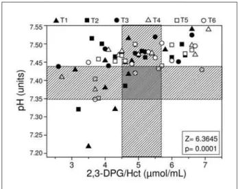
Correlation between the pH and the concentra-tion of 2,3-DPG/Hct – A positive correlation between the mean arterial pH and the concentration of 2,3-DPG/Hct was demonstrated through GEE analysis (p=0.0001) (Fig 1).
Blood transfusion, hypophosphatemia and
2,3-DPG/Hct – The concentration of 2,3-DPG/Hct was
lower when the patients received stocked red blood cell transfusion within the 24 hours that preceded blood collection (p=0.001) and/or presented a serum phosphate below 2.0 mg/dL (hypophosphatemia) (p= 0.005). The interference of these factors on the cor-relation between pH and the concentration of 2,3-DPG/Hct can be appreciated in Fig 2.
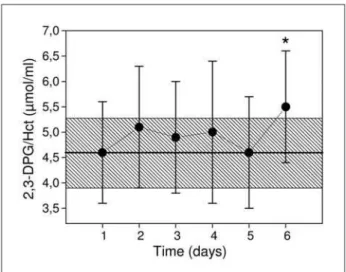
2,3-DPG/Hct in relation to the time – The concen-tration of 2,3-DPG/Hct, remained normal from the first to the sixth day with a slight, non significant rise at day 6 (Fig 3) when the value of reference was the mean of the first day (p=0.05) or the mean of the control group (p=0.09).
Correlation between the concentration of 2,3-DPG/Hct and the P50st – As expected, a significant correlation was found comparing the concentration of 2,3-DPG/Hct and P50st (p=0.0001).
DISCUSSION
The data of the present study show a positive cor-relation between the arterial blood pH and the con-centration of 2,3-DPG/Hct in patients with severe TBI artificially ventilated (Fig 1). Changes in 2,3-DPG as a response to pH variations had previously been report-ed in other physiological and clinical situations. In el-evated altitudes, the increase of the concentration of 2,3-DPG in the erythrocytes has been interpreted as a compensating mechanism for oxygen transport at low barometric pressure11
. In this situation, respira-tory alkalosis seems to be the main cause to increase the concentration of 2,3-DPG in the red cells. Cor-responding change in 2,3-DPG concentration in the red cells secondary to alteration in arterial blood pH, either as metabolic or respiratory alkalosis or acido-sis, have been reported in some clinical situations12,13
. As a matter of fact, Bellingham et al.14 confirmed
experimentally this correlation, inducing metabolic alkalosis or acidosis in healthy volunteers.
The time and the depth of the alteration of pH to cause a significant response in the concentration of 2,3-DPG are not clear-cut in the literature. Venuxem et al.13 demonstrated changes in the concentration
of 2,3-DPG only in chronic states of acidosis or alka-losis, because in acute situations (pH changes within
Fig 2. Correlation between the concentration of 2,3-DPG/Hct and the mean arterial pH for each patient. The solid circles (NPT) are measures in patient not transfused in the previous day and/or a phosphatemia above 2.0 mg/dL. The open circles are samples collected from patients transfused in the previous day and/or with hypophosphatemia (Pi<2.0 mg/dL). The dotted line represents the correlation of the PT samples and the con-tinuous line the NPT samples. The tread area delimits the nor-mal values of the concentration of 2,3-DPG/Hct (3.9–5.3 mmol/ ml) and/or pH (7.35–7.45).
Fig 3. Mean and standard deviation of 2,3-DPG/Hct concen-tration over time for all patients. The tread area delimits the mean and standard deviation of the control group (4.6±0.7
24 to 48 hours), the concentration of 2,3-DPG was normal. On the other hand, Bellingham et al.14
dem-onstrated that the concentration of 2,3-DPG does not change within the first four hours of metabolic aci-dosis or metabolic alkalosis. However, a significant response was shown to occur after 48 hours. Respira-tory alkalosis induced by hyperventilation in healthy volunteers, did not exhibit deviation in the red cell 2,3-DPG concentration15
. However, in these subjects hyperventilation was not sustained for more than twenty minutes. In the current study, the time and the depth of the hyperventilation varied, from min-utes to hours, according to the attending physician’s orders based on the clinical situation of the patient. Nevertheless, the mean daily arterial pH remained elevated (moderate alkalosis) during the six days of the study in the majority of the patiente. Yet, the concentration of 2,3-DPG/Hct, exhibited great daily variation from patient to patient, with a mean, non significant rise by the sixth day (Fig 3). We attribute this finding to the intermittent and non sustained hyperventilation applied to these patients with se-vere TBI based on a previously established protocol of optimized ventilation.
For many years, systematic and indiscriminate hy-perventilation has been used to prevent intracranial hypertension in patient victims of severe TBI16. PaCO
2
was, systematically, kept between 20 and 30 mmHg. From the decade of 1980, the indiscriminate use of hyperventilation started to be criticized, because the reduction of cerebral blood flow induced by hy-pocapnia could lead, or exacerbate, post-traumatic brain ischemia. Currently, hyperventilation is no lon-ger used prophylactically in the treatment of severe TBI17. However, in refractory intracranial
hyperten-sion, hyperventilation may be cautiously applied un-der strict evaluation of cerebral oxygen extraction. This is currently done measuring the hemoglobin oxygen saturation in the jugular bulb, indirectly ex-pressing cerebral blood flow. Hyperventilation is then titrated to keep oxygen extraction within safe limits, a practice called optimized ventilation6,7.
As respiratory alkalosis displaces the oxygen dis-sociation curve to the left (low P50st), the increased hemoglobin affinity for oxygen may hamper tissue availability. Therefore, the expected physiological re-sponse would be the return of the dissociation curve to normal (P50 normal, around 26.6 mmHg). In the current study, the expected gradual increase in the concentration of 2,3-DPG/Hct in response to hyper-ventilation (respiratory alkalosis) was not observed. This was attributed to the fact that hyperventilation
was applied intermittently and based on an estab-lished protocol of optimized ventilation.
Other factors that may have contributed to the lack of response of 2,3-DPG/Hct must be mentioned. First, it is known that preserved blood has a progres-sive reduction in the levels of 2,3-DPG/Hct related to the time of Blood Bank storage. Five of the eleven patients in the present study received blood transfu-sion. All together they received 18 units of packed red cell. These units had been stocked for more than six days; two units were stocked for 18 days and 11 units for more than 20 days in the Hospital Blood Bank. Each patient transfused, received two or more units of packed red cells. These units were preserved with citrate-phosphate-dextrose-adenine (CPDA-1), that permits blood transfusion after 35 days of Blood Bank storage. Measured concentration of 2,3-DPG in blood preserved with the CPDA-1 show discrepan-cies on the weekly reduction of 2,3-DPG. A loss of 2,3-DPG around 40% to 60% occurs in the second week, from 70% to 90% in the third week and great-er than 90% from the fourth week on18-21
. In healthy volunteers, Heaton et al. evaluated, the restoration of 2,3-DPG in erythrocytes after blood transfusion, with Blood Bank storage of 35 days. When CPDA-1 was used the as conservative, they describe an impor-tant fall in the concentration of 2,3-DPG/Hb22. After
a period of seven hours, 50% of the concentration of 2,3-DPG already had been restored, but the complete restoration was seen only after 48 hours.
A second factor that may interfere with 2,3-DPG production is hypophosphatemia. The reduction of serum phosphate can cause a fall in 2,3-DPG because of decreased 1,3-DPG, as well as increased degrada-tion of 2,3-DPG22. The normal levels of serum
phos-phate lay between 2.5 and 4.5 mg/dL. The clinical symptoms of hypophosphatemia are observed when serum phosphate falls below of 2.0 mg/dL23. In the
analysis of our data, hypophosphatemia was defined when serum phosphate was under 2.0 mg/dL. Hypo-phosphatemia has been described in patients with TBI24-26
. In the current study, the concentration of 2,3-DPG/Hct was lower than normal when hypophospha-temia was present. Pas’ko and Volosheniuk25
also cor-related serum phosphate, with the concentration of 2,3-DPG and ATP in the red cell. Hypophosphatemia may occur for several reasons in critical patients when associated to conditions such as alkalosis, the use of diuretics, volume expansion and sepsis27-29
the blood to the cell across the cell membrane is at-tributed the two mechanisms. The first is the increase in glycolitic activity in response to the increase of in-tracellular pH, induced by respiratory alkalosis. The second is the level of insulin in the bloodstream in re-sponse to glucose infusion, promoting greater migra-tion of phosphate into the cell27. Recently, in healthy
volunteers, it has been demonstrated that the longer the time of hyperventilation, or the deeper the hy-pocapnia, greater is the fall in serum phosphate15
. In patients with severe TBI, Pas’ko and Volosheniuk25
had demonstrated that serum phosphate was lower in patients with hypocapnia. Hypophosphatemia in these patients was attributed to the insufficient re-placement of phosphate in parenteral nutrition, as well as a loss of phosphate in the urine. Sepsis is an-other well described cause of hypophosphatemia and is associated to increased risk of death29,30. We
report-ed that in 14 patients with severe TBI, a preliminary and partial analysis revealed an inverse relationship between the pH and serum phosphate. Furthermore, the most important factor leading to hypophospha-temia was sepsis. In addition, it was observed that serum phosphate was significantly lower in patients that died compared to survivors30
.
Concluding, the variations in the concentration of 2,3-DPG/Hct observed in this group of patients with severe TBI exhibited a significant correlation with P50st (p<0.05). This correlation of two independently measured variables certifies the reliability and the consistency of our data. It was not possible to dem-onstrate a gradual increase of the concentration of 2,3-DP/Hct in the course of the treatment with opti-mized hyperventilation. Despite the daily averages of all patients, pointing to respiratory alkalosis (high pH and low PaCO2) alkalosis was not sustained during all
the period of study.
One limitation of this study is the absence of a comparative group with systematic and continuous hyperventilation. It is quite possible that the expect-ed increase in 2,3-DPG/Hct could then be demonstrat-ed. However, with basis on the present knowledge and current practice, it would have been unethical a study design including this group. Despite this con-straint, our data suggest that optimized ventilation is a safe practice that does not interfere significantly, with P50st and ultimately, with oxygen delivery. Fur-thermore, it was possible to demonstrate a statistical correlation between arterial pH and the concentra-tion of 2,3-DPG/Hct. Finally, we were able to demon-strate that blood transfusion and hypophosphatemia are adjuvant factors that may contribute to hinder the rise of 2,3-DPG/Hct.
REFERENCES
1. Benesch R, Benesch RE. Intracellular organic phosphates as regulators of oxigen release by hemoglobin. Nature 1969;221:618-622.
2. Chanutin A, Curnish RR. Effect of organic and inorganic phophastes on the oxygen equilibrium of human erytrocyte. Arch Biochem Biophys 1967;121:96.
3. Duhm J, Gerlach E. On the mechanisms of the hypoxia-induced increase of 2,3-diphosphoglycerate in erythrocytes: studies on rat erythrocytes in vivo
and on human erythrocytes in vitro. Plugers Arch 1971;326:254-269. 4. Lundberg N, Kjallquist A, Bien C. Reduction of increased intracrani
-al pressure by hyperventilation: a therapeutic aid in neurologic-al
sur-gery. Acta Psychiatr Scand 1959;34(Suppl 139):S1-S64.
5. Muizelaar JP, Marmarou A, Ward JD, et al. Adverse effects of prolonged hyperventilation in patients with severe head injury: a randomized clin
-ical trial. J Neurosurg 1991;75:731-739.
6. Cruz J. Relevance of ventilatory optimization in acute intracranial hy -pertension: a clinical, physiological, and therapeutic approach. Arq
Neuropsiquiatr 1995;53:131-140.
7. Falcao A, Araujo S, Dragosavac D, et al. Cerebral hemometabolism:
variability in the acute phase of traumatic coma. Arq Neuropsiquiatr
2000;58:877-882.
8. Teasdale G, Jennet B. Assessment of coma and impared consciosness.
Lancet 1974;7:81-84.
9. Marshall LF, Marshall SB, Klauber MR et al. A new classiication of head injury based on computerized tomography. J Neurosurg 1991;75(Sup
-pl):S14-S20.
10. Zeger SL, Liang KY. Longitudinal data analysis for discrete and contin
-uous outcomes. Biometrics 1986;42:121-130.
11. Samaja M, Brenna L, Allibardi S, Cerretelli P. Human red blood cell aging at 5,050-m altitude: a role during adaptation to hypoxia. J Appl Physiol 1993;75:1696-1701.
12. Carlone S, Serra P, Farber MO, et al. Red blood cell alkalosis and de
-creased oxyhemoglobin afinity. Am J Med Sci 1982;284:8-16.
13. Vanuxem D, Fornaris E, Delpierre S, Grimaud C. Role of the acid-base
sta-tus on the changes of haemoglobin oxygen afinity in arterial hypoxemia (author’s transl). Bull Physiopathol Respir (Nancy) 1975;11:305-314. 14. Bellingham AJ, Detter JC, Lenfant C. Regulatory mechanisms of he
-moglobin oxygen affinity in acidosis and alkalosis. J Clin Invest 1971;50:700-706.
15. Paleologos M, Stone E, Braude S. Persistent, progressive hypophosphatae
-mia after voluntary hyperventilation. Clin Sci (Lond) 2000;98:619-625. 16. Backer DP, Gardner S. Intensive management of head injury. In Wilkins
RH, Rengachary SS (Eds). Neurosurgery. New York: McGraw-Hill, 1985: 1593-1599.
17. Guidelines for the management of severe head injury. Brain Trauma Foun -dation, American Association of Neurological Surgeons, Joint Section on
Neurotrauma and Critical Care. J Neurotrauma 1996;13:641-734. 18. Kreuger A, Akerblom O, Hogman CF. A clinical evaluation of
citrate-phosphate-dextrose-adenine blood. Vox Sang 1975;29:81-89. 19. Moore GL, Ledford ME, Brummell MR. Red cell ATP and 2,3-diphos
-phoglycerate concentrations as a function of dihydroxyacetone
supple-mentation of CPD adenine. Vox Sang 1981;41:11-17.
20. Moroff G, Dende D. Characterization of biochemical changes occurring
during storage of red cells. Comparative studies with CPD and
CPDA-1 anticoagulant-preservative solutions. Transfusion CPDA-1983;23:484-489. 21. Heaton A, Keegan T, Holme S. In vivo regeneration of red cell 2,3-di
-phosphoglycerate following transfusion of DPG-depleted AS-1, AS-3
and CPDA-1 red cells. Br J Haematol 1989;71:131-136.
22. Larsen VH, Waldau T, Gravesen H, Siggaard-Andersen O. Erythrocyte
2,3-diphosphoglycerate depletion associated with hypophosphatemia
detected by routine arterial blood gas analysis. Scand J Clin Lab Invest Suppl 1996;224:83-87.
23. Aubier M, Murciano D, Lecocguic Y, et al. Effect of hypophosphatemia
on diaphragmatic contractility in patients with acute respiratory
fail-ure. N Engl J Med 1985;313:420-424.
24. King LR, Knowles HC Jr, Mclaurin RL. Calcium, phosphorus, and magne
-sium metabolism following head injury. Ann Surg 1973;177:126-131. 25. Pas’ko SA, Volosheniuk TG. [Disordered phosphorus metabolism and
its correction in the acute period of severe craniocerebral trauma]. Zh Vopr Neirokhir Im N N Burdenko 1990;3:14-16.
26. Polderman KH, Bloemers FW, Peerdeman SM, Girbes AR. Hypomag -nesemia and hypophosphatemia at admission in patients with severe
head injury. Crit Care Med 2000;28:2022-2025.
27. Brautbar N, Leibovici H, Massry SG. On the mechanism of hypophos -phatemia during acute hyperventilation: evidence for increased
mus-cle glycolysis. Miner Electrolyte Metab 1983;9:45-50.
28. Weisinger JR, Bellorin-Font E. Magnesium and phosphorus. Lancet. 1998;352:391-396.
29. Barak V, Schwartz A, Kalickman I, et al. Prevalence of hypophospha
-temia in sepsis and infection: the role of cytokines. Am J Med 1998;104: 40-47.
30. Torres RB, Terzi RGG, Falcão ALE, Hôer NF, Dantas Filho VP. Hypo