Arq Neuropsiquiatr 2009;67(4):1037-1044
Hyponatremia and brain injury
Absence of alterations of serum
brain natriuretic peptide and vasopressin
Karina Nascimento Costa¹, Helen Mayumi Nakamura², Leonardo Rodrigues da Cruz²,
Lucas Sampaio Valente Fernandes de Miranda², Rubens Carneiro dos Santos-Neto²,
Susyanne de Lavor Cosme², Luiz Augusto Casulari³
abstract – Objective: To study any possible relation between hyponatremia following brain injury and the presence of cerebral salt-wasting syndrome (CSWS) or the syndrome of inappropriate secretion of antidiuretic hormone (SIADH), and if vasopressin, brain natriuretic peptide (BNP) and aldosterone have a role in its mechanism. Method: Patients with brain injury admitted to the intensive care unit were included and had their BNP, aldosterone and vasopressin levels dosed on day 7. Results: Twenty six adult patients were included in the study. Nine (34.6%) had hyponatremia and presented with a negative water balance and higher values of urinary sodium, serum potassium and diuresis than patients with normonatremia. The serum levels of BNP, aldosterone, and vasopressin were normal and no relation was observed between plasma sodium and BNP, aldosterone or vasopressin. Conclusion: The most likely cause of hyponatremia was CSWS and there was no correlation between BNP, aldosterone and vasopressin with serum sodium level.
Key WorDS: hyponatremia, brain injury, natriuretic peptide, aldosterone, vasopressin.
Hiponatremia e traumatismo cranioencefálico: ausência de alteração sanguínea do peptídeo natriurético cerebral e hormônio antidiurético
resumo – Objetivo: estudar a possível relação entre a hiponatremia seguindo traumatismo cranioencefálico e a presença da síndrome cerebral perdedora de sal (SCPS) ou a síndrome da secreção inapropriada do hormônio antidiurético (SSIHAD), e se a vasopressina, peptídeo natriurético cerebral (BNP) e aldosterona têm um papel nesse mecanismo. Método: Foram incluídos pacientes com traumatismo cranioencefálico admitidos na unidade de terapia intensiva e foram dosados no sétimo dia seguindo o trauma, BNP, aldosterona e vasopressina. Resultados: Vinte e seis pacientes foram incluídos no estudo. Nove (34,6%) tiveram hiponatremia e apresentaram um balanço hídrico mais negativo e altos valores de sódio urinário, potássio sérico e diurese quando comparados com o grupo que apresentou normonatremia. os níveis séricos de BNP, aldosterona e vasopressina foram normais e não foi observada relação entre o sódio sérico e BNP, aldosterona e vasopressina. Conclusão: A causa mais provável da hiponatremia foi a SCPS e não houve correlação entre BNP, aldosterona e vasopressina com o nível sérico de sódio.
PAlAVrAS-CHAVe: hiponatremia, traumatismo cranioencefálico, peptídeo natriurético, aldosterona, vasopressina.
Intensive Care Unit of the Hospital de Base do Distrito Federal, Brasília DF - Brazil: 1 Doctor, Intensive Care Unit of the Hospital de Base do Distrito
Federal, Brasilia DF, Brazil; 2 Student, escola Superior em Ciências da Saúde, Brasília DF, Brazil; 3 Professor, Division of endocrinology, University of
Bra-sília, Brasília DF, Brazil.
This research received support from the Fundação de Apoio à Pesquisa do Distrito Federal, with resources from the Programa de Apoio a Jovens Pes-quisadores – Programa Primeiros Projetos (Process number 193000192/2004).
received 1 April 2009, received in inal form 21 July 2009. Accepted 5 August 2009.
Hyponatremia is common in patients with brain inju-ry and it is associated with high morbidity and mortali-ty rates1. Its physiopathology is not well described, but
the cerebral salt-wasting syndrome (CSWS)2 and the
syn-drome of inappropriate secretion of antidiuretic hormone (SIADH)3 have been considered causes of this disturbance.
CSWS is the renal loss of sodium during intracranial ill-ness, leading to hyponatremia, decrease of extracellular volume and dehydration4. Brain (BNP) and atrial (ANP)
na-triuretic peptides can be involved in the reduction of al-dosterone and, thus, the renal loss of sodium5. SIADH is
marked by the excess of vasopressin production, which causes water retention, accumulation of water in extracel-lular liquid and, as a consequence, dilution hyponatremia. We have previously published a detailed discussion of the differential diagnosis between the two syndromes1.
The mechanisms involved in its pathophysiology are not known6,7.
The objective of this work is to study the presence of SIADH or CSWS in hyponatremia following brain inju-ry and if vasopressin, BNP and aldosterone are related to the lower sodium.
metHod
This was an observational, analytical and prospective study involving patients with brain injury admitted to the Trauma In-tensive Care Unit of the Hospital de Base do Distrito Federal in Brazil (HBDF), from December 2004 to July 2005.
The project was approved by the research ethics Commit-tee of the State Health Department of the Federal District Gov-ernment (Project number 171/2004). A legal representative of the patients signed the informed consent for their participa-tion in the study.
Subjects with brain injuries who scored lower than 9 points at the Glasgow Coma Scale (GCS) were included8.
Patients were excluded when with cardiopathy (heart fail-ure, congenital cardiopathy), acute or chronic renal failfail-ure, clin-ical signs of brain death upon admission, admission to the Trau-ma Intensive Care unit after more than one day of the trauTrau-ma event, received glucocorticoid and diuretics during the study period, stayed in the Trauma Intensive Care Unit for less than 10 days and remained in the emergency room, because these pa-tients did not have conditions of proper supervision, thus com-promising the sampling of the research data.
Delineation of the research
The patients included in the study were evaluated accord-ing to age, sex, weight, cause of brain and associated injuries, the GCS upon admission, and the brain CT scan indings upon hos-pital admission.
For the ten days following the trauma, the serum sodium and potassium, hematocrit, and urinary sodium were assessed daily. Serum uric acid and osmolality were determined on days
1st, 5th, 7th, and 10th following the trauma. Serum BNP,
aldoster-one, and vasopressin were assessed on the seventh day. That day was chosen because CSWS more frequently occurs at this pe-riod1. Urinary debit and water balance control was performed
every 12 hours.
The hemodynamic monitoring included non-invasive mea-surement of blood pressure and cardiac frequency, using DIXTAl– DX 2710 monitor. The normal values for patients above 18 years old are: systolic blood pressure <130 mmHg and diastolic blood pressure <85 mmHg; cardiac frequency between 60 and 100 bpm9.
The daily amount of sodium supplied, in meq/day, included the offer of sodium from both venous hydration and diet. Pa-tients with hyponatremia were treated with increased daily infu-sion of intravenous sodium with physiological 0.9% saline or so-dium chloride at 20%. Volume replacement, whenever indicated, was done with either crystalloid (physiological solution at 0.9%, ringer or ringer lactate) or colloid (albumin at 20%).
Blood samples were collected between 7 and 9 am, centri-fuged at 3000 rpm for 10 minutes and the serum stored at –20oC
until assayed. For the dosages of BNP whole blood was drawn in a cooled syringe and in a frozen blood collecting tube with eDTA (ethylene-diamine-tetra-acetic acid) and kept on ice un-til submitted to centrifugation; the chemoluminescence meth-od was used, with the kit – BNP DVIA Centaur® (Bayer Diagnos-tics Corporation, Tarrytown, Ny, USA); to measure aldosterone whole blood was drawn without anticoagulant and radioimmu-noassay method was used, with the kit – CoAT-A-CoAT® Aldos-terone (DPC Diagnostic Products Corporation, los Angeles, CA, USA); as to vasopressin measurements, whole blood was drawn on blood collecting tube containing eDTA and radioimmunoas-say was applied with the kit – VASoPreSSIN125 I (INCSTAr
Cor-poration - Stillwater, Minnesota, USA). Serum osmolality was assessed by Advanced Digimatic osmometer model 3D2 (Ad-vanced Instruments, Inc.).
Statistical analysis
Multiple linear regression model was used to study the rela-tionship between plasma sodium and the variables BNP, aldos-terone, vasopressin, diuresis, osmolality, sodium and urinary so-dium levels on the seventh day of study.
The analysis of longitudinal data detailed the behavior of the variables urinary and plasma sodium, diuresis, and sodium level during the ten days of study in the two groups (hypona-tremia and normona(hypona-tremia). For that study, a model of mixed ef-fects linear regression was used.
Pearson’s coeficient of linear correlation was applied to ver-ify the correlation between the variables BNP and aldosterone, plasma sodium and urinary sodium on the seventh day of study. Student’s t test was used to compare variables between nor-monatremia and hyponatremia groups. A level of signiicance of p<0.05 was considered. The results are presented as average ±
reSuLtS
During the study, 59 patients with severe brain injury were admitted to the intensive care unit, but 33 patients were excluded for the following reasons: admission to the unit with more than one day of trauma (16), death with less than 10 days of study (7), medical discharge from the unity in less than 10 days (3), brain death with less than 10 (5), acute renal failure (1) and previous cardiopathy (1). Twenty-six patients were then included in the study.
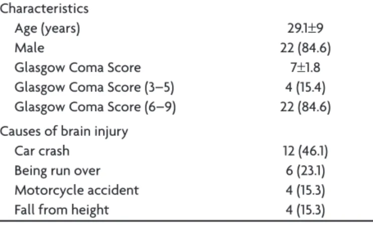
As presented on Table 1, most patients were young adults (18 to 54 years old), males and the most frequent causes of brain injury were car crash and being run over.
The most frequent injuries observed in the brain CT scan were brain swelling in 17 patients (65.3%), intracranial bleeds in 13 (50%), and brain contusion in 8 (30.7%).
The associated injuries were bone fracture in 11 pa-tients (42.3%), pulmonary contusion in 3 (11.5%), hemotho-rax and pneumothohemotho-rax each in 2 subjects (7.7%), liver inju-ry, urethra injuinju-ry, spinal trauma and nasal trauma, one case each (3.8%). Three deaths occurred (11.5%) and the causes were sepsis in two patients and brain death in one.
Six surgical procedures were performed (in parenthe-ses the number of procedures): craniotomy for
extradur-al hematoma draining (3), decompressive craniotomy (1), correction of open fracture (1), and amputation of right inferior limb (1).
The hemodynamic parameters in the seventh day fol-lowing the trauma are presented on Table 2. Cardiac fre-quency and maximum systolic and diastolic blood pres-sure above normal limits in all cases. The patients in the hyponatremia group showed systolic blood pressure low-er than the normonatremia group (p<0.05). The hypona-tremics, when compared with the normonatremia group, also showed higher serum potassium (p<0.01), higher di-uresis (p=0.01) and negative water balance (p<0.01) on days 6 and 7 following the trauma (Table 3).
Laboratory variable analysis
Hyponatremia (sodium ≤ 130 meq/l) was detected in 9 (34.6%) patients, totaling 20 episodes. It occurred between the second and the tenth day of trauma, more frequently on the 6th (four patients) and the 7th (ive patients).
Plas-ma sodium values lower than 125 meq/l were observed in three patients between days 5 and 9 following the trauma. The values of serum uric acid and osmolality were nor-mal, but hematocrit values on day 7 following the trauma were below normal (Table 3).
The values of BNP, aldosterone, vasopressin, plasma and urinary sodium, on the seventh day of study, were normal (Table 3).
Through multiple linear regression, a positive and sig-niicant relation was observed (p=0.01) between the offer of sodium and plasma sodium, that is, for each one unit increase of sodium offered there was an addition of 0.011 units in the average plasma sodium. Serum osmolality also showed a positive and signiicant correlation with plasma sodium (p<0.01), and each increase of a unit of osmolal-ity led to an addition of 0.45 units in the average plasma sodium. No relation was observed between plasma sodi-um and BNP, aldosterone, vasopressin, and diuresis or be-tween urinary sodium.
Table 1. Demographic characteristics of the 26 patients who have suffered brain injury. Percentages are presented in parentheses (%).
Characteristics Age (years) Male
Glasgow Coma Score Glasgow Coma Score (3–5) Glasgow Coma Score (6–9)
29.1±9 22 (84.6)
7±1.8 4 (15.4) 22 (84.6) Causes of brain injury
Car crash Being run over Motorcycle accident Fall from height
12 (46.1) 6 (23.1) 4 (15.3) 4 (15.3)
Table 2. Hemodynamic parameters in the seventh day following trauma in the 26 patients who have suffered brain injury.
Characteristics
Total n=26
Hyponatremia n=9
Normonatremia n=17 Maximum cardiac frequency (bpm) 119.8±17.8 117±14.5 121.2±19.6 Minimum cardiac frequency (bpm) 77.6±14.4 75.3±13.5 78.8±15.1 Maximum systolic blood pressure (mmHg) 154.5±18.7 145±16.3 159.3±18.5 a Minimum systolic blood pressure (mmHg) 112.3±15.5 110.8±12.3 113±17.2 Maximum diastolic blood pressure (mmHg) 95.9±18.3 91±15.4 98.5±19.5 Minimum diastolic blood pressure (mmHg) 61.4±11.8 59.7±8.5 62.2±13.5
Table 3. Dosages of brain natriuretic peptide, aldosterone, vasopressin, serum potassium, and hematocrit (7th day following trauma); uric
acid, and osmolality (1st, 5th, 7th and 10th day following trauma); diuresis, offer of sodium, water and sodium balance, plasma and urinary
sodium (6th and 7th day following trauma) in all 26 patients, the hyponatremia (n=9) and normonatremia (n=17) groups.
Variable
Total n=26
Hyponatremia n=9
Normonatremia n=17 Brain natriuretic peptide (pg/ml) (Vr: < 100 pg/ml) 17.7±16.1 21±20.3 16±13.8 Aldosterone (ng/ml) (Vr: 1–16 ng/100 ml) 7.5±11.6 8.6±10.9 6.9± 12.2 Vasopressin (pg/ml) (Vr: 0.4–2.4 pg/ml osm <285mosm/kg) 1.9±0.7 1.9±0.3 1.8±0.9 (Vr: 2 a 12 pg/mml osm>285 mosm/kg)
Potassium (meq/l) (Vr: 3.5–4.5meq/l) 4.1±0.4 4.4±0.3 3.9±0.2b
Plasma sodium (meq/l) (Vr: 136–145 meq/l) 137.2±6.7 128.4±3.2 141±4.6 Urinary sodium (meq/l) (Vr: 100–200 meq/l) 139.6±69.6 134.1±33 129±65 Uric acid (mg/dl) (Vr: < 7 mg/100dl (male) and < 6 mg/100dl (female)
Days
1 2.3±1.3 1.9±1.5 2.5±1.2
5 1.3±0.6 1.2±0.7 1.4±0.5
7 1.6±0.8 1.4±1 1.6±0.6
10 2.1±2.0 2.7± 3.2 1.8±1
osmolality (mosm/kg) (Vr: 290–313 mosml) Days
1 303.4±12.5 302±11 304±13.4
5 298±8.5 293.3±6.2 301±8.6
7 295.6±8.3 291.4±6.5 298±8.5
10 303.3±15.8 301±9.4 305±19.1
Hematocrit (Vr: male–47±7%. female–42±5%) 32.0±3.1 32.1±1.7 32±3.6 Diuresis (ml/h) 158.4±73.7 215.4±73.1 150.6±72a
Water balance (ml/24h) 394.1±1937.1 –1238±1493.3 663.4±1961b
Sodium balance (meq/day) 305.2±189.8 242.2±89.2 314.7±199 offer of sodium (meq/day) 444.7±171 376.3±87.8 444±187
Hyponatremia and normonatremia group: ap=0.01; bp<0.01
We did not observe a correlation between BNP and the following variables: aldosterone, plasma sodium and urinary sodium using Pearson’s linear correlation.
We observed that, with regard to the behavior of plas-ma sodium throughout the 10 days of study, patients from the hyponatremia group showed a decreasing linear trend
whereas the normonatremia group tended towards a con-stant behavior, as presented on Fig 1.
Figure 2 shows that there was basically no difference between the two groups in the behavior of diuresis that is, the values tended to increase at irst and to fall towards the end. However, average urinary volume in the
hypona-Fig 2. Diuresis during the 10 days of study in the group with hyponatremia (9) and normonatremia (17).
tremia group was clearly superior to the average values of the normonatremia group.
As for urinary sodium (Fig 3), the hyponatremia group averaged 177.2 meq/l in the irst day, whereas the patients with normonatremia presented 127.7 meq/l, a statistical-ly signiicant difference (p<0.05). The behavior of the two groups practically did not differ, but the hyponatremia group showed higher values compared to the normona-tremics.
The hyponatremic group showed a sodium average level slightly superior to the group with normonatremia throughout the 10 days, but that was not statistically sig-niicant (p=0.86). It was also observed that the values of this variable remained constant throughout the whole pe-riod of evaluation at both groups.
diSCuSSion
This study demonstrated that patients with severe brain injury who showed hyponatremia had superior val-ues of diuresis and natriuresis when compared to the nor-monatremia group, despite showing similar evolution of these variables throughout the 10 days of study. To our knowledge, this is the irst time that the behavior of na-triuresis and diuresis in patients who have suffered brain injury is detailed described.
Individuals that received an increased amount of so-dium also presented this response, with renal excretion of sodium rising for 4 to 5 days before matching the of-fer10. It is possible that the behavior of natriuresis was a
consequence to the high amount of sodium supplied for the patients in this study, a measure to prevent hypergly-cemia, which can itself worsen the prognosis of patients with brain injury11.
Diuresis had similar evolution in the two groups, that is, it tended to grow initially and to fall towards the end. Natriuresis started with high values, remained high and later fell, especially in the hyponatremia group. This be-havior was also described by other authors12. It can be
ex-plained because the reabsorption of water is linked to the reuptake of sodium. Thus, more reabsorption of sodium raises that of water in the proximal tubule, hence increas-ing urinary volume7.
We also observed hyponatremia (sodium <130 meq/l), in 9 (34.6%) patients. Hyponatremia following brain injury is frequent13 and in this study it occurred between the
sec-ond and the tenth day of trauma, more frequently on the sixth and seventh days as observed by other authors14.
The nine hyponatremic patients had the CSWS as a probable etiology because of brain injury, natriuresis and hyponatremia7. They showed higher urinary sodium and
volume, and a negative average water balance when hy-ponatremia occurred. The loss of water and natriuresis suggest a reduction on extracellular volume and, thus,
CSWS15. However, these patients did not present clinical
indings of dehydration, and that was not found by other authors either16, probably because of water balance
con-trol and replacement of losses. The sodium balance aver-aged positive in all patients of the group and can be ex-plained by the higher offer of sodium received17.
Hyponatremia following severe brain injury has been associated with high levels of BNP and ANP18. The increase
of these peptides must be a result of intracranial and sub-arachnoid hemorrhages and brain swelling18. However,
despite the fact that the majority of our patients have showed these injuries, an increase of BNP on the seventh day after severe brain injury was not observed. It is pos-sible that a single dosage during the 10 days did not re-lect the previously described evolution of a gradual in-crease in the concentration of BNP found on victims of severe brain injury19.
Two other possibilities can justify the lack of increase of BNP. The irst one would be that the offer of liquid and sodium was not enough to stimulate its secretion. Patients with subarachnoid hemorrhage who receive increased of-fer of liquid and sodium to prevent vasospasm18 showed
greater release of natriuretic peptides. Although our pa-tients showed positive water balance of 394.1±1,937.1 ml/ day, they did not receive such a high daily offer of liquids.
The second possibility would be that our patients were submitted to mechanical ventilation whose level of pos-itive end-expiratory pressure led to higher transpulmo-nary cardiac pressure, compromising atrial distension and, thereafter, the release of ANP20. Such mechanism would
be applied to BNP for the impairment of the cardiac ven-tricles distension21.
Besides the fact we did not observe an increase in the levels of BNP, it also did not correlate with the levels of sodium serum, and therefore did not correlate with CSWS. This data is in accordance with other authors who have also been unable to ind an association between levels of BNP or ANP and the presence of CSWS22.
We observed that the urinary sodium concentration was high, an average of 137.2±6.7 meq/l, and this could lead to the generation of electrolyte-free water23. Thus
the possible cause of hyponatremia presented in our pa-tients could be the free water generated by the kidneys, caused by the excretion of hypertonic urine, the so-called desalination process. This water then would be incorpo-rated into the intravascular volume, leading, thereafter, to hyponatremia. The antidiuretic hormone would be neces-sary for such incorporation.
lev-els24. Dopamine acts directly impairing the absorption of
NaCl and water in renal tubule, more precisely inhibiting the Na+-K+–ATPase, with its effect mediated by the D1 re-ceptor25. The renal production of dopamine increases with
a sodium-rich diet. Although our patients did not have the levels of dopamine measured, there is a possibility that they could have shown increased levels of dopamine caused by brain injury and by the increased amounts of sodium, leading to renal vasodilatation by the kidney and, as a consequence, natriuresis26.
Moreover, the patients received an increased infusion of sodium, which can also raise tensional levels and car-diac frequency. It is also possible that the observed in-crease in blood pressure in the present study could have contributed for the increase of natriuresis. In an exper-imental work, it was demonstrated that an increase in blood pressure leads to pressure natriuresis and diuresis, without, however, leading to changes in renal blood low or the rate of glomerular iltration27. Natriuresis occurs
due to decreased reabsorption of sodium in the proxi-mal tubule, through the internalization by endocytosis of Na+–H+ contra transporters from apical membrane and from Na+–K+–ATPase sodium pumps from basolat-eral membrane of the proximal tubule27. It was suggested
that in CSWS this process of pressure natriuresis, togeth-er with BNP or othtogeth-er existing natriuretic agents, can lead to the increase of urinary sodium concentration22.
It is worth mentioning that the patients with hypona-tremia showed lower values of systolic blood pressure when compared with the normonatremia group. This fact could have occurred due to the negative water balance, which could also explain the higher potassium levels ob-served in those cases.
our patients showed severe brain injury that could have led to injuries of the pituitary with consequent re-duction of growth hormone levels. This hormone acti-vates the renin-aldosterone system in children with low stature28, thus showing an anti-natriuretic function. The
possible growth hormone deiciency could have lead to changes in the renin-aldosterone system, eventualy result-ing in natriuresis, increased diuresis and hyponatremia.
The levels of uric acid were lower than 3 mg/dl, a val-ue that can be considered as hypouricemia10. A possible
explanation for the reduction of uric acid reabsorption is the fact that this, along with sodium, is reabsorbed proxi-mally in the nephron, and because the reabsorption of the latter is altered, uric acid would be more excreted15.
The average hematocrit was below normal because our patients are trauma victims29. The increased level of
sodium and positive water balance can also have contrib-uted for that inding7.
Considering that the patients of this study showed normal average values of osmolality and that the values
of vasopressin were normal, we can conclude that the pa-tients did not show SIADH. The same result was observed by Moro et al.7.
Plasma sodium showed a positive and signiicant re-lation with osmolality and offer of sodium. The positive correlation of these two variables is justiied by the fact that sodium, along with glucose and urea, contributes for the osmolality value30.
In conclusion, hyponatremia was more frequent in the sixth and seventh day after trauma and it was most like-ly caused by CSWS. The group that presented hypona-tremia had higher values of natriuresis and diuresis that could have been caused by pressure natriuresis and de-salination. There was no correlation between brain natri-uretic peptide, aldosterone and vasopressin with serum sodium level.
reFerenCeS
1. Casulari LA, Costa KN, Albuquerque RC, et al. Differential diagnosis and treatment of hyponatremia following pituitary surgery. J Neuro-surg Sci 2004;48:11-18.
2. Peters JP, Welt LG, Sims EAH, et al. A salt-wasting syndrome associat-ed with cerebral disease. Trans Assoc Am Physicians 1950;63:57-64. 3. Schwartz WB, Bennett W, Curelop S. A syndrome of renal sodium loss
and hyponatremia probably resulting from inappropriate secretion of antidiuretic hormone. Am J Med 1957;23:529-542.
4. Harrigan MH. Endocrine and metabolic dysfunction syndromes in the critically ill. Cerebral salt wasting syndrome. Crit Care Clin 2001;17: 125-138.
5. Sviri GE, Soustiel JF, Zaaroor M. Alteration in brain natriuretic peptide (BNP) plasma concentration following severe traumatic brain injury. Acta Neurochir (Wien) 2006;148:529-533.
6. Casulari LA, Borba AM, Lima BO, et al. Hiponatremia prolongada após traumatismo cerebral tratada com ludrocortisona. Brasília Med 2006;43:63-68.
7. Moro N, Katayama Y, Igarashi T, et al. Hyponatremia in patients with traumatic brain injury: incidence, mechanism, and response to sodium supplementation or retention therapy with hydrocortisone. Surg Neu-rol 2007;68:387-393.
8. Jennet B, Bond M. Assessment of outcome after severe brain damage. A practical scale. Lancet 1975;1:480-484.
9. Porto CC. Semiologia médica. 4. Ed. Rio de Janeiro, Guanabara Koogan, 2001.
10. Maesaka JK, Gupta S, Fishbane S. Cerebral salt wasting syndrome: Does it exist? Nephron 1999;82:100-109.
11. Vespa P, Boonyaputthikul R, McArthur DL, et al. Intensive insulin ther-apy reduces microdialysis glucose values without altering glucose uti-lization or improving the lactate/pyruvate ratio after traumatic brain injury. Crit Care Med 2006;34:850-856.
12. Mori T, Katayama Y, Kawamata T, et al. Improved eficiency of hy
-pervolemic therapy with inhibition of natriuresis by ludrocortisone in patients with aneurysmal subarachnoid hemorrhage. J Neurosurg 1999;91:947-952.
13. Donati-Genet PCM, Dubuis JM, Girardin E, et al. Acute symptomatic hyponatremia and cerebral salt wasting after head injury: an important clinical entity. J Pediatr Surg 2001;36:1094-1097.
14. Virgerhoets F, Tribolet N. Hyponatremia hypo-osmolarity in neurosur-gical patients “appropriate secretion of ADH” and “cerebral salt wast-ing syndrome”. Acta Neurochir (Wien) 1988;91:50-54.
syn-drome in children with acute central nervous system injury. Pediatr Neurol 2006;35:261-263.
17. Wijdicks EFM, Ropper AH, Hunnicutt EJ, et al. Atrial natriuretic factor and salt wasting after aneurismal subarachnoid hemorrhage. Stroke 1991;22:1519-1524.
18. Fukui S, Katoh H, Tsuzuki N, et al. Focal brain edema and natriuret-ic peptides in patients with subarachnoid hemorrhage. J Clin Neuros-ci 2004;11:507-511.
19. Kirchhoff C, Stegmaier J, Bogner V, et al. Intratecal and systemic con-centration of NT-proBNP in patients with severe traumatic brain inju-ry. J Neurotrauma 2006;23:943-949.
20. Costa KN, Carvalho WB, Kopelman BI, et al. Dosagem do fator natriu-rético atrial em pacientes pediátricos submetidos à ventilação mecâni-ca. Rev Ass Med Bras 2000;46:320-324.
21. Denus S, Pharand C, Williamson DR. Brain natriuretic peptide in the management of heart failure. Chest 2004;125:652-668.
22. Singh S, Bohn D, Carlotti AP, et al. Cerebral salt wasting: truths, falla-cies, theories, and challenges. Crit Care Med 2002;30:2575-2579. 23. Steele A, Gowrishankar M, Abrahamson S, et al. Postoperative
hypona-tremia despite near-isotonic saline infusion: A phenomenon of desali-nation. Ann Intern Med 1997;126:20-25.
24. Clifton GL, Ziegler MG, Grossman RG. Circulating catecholamines and sympathetic after head injury. Neurosurgery 1981;8:10-14.
25. Aperia A. Renal dopamine system and salt balance. Am J Kidney Dis 1998;31:xliii-xlv.
26. Israel A, Torres M, Cierco M, et al. Further evidence for a dopaminer-gic involvement in the renal action of centrally administered atrial na-triuretic peptide in rats. Brain Res Bull 1991;27:739-742.
27. Zhang Y, Mircheff AK, Hensley CB, et al. Rapid redistribution and in-hibition of renal sodium transporters during acute pressure natriure-sis. Am J Physiol 1996;270:F1004-F1014.
28. Papadimitriou DT, Spiteri A, Pagnier A, et al. Mineralocorticoid dei -ciency in post-operative cerebral salt wasting. J Pediat Endocrinol Me-tab 2007;20:1145-1150.
29. Betjes MGH. Hyponatremia in acute brain disease: the cerebral salt wasting syndrome. Eur J Intern Med 2002;13:9-14.